. 2018 Mar 16;13(3):035013.
doi: 10.1088/1748-605X/aaaa29.

Self-neutralizing PLGA/magnesium composites as novel biomaterials for tissue engineering

Affiliations

Self-neutralizing PLGA/magnesium composites as novel biomaterials for tissue engineering

Thomas O Xu et al. Biomed Mater. .

Abstract

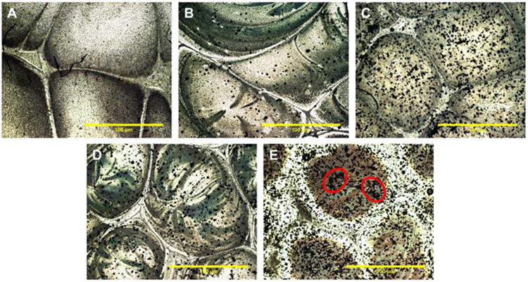
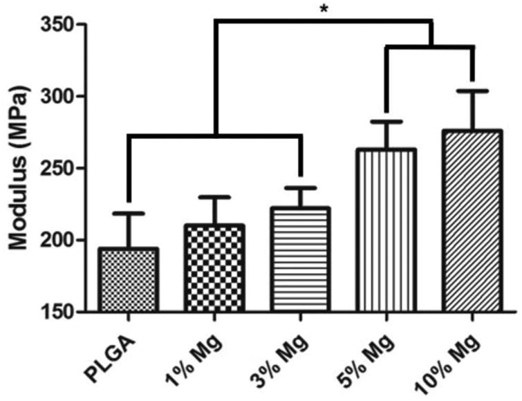
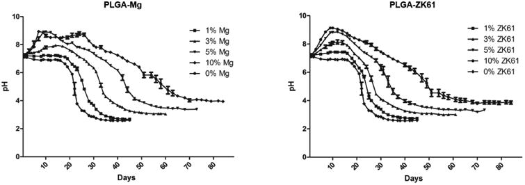
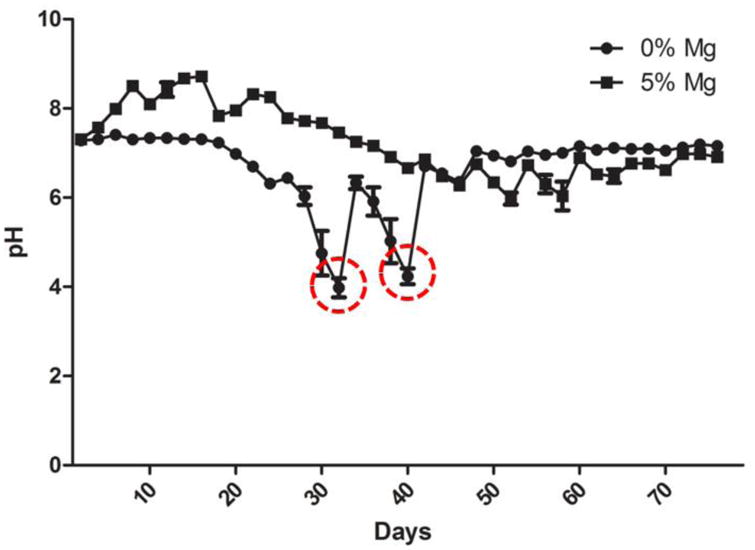
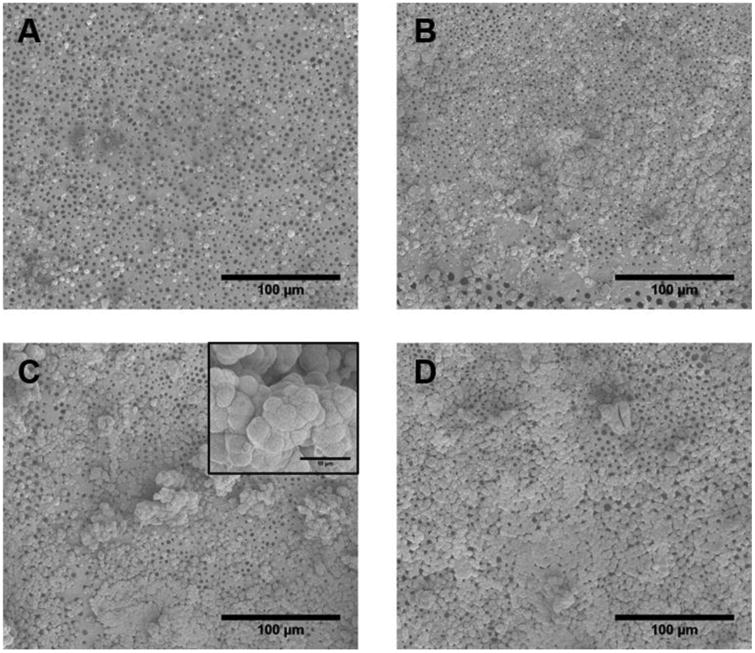
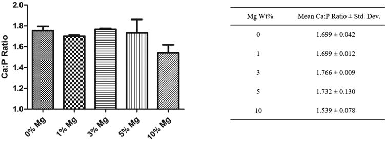
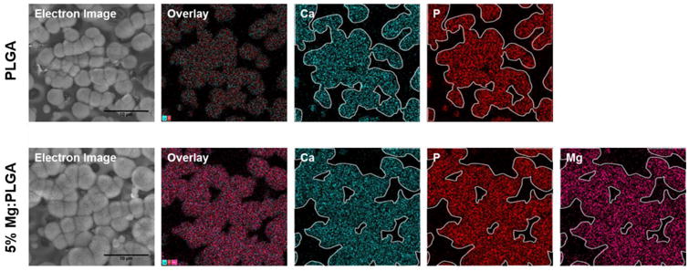
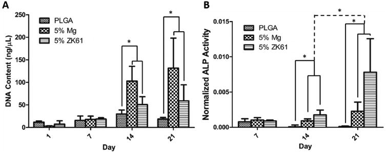
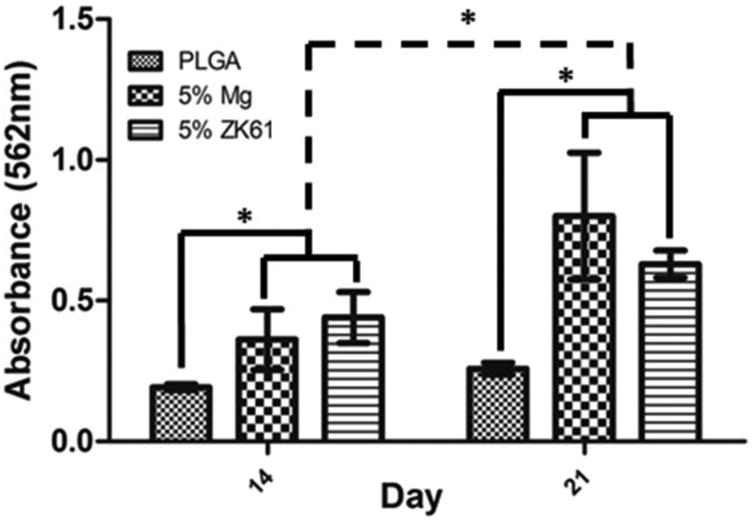
Controlling acidic degradation of biodegradable polyesters remains a major clinical challenge. This work presents a simple and effective strategy of developing polyester composites with biodegradable magnesium metal or alloys. PLGA samples with compositions of 1, 3, 5, and 10 wt% magnesium were produced using a simple solvent-casting method, which resulted in composite films with near uniform Mg metal/alloy particle dispersion. Degradation study of the composite films showed that all compositions higher than 1 wt% magnesium were able to extend the duration of degradation, and buffer acidic pH resulting from PLGA degradation. PLGA composite with 5 wt% of magnesium showed near-neutral degradation pattern under sink conditions. Magnesium addition also showed improved mechanical characteristics in terms of the tensile modulus. In vitro experiments conducted by seeding PLGA composites with MC3T3-E1 pre-osteoblasts demonstrated increased ALP expression and cellular mineralization. The established new biodegradable polymer-metal system provides a useful biomaterial platform with a wide range of applications in biomedical device development and scaffold-based tissue engineering.


Figures

Figure 1
Figure 1
Magnesium particle dispersion in PLGA-Mg composite films. Representative optical micrographs of (A) PLGA only, and PLGA-Mg composites with (B) 1 wt% Mg, (C) 3 wt% Mg, (D) 5 wt% Mg, and (E) 10 wt% Mg. Mg particles, seen in black, are evenly dispersed throughout the film in concentration ranging from 0-5 wt%. Traces of Mg particle agglomeration was seen in PLGA-Mg composite films with 10wt% of Mg (circled regions in E show Mg particle agglomeration).
Figure 2
Figure 2
Effect of Mg loading on tensile modulus of the composite films. PLGA-Mg composite films with varying Mg wt% loading were subjected for mechanical strength measurement in tensile mode. An increase in tensile modulus of the composites was seen with increase in Mg loading. The increase in the modulus is statistically significant for the composite groups of 3 and 5 wt% of Mg (n=6; * = p <0.05).
Figure 3
Figure 3
In vitro degradation profile of composite films. PLGA-Mg films of different Mg concentrations were subjected to degradation at elevated temperature 47 C in PBS. (A) degradation profile of composites using pure Mg particles. (B) Degradation profile of films containing with the Zn-Mg alloy ZK61. For both pure Mg and Zn-Mg alloys, near neutral or above pH 7 conditions were maintained for longer than the pure PLGA samples and extended the duration of degradation. Initial increase in pH was also observed in samples containing Mg or ZK61 alloy, that varied in intensity relative to the concentration of Mg or Zn-Mg alloy.
Figure 4
Figure 4
Dynamic or sink condition degradation profile of PLGA-Mg films. In vivo fluid clearance is simulated in vitro for the degradation of the PLGA-Mg composites at 47 C in PBS. Degradation media was changed every 7 days to simulate the normal sink conditions in vivo. The PLGA-Mg composites displayed a steady near neutral conditions throughout the duration of the study. The PLGA alone group displayed sharp decreases in pH (highlighted within red circles).
Figure 5
Figure 5
In vitro biomineralization of the PLGA-Mg composites. The samples were immersed in 1.5 × for 14 days. Representative SEM micrographs show the deposited biomineral for (A) PLGA, and (B-D) for composites with Mg content of 1, 3, and 5 wt%, respectively. Insert in C shows representative close-up of apatite nucleates (Scale bar 10 μM). PLGA sample showed small isolated nucleation of calcium apatite. The amount of apatite nucleation increased with Mg concentration in composites, with 5 wt% samples displaying the greatest amount of apatite nucleates with greater amount of surface coverage.
Figure 6
Figure 6
Biomineral composition and Ca to P ratio analysis. Mean Ca to P atomic ratio was obtained from EDS analysis of the precipitates on the composite films. PLGA and PLGA-Mg samples showed similar Ca:P ratio and did not differ with the amount of Mg loading. (n=3).
Figure 7
Figure 7
Effect of Mg composition on in vitro biomineralization. EDS elemental analysis of the PLGA and PLGA-Mg composites after 14 day immersion in 1.5× SBF. The presence of Ca (outlined in white and overlaid in other groups) and P groups were detected in the nucleates for both polymer and composite groups, indicating formation of calcium apatite. The precipitation of calcium apatite coincided with the presence of Mg in the PLGA-Mg composite films, indicating a relationship between the increased local Mg concentration and the calcium apatite nucleation and growth.
Figure 8
Figure 8
In vitro biocompatibility. Mouse pre-osteoblastic (MC3T3-E1) cell performance and osteogenic differentiation on PLGA, PLGA-Mg, and PLGA-ZK61 was assessed through DNA quantification and ALP expression. (A) Cellular proliferation was analyzed using Picogreen dsDNA assay. Increased cell proliferation for Mg and ZK61 samples was observed at days 14 and 21, with little change in DNA concentration in PLGA group throughout the study. (B) ALP expression was analyzed using an ALP assay kit and normalized to DNA concentration. Increased ALP expression was seen between PLGA and the composite groups at days 14 and 21. Significant increase in ALP expression between day 14 and 21 was also seen for the PLGA-Mg and PLGA-ZK61 samples (n=3* = p <0.05).
Figure 9
Figure 9
In vitro mineralization via Alizarin red staining and quantification. MC3T3-E1 pre-osteoblastic cell induced mineralization for the PLGA and PLGA-Mg composite groups after 14 and 21 days in vitro. Mg or ZK61 alloy composite groups displayed significant increases in mineral formation in comparison to PLGA alone group at 14 and 21 days. The PLGA-Mg and PLGA-ZK61 samples also showed significant increase in mineralization between day 14 and 21 (n=3* = p<0.05).

References

    1. Kumbar SG, Laurencin CT, Deng M. Natural and Synthetic Biomedical Polymers. Oxford: Elsevier; 2014.
    1. Gunatillake PA, Adhikari R. Biodegradable synthetic polymers for tissue engineering. Eur Cell Mater. 5:1–16. discussion 16, May 2003. - PubMed
    1. Nukavarapu SP, Kumbar SG, Brown JL, Krogman NR, Weikel AL, Hindenlang MD, Nair LS, Allcock HR, Laurencin CT. Polyphosphazene/nano-hydroxyapatite composite microsphere scaffolds for bone tissue engineering. Biomacromolecules. 2008 Jul;9(7):1818–1825. - PMC - PubMed
    1. Hutmacher DW. Scaffolds in tissue engineering bone and cartilage. Biomaterials. 2000 Dec;21(24):2529–2543. - PubMed
    1. Nukavarapu SP, Freeman J, Laurencin CT. Regenerative Engineering of Musculoskeletal Tissues and Interfaces. Elsevier Science & Technology; 2015.

Publication types

MeSH terms

LinkOut - more resources