. 2018 Jan 24:24:453-460.
doi: 10.12659/msm.905319.

Role of Xeroderma Pigmentosum Group D in Cell Cycle and Apoptosis in Cutaneous Squamous Cell Carcinoma A431 Cells

Affiliations

Role of Xeroderma Pigmentosum Group D in Cell Cycle and Apoptosis in Cutaneous Squamous Cell Carcinoma A431 Cells

Ou-Gen Liu et al. Med Sci Monit. .

Abstract

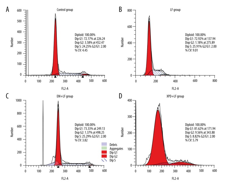
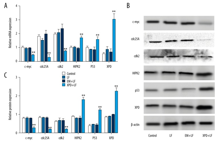
BACKGROUND Cutaneous squamous cell carcinoma (cSCC) is the second most widespread cancer in humans and its incidence is rising. Novel therapy with better efficacy is needed for clinical treatment of cSCC. Many studies have shown the importance of DNA repair pathways during the development of cancer. A key nucleotide excision repair (NER) protein, xeroderma pigmentosum group D (XPD), is responsible for the excision of a large variety of bulky DNA lesions. MATERIAL AND METHODS To explore the role of XPD in A431 cells, we overexpressed XPD in A431 cells and performed MTT assay, flow cytometry, and Western blot analysis to examine cell proliferation, cell apoptosis, and genes expression. RESULTS We found that the overexpression of XPD suppressed cell viability, induced cell cycle arrest at G1 phase, and promoted cell apoptosis. Additionally, XPD blocked the expression of c-myc, cdc25A, and cdk2, and improved the levels of HIPK2 and p53. CONCLUSIONS These results provide new evidence to reveal the role of XPD in cSCC A431 cells and suggest that XPD may serve as an anti-oncogene during cSCC development.


Conflict of interest statement

Conflicts of interest

None.

Figures

Figure 1
Figure 1
XPD repressed cell proliferation. A431 cells were divided into 4 groups, and the control group was administered the same amount of medium (Control group). The other 3 groups were transfected with Lipofectamine (LF group), pEGFP-N2 (empty vector) + Lipofectamine (EM+LF group), or pEGFP-N2-XPD + Lipofectamine (XPD+LF group). (A) The signals of XPD were detected after pEGFP-N2-XPD transfection. After cell transfection, A431 cells were observed with an inverted fluorescence microscope. Scale bar=100 μm. (B) The mRNA level of XPD was increased in the XPD+LF group. Total RNA was isolated from 4 groups for QRT-PCR analysis. * P<0.05, ** P<0.01. (C) The protein level of XPD was enhanced in the XPD+LF group. After cell transfection, proteins were extracted for Western blot analysis. * P<0.05, ** P<0.01. (D) The overexpression of XPD blocked cell proliferation. After cell transfection, cell proliferation was examined by MTT assay. * P<0.05, ** P<0.01.
Figure 2
Figure 2
XPD induced cell cycle arrest in G1 phase. A431 cells were transfected with the same amount of medium (Control, A), Lipofectamine (LF, B), pEGFP-N2 (EM) + Lipofectamine (EM+LF group, C), or pEGFP-N2-XPD + Lipofectamine (XPD+LF group, D). Cell cycle distribution was measured using flow cytometry assay.
Figure 3
Figure 3
The overexpression of XPD promoted cell apoptosis. A431 cells were transfected with the same amount of medium (Control, A), Lipofectamine (LF, B), pEGFP-N2 (EM) + Lipofectamine (EM+LF, C), or pEGFP-N2-XPD + Lipofectamine (XPD+LF, D). Cells that stained positive for early apoptosis markers (annexin V-FITC stained only) and for late apoptosis markers (annexin V-FITC and PI stained) were combined for analysis.
Figure 4
Figure 4
XPD monitored the related genes expression. A431 cells were transfected with the same amount of medium (Control), Lipofectamine (LF), pEGFP-N2 (EM) + Lipofectamine (EM+LF), or pEGFP-N2-XPD + Lipofectamine (XPD+LF). (A) The overexpression of XPD affected the mRNA levels of related genes for cell proliferation and cell apoptosis. After cell transfection, cells were collected and total RNA was isolated for qRT-PCR. (B) The effect of XPD overexpression on related proteins expression was analyzed by Western blot. (C) The target bands were normalized to β-actin using Quantity One software. * P<0.05, ** P<0.01.

References

    1. Youssef KK, Van Keymeulen A, Lapouge G, et al. Identification of the cell lineage at the origin of basal cell carcinoma. Nat Cell Biol. 2010;12:299–305. - PubMed
    1. Zhang SH, Li ZY, Liu ZJ, et al. MicroRNA15b regulates apoptosis of cutaneous squamous cell carcinoma SCL-1 cell line: A mechanism study. Eur Rev Med Pharmacol Sci. 2017;21:227–33. - PubMed
    1. Li X, Huang K, Yu J. Inhibition of microRNA-21 upregulates the expression of programmed cell death 4 and phosphatase tensin homologue in the A431 squamous cell carcinoma cell line. Oncol Lett. 2014;8:203–7. - PMC - PubMed
    1. Bertero T, Bourget-Ponzio I, Puissant A, et al. Tumor suppressor function of miR-483-3p on squamous cell carcinomas due to its pro-apoptotic properties. Cell Cycle. 2013;12:2183–93. - PMC - PubMed
    1. Ravegnini G, Nannini M, Simeon V, et al. Polymorphisms in DNA repair genes in gastrointestinal stromal tumours: Susceptibility and correlation with tumour characteristics and clinical outcome. Tumour Biol. 2016;37:13413–23. - PubMed

MeSH terms

Supplementary concepts