. 2018 Jan 23;9(2):81.
doi: 10.1038/s41419-017-0145-x.

Mitochondria are transported along microtubules in membrane nanotubes to rescue distressed cardiomyocytes from apoptosis

Affiliations

Mitochondria are transported along microtubules in membrane nanotubes to rescue distressed cardiomyocytes from apoptosis

Jing Shen et al. Cell Death Dis. .

Abstract

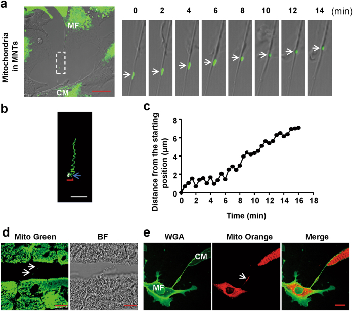
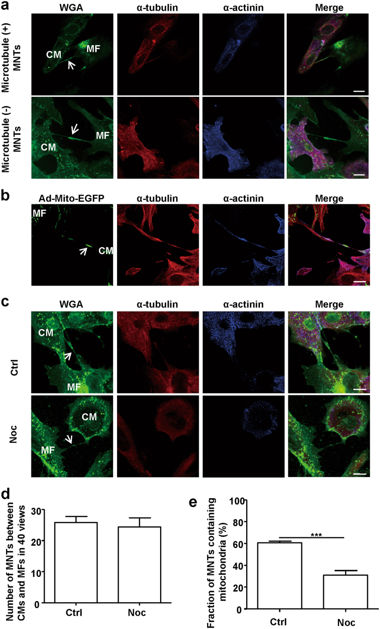
Membrane nanotubes (MNTs) act as "highways" between cells to facilitate the transfer of multiple signals and play an important role in many diseases. Our previous work reported on the transfer of mitochondria via MNTs between cardiomyocytes (CMs) and cardiac myofibroblasts (MFs); however, the elucidation of the underlying mechanism and pathophysiological significance of this transfer requires additional study. In this study, we determined that the mean movement velocity of mitochondria in MNTs between CMs and MFs was approximately 17.5 ± 2.1 nm/s. Meanwhile, treatment with microtubule polymerisation inhibitors nocodazole or colcemid in cell culture decreased mitochondrial velocity, and knockdown of the microtubule motor protein kinesin family member 5B (KIF5B) led to a similar effect, indicating that mitochondrial movement was dependent on microtubules and the motor protein KIF5B. Furthermore, we showed that hypoxia/reoxygenation-induced CM apoptosis was attenuated by coculture with intact or hypoxia/reoxygenation-treated MFs, which transferred mitochondria to CMs. This rescue was prevented either by separating the cells using Transwell culture or by impairing mitochondrial transfer with nocodazole or colcemid treatment. In conclusion, as a novel means of intercellular communication, MNTs rescue distressed CMs from apoptosis by transporting mitochondria along microtubules via KIF5B.


Conflict of interest statement

The authors declare that they have no conflict of interest.

Figures

Fig. 1
Fig. 1. Mitochondrial movement in MNTs between neonatal rat ventricular CMs and MFs
a A series of confocal images from a time-lapse movie (Online supplementary material, movie 1) show the movement of the EGFP-labelled mitochondrion (Ad-Mito-EGFP, arrows) between two cells via a MNT. Scale bar: 20 μm. b The mobile trajectory of a representative mitochondrion in a MNT in (a). The arrow points to the start point, and the red number “1” was automatically generated by the analysis software to specify the analysed mitochondrion. Scale bar: 5 μm. c Distance changes over time (relative to the starting point) of the mitochondrion in (a). d Confocal micrographs of adult rat heart frozen sections stained with MitoTracker Green (Mito Green). White arrows indicate the mitochondria within MNTs. BF, bright field. N = 5. Scale bar: 10 μm. e Cocultured adult CMs and MFs. Membrane and mitochondria were labelled by Alexa Fluor 488-conjugated WGA (WGA, green) and MitoTracker Orange (Mito Orange), respectively. White arrows indicate the mitochondria within the MNTs. Scale bar: 20 μm
Fig. 2
Fig. 2. Microtubules are required for mitochondrial localisation in MNTs between CMs and MFs
a Cocultured CM and MF membranes were labelled by WGA (green), microtubules were labelled by α-tubulin antibodies (red) and CMs were labelled by CM-specific sarcomeric α-actinin antibodies (blue). Arrows indicate MNTs with (+) and without (−) microtubules. N = 5. Scale bar: 10 μm. b Cocultured CMs and MFs were transfected with Ad-Mito-EGFP (green) and then stained with antibodies against α-tubulin (red) and α-actinin (blue). Microtubules were observed in all mitochondria-containing MNTs (n = 36 from 3 independent experiments). The arrow points to a mitochondrion in the MNT. Scale bar: 20 μm. c Cocultured CMs and MFs were triple labelled by WGA, α-tubulin and α-actinin and then treated with or without the microtubule polymerisation inhibitor nocodazole (Noc). WGA-labelled MNTs were still detectable (arrows) in the cells treated with nocodazole. Ctrl, control. Scale bar: 10 μm. d Quantification of 40 randomly selected fields showing a difference in the number of MNTs between CMs and MFs in the control and nocodazole treatment groups (N = 5). e A fraction of mitochondria-containing MNTs between CMs and MFs was significantly decreased when the cells were treated with nocodazole (N = 5). Data are shown as the mean ± S.E.M. ***P < 0.001 using Student’s unpaired two-tailed t-test
Fig. 3
Fig. 3. Microtubules are essential for mitochondrial movement in MNTs between CMs and MFs
a A series of confocal images from time-lapse movies (Online supplementary material, movies 2 and 3) show the movement of Ad-Mito-EGFP-labelled mitochondria (arrows) in MNTs between CMs and MFs, with or without nocodazole treatment. Scale bar: 20 μm. b Mean movement velocities of mobile mitochondria in MNTs, with or without nocodazole treatment (n = 13 mitochondria from 3 independent experiments). c The mobile trajectories of mitochondria in MNTs (in (a)) were shorter after nocodazole treatment. The trajectories are represented by green lines, and the arrows point to the starting points. Scale bar: 7.5 μm. d Distance changes over time (relative to the starting point) of the mitochondria are shown in (a). Data are shown as the mean ± S.E.M. ***P < 0.001 using Welch’s t-test
Fig. 4
Fig. 4. KIF5B is the motor protein involved in the transfer of mitochondria in MNTs
a Cocultured CMs and MFs were triple labelled for KIF5B (red), WGA (membrane marker, green) and α-actinin (CM marker, blue). KIF5B was observed in CMs and MFs, as well as in MNTs between the two cell types (arrow). Scale bar: 50 μm. b A series of confocal images from the time-lapse movies (Online supplementary material, movies 4-6) show the movement of Ad-Mito-EGFP-labelled mitochondria in MNTs between cells treated with scramble siRNA (Scr) or KIF5B siRNA1/KIF5B siRNA2. RFP-positive cells (red) indicate the successful transfection of lentivirus carrying siRNA, and EGFP-positive bodies (green) are mitochondria. Scale bar: 20 μm. c Quantification of the mean movement velocities of Ad-Mito-EGFP-labelled mitochondria in MNTs in cells, with or without KIF5B depletion (Scr: n = 7 mitochondria; siRNA1: n = 5 mitochondria; and siRNA2: n = 13 mitochondria, from 3 independent experiments). d The mobile trajectories of mitochondria in MNTs (in (b)) were shorter after KIF5B depletion. The trajectories from different mitochondria are represented as different coloured lines, and the red number was automatically generated by the analysis software to specify the mitochondrion analysed. Scale bar: 5 μm. e Distance changes over time (relative to the original point) of mitochondria are shown in (b). Data are shown as the mean ± S.E.M. *** P < 0. 001 using one-way ANOVA with Tukey’s post hoc test
Fig. 5
Fig. 5. H/R-induced CM apoptosis can be rescued by the MNT-mediated transfer of mitochondria from intact MFs
a With or without prior hypoxia treatment for 6 h, CMs were cocultured with Mito Orange (red)-labelled MFs for 6 h under normoxic conditions. Cocultured CMs and MFs were stained with WGA (membrane marker, green) and α-actinin (CM marker, blue). Mito Orange staining was observed in CMs, suggesting mitochondrial transfer from MFs to CMs. Scale bar: 50 μm. b CMs treated with hypoxia for 6 h were then cocultured with Ad-Mito-EGFP-transfected MFs. A series of confocal images show the transfer of mitochondria (arrows) from MFs to CMs within MNTs. Scale bar: 10 μm. c Schematic representation of the cell treatments. d H/R-induced CM apoptosis was identified by TUNEL staining (green) and high-content screening imaging. CMs were differentiated by α-actinin staining (red). CoC, coculture. Scale bar: 100 μm. e Quantification of the fraction of apoptotic CMs by high-content screening (N = 5). Data are shown as the mean ± S.E.M. **P < 0. 01 and ***P < 0. 001 using one-way ANOVA with Tukey’s post hoc test
Fig. 6
Fig. 6. H/R-induced CM apoptosis can be rescued by the MNT-mediated transfer of mitochondria from H/R-treated MFs
a H/R-induced apoptotic CMs and MFs were identified by TUNEL staining (green) and high-content screening imaging. Scale bar: 100 μm. b Quantification of the apoptosis ratio of CMs by high-content screening (N = 6). c Quantification of the apoptosis ratio of MFs by high-content screening (N = 6). d Schematic representation of the cell treatments. MFs were cocultured with CMs 16 h prior to the H/R treatment. e H/R-induced apoptotic CMs were identified by TUNEL staining (green), and CMs were differentiated by α-actinin staining (red) and high-content screening imaging. Scale bar: 100 μm. f Quantification of the apoptosis ratio of CMs by high-content screening (N = 6). Data are shown as the mean ± S.E.M. *P < 0. 05 and ***P < 0. 001 using Student’s unpaired two-tailed t-test or one-way ANOVA with Tukey’s post hoc test
Fig. 7
Fig. 7. A model of mitochondrial transfer in MNTs between CMs and MFs
MNTs physically connect CMs and MFs. Mitochondria are transported along microtubules by the motor protein KIF5B in MNTs and can rescue H/R-induced CM apoptosis. F-actin provides the basic structure of MNTs

References

    1. Baudino TA, et al. Cell patterning: interaction of cardiac myocytes and fibroblasts in three-dimensional culture. Microsc. Microanal. 2008;14:117–125. doi: 10.1017/S1431927608080021. - DOI - PubMed
    1. Porter KE, Turner NA. Cardiac fibroblasts: at the heart of myocardial remodeling. Pharmacol. Ther. 2009;123:255–278. doi: 10.1016/j.pharmthera.2009.05.002. - DOI - PubMed
    1. Zhang J, Zhang Y. Membrane nanotubes: novel communication between distant cells. Sci. China Life Sci. 2013;56:994–999. doi: 10.1007/s11427-013-4548-3. - DOI - PubMed
    1. Burtey A, et al. Intercellular transfer of transferrin receptor by a contact-, Rab8-dependent mechanism involving tunneling nanotubes. FASEB J. 2015;29:4695–4712. doi: 10.1096/fj.14-268615. - DOI - PubMed
    1. Rustom A, Saffrich R, Markovic I, Walther P, Gerdes HH. Nanotubular highways for intercellular organelle transport. Science. 2004;303:1007–1010. doi: 10.1126/science.1093133. - DOI - PubMed

Publication types

LinkOut - more resources