. 2018 Jan 23;9(1):341.
doi: 10.1038/s41467-017-02661-3.

Lack of beta-arrestin signaling in the absence of active G proteins

Affiliations

Lack of beta-arrestin signaling in the absence of active G proteins

Manuel Grundmann et al. Nat Commun. .

Abstract

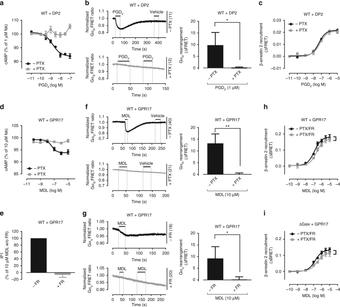
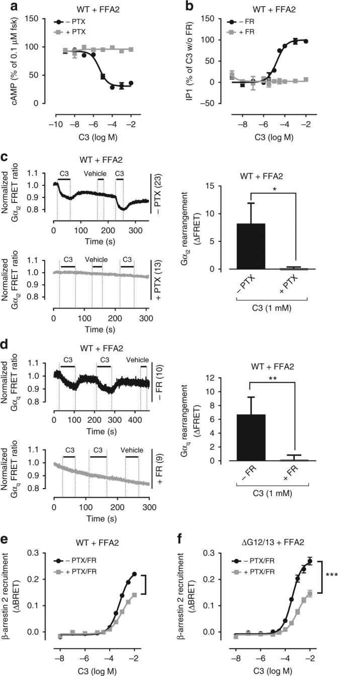
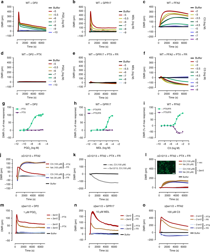
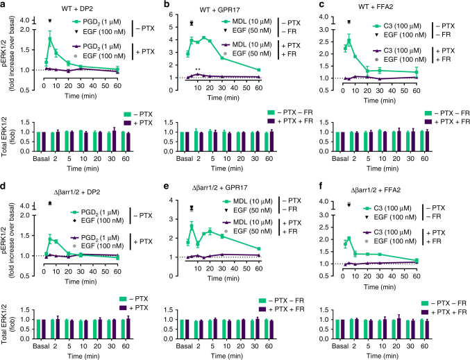
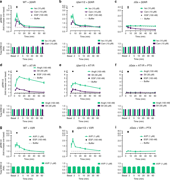
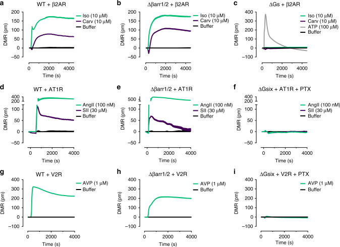
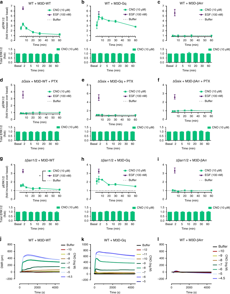
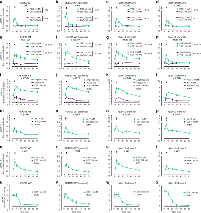
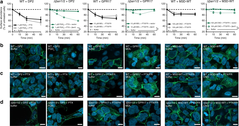
G protein-independent, arrestin-dependent signaling is a paradigm that broadens the signaling scope of G protein-coupled receptors (GPCRs) beyond G proteins for numerous biological processes. However, arrestin signaling in the collective absence of functional G proteins has never been demonstrated. Here we achieve a state of "zero functional G" at the cellular level using HEK293 cells depleted by CRISPR/Cas9 technology of the Gs/q/12 families of Gα proteins, along with pertussis toxin-mediated inactivation of Gi/o. Together with HEK293 cells lacking β-arrestins ("zero arrestin"), we systematically dissect G protein- from arrestin-driven signaling outcomes for a broad set of GPCRs. We use biochemical, biophysical, label-free whole-cell biosensing and ERK phosphorylation to identify four salient features for all receptors at "zero functional G": arrestin recruitment and internalization, but-unexpectedly-complete failure to activate ERK and whole-cell responses. These findings change our understanding of how GPCRs function and in particular of how they activate ERK1/2.


Conflict of interest statement

The authors declare no competing financial interests.

Figures

Fig. 1
Fig. 1
G protein activation and βarr2 recruitment in absence and presence of active G proteins. a PGD2-mediated depression of cAMP production in DP2-HEK293 cells, in the absence or presence of PTX. b Representative real-time FRET ratios and summary of Gi2 protein rearrangement after stimulation with PGD2 in DP2 expressing wild-type (WT) cells in absence and presence of PTX. c β-arrestin2 recruitment to PGD2-stimulated DP2 receptors, in the absence or presence of PTX. d Effect of MDL on the production of cAMP by Gi/q-linked GPR17, in the absence or presence of PTX. e IP1 accumulation of MDL-activated GPR17, in the presence or absence of FR. fg Gi2 (f) and Gq (g) protein rearrangement in GPR17 expressing wild-type (WT) cells stimulated with MDL in absence and presence of G protein inhibitors. h, i β-arrestin2 recruitment to MDL-stimulated GPR17 when Gi/q is inhibited with PTX and FR (h) or when the entire complement of cellular Gα proteins (ΔGsix+PTX) is inactivated (i). Shown are representative FRET traces with the indicated number of total cells per condition (b, f, g) and bar diagrams are averages from three independent experiments. Data are mean ± s.e.m. (a, c, d, h, i) or + s.d. (b, e, f, g) of three (ag, i) and four (h) experiments (three technical replicates each). For statistical analysis, two-tailed unpaired t-test (b, f, g) and two-sample paired Wilcoxon test (h, i) was applied to paired points at different concentrations. *P < 0.05; **P < 0.01; not significant where no asterisk
Fig. 2
Fig. 2
G protein activation and βarr2 recruitment in absence and presence of active G proteins. a Inhibition of forskolin-mediated cAMP accumulation by C3-activated FFA2 in FFA2-HEK293 cells, in the presence or absence of PTX. b IP1 accumulation of C3-stimulated FFA2, in the presence or absence of FR. c, d FRET measurements of Gi (c) and Gq (d) activation-induced conformational changes in wild-type (WT) HEK293 cells expressing FFA2 after stimulation with C3 in absence and presence of G protein inhibitors. e, f C3-induced β-arrestin2 recruitment to FFA2 in wild-type (WT) (e) or ΔG12/13 HEK293 cells (f), in the absence and presence of PTX and FR. Shown are representative FRET traces with the indicated number of total cells per condition (c, d) and bar diagrams are averages from three independent experiments. Data are mean ± s.e.m. (a, b, e, f) or + s.d. (c, d) of three experiments each performed in triplicate. For statistical analysis, two-tailed unpaired t-test (c, d) and two-sample paired Wilcoxon test (e, f) was applied to paired points at different concentrations. *P < 0.05; **P < 0.01; ***P < 0.001; not significant where no asterisk
Fig. 3
Fig. 3
DMR biosensing of G protein- and arrestin-mediated cellular responses. DMR recordings in absence (ac) and presence of G protein inhibitors (df) of agonist-mediated DP2 (a, d), GPR17 (b, e) and FFA2 (c, f) in wild-type (WT) HEK293 cells. Concentration-response-curves of DMR peak values in absence and presence of G protein inhibitors of DP2 (g), GPR17 (h), and FFA2 (i) response in wild-type HEK293 cells. j C3-mediated and forskolin-mediated DMR response of FFA2 in ΔG12/13 cells in the absence and presence of PTX/FR. k C3-induced DMR response of FFA2 in vector- or Gα12/13-transfected ΔG12/13 cells. l C3- or forskolin-mediated DMR response of FFA2 in vector- or β-arrestin2-transfected ΔG12/13 cells in the presence of PTX/FR. Inset shows βarr2-GFP transfected ΔG12/13 cells, scale bar 50 µm. mo Agonist-induced DMR response of DP2 (m), GPR17 (n), or FFA2 (o) in vector- or β-arrestin2-transfected Δβarr1/2 cells in absence and presence of G protein inhibitors. af, jo Shown are representative traces (mean + s.e.m.) of three independent experiments, each performed in triplicate. gi Data are mean ± s.e.m. of three independent experiments (three technical replicates)
Fig. 4
Fig. 4
G protein vs. βarr1/2 dependence of ligand-stimulated ERK1/2 phosphorylation. ac Kinetic pERK1/2 and total ERK1/2 profile of PGD2-stimulated DP2 (a), MDL-activated GPR17 (b), and C3-stimulated FFA2 (c) in the absence and presence of PTX and FR in wild-type (WT) HEK293 cells. df Temporal pattern of pERK1/2 and total ERK1/2 for PGD2-stimulated DP2 (d), MDL-stimulated GPR17 (e), and C3-stimulated FFA2 (f) in Δβarr1/2 cells in the absence and presence of PTX and FR. Data are mean +/± s.e.m. of three (b, c, f) and four (a, d, e) independent experiments (3 technical replicates each). For statistical analysis, one-sample t-test (b) was performed. **P < 0.01
Fig. 5
Fig. 5
pERK1/2 profiles of ligand-activated β2AR, AT1R, and V2R in WT and Δβarr1/2 cells. ac Temporal pattern of ERK1/2 phosphorylation and total ERK1/2 by β2AR in wild-type (a), Δβarr1/2 (b), and ΔGs (c) HEK293 cells treated with the indicated ligands. df Temporal pattern of ERK1/2 phosphorylation and total ERK1/2 by ligand-activated AT1R in wild-type (d), Δβarr1/2 (e), and ΔGsix cells+PTX (f). gi Kinetic pERK1/2 and total ERK1/2 profile of V2R in wild-type (g), Δβarr1/2 (h), and ΔGsix cells+PTX (i). Data are mean +/± s.e.m. of three (g, h, i), four (ad, f) and five (e) experiments each performed in triplicate
Fig. 6
Fig. 6
DMR profiles of ligand-activated β2AR, AT1R, and V2R in WT and Δβarr1/2 HEK cells. ac DMR recordings of β2AR in wild-type (a), Δβarr1/2 (b), and ΔGs (c) cells treated with Isoproterenol (Iso) or Carvedilol (Carv). df DMR recordings of AT1R wild-type (d), Δβarr1/2 (e) and ΔGsix cells+PTX (f) treated with AngII or SII. gi DMR phenotyping of V2R in wild-type (g), Δβarr1/2 (h), and ΔGsix cells+PTX (i). DMR traces shown are representative (mean + s.e.m.) for one out of three independent experiments, each performed in triplicate
Fig. 7
Fig. 7
Designer receptor-mediated ERK1/2 activation and DMR response. ac Clozapine-N-oxide (CNO)- and epidermal growth factor (EGF)-mediated ERK1/2 phosphorylation and total ERK1/2 in wild-type HEK293 cells (WT) expressing M3D-WT (a), M3D-Gq (b) or M3D-βArr (c). df CNO- and EGF-mediated ERK1/2 phosphorylation and total ERK1/2 in G protein-deficient (ΔGsix+PTX) HEK293 cells by M3D-WT (d), M3D-Gq (e) or M3D-β-arr (f). gi CNO-mediated and EGF-mediated ERK1/2 phosphorylation and total ERK1/2 in β-arrestin-deficient (Δβarr1/2) cells by M3D-WT (g), M3D-Gq (h) or M3D-βArr (i). jl Real-time DMR traces in CNO-stimulated cells expressing M3D-WT (j), M3D-Gq (k) or M3D-βArr (l). ai Data are mean +/± s.e.m. of three independent experiments (three technical replicates). jl Shown are representative traces (mean + s.e.m.) of three independent experiments, each measured in triplicates. For statistical analysis, two-sample paired Wilcoxon test (g) was applied to paired points at different times. *P < 0.05
Fig. 8
Fig. 8
Ligand-stimulated pERK1/2 profiles in wild-type HEK293 and Δβarr1/2 cells. Kinetic pERK1/2 profiles of agonist-stimulated DP2 (ad), M3D-WT (eh), AT1R (il), β2AR (mp), V2R (qt), and GPCR-independent MAPK pathway activation by EGF (ux) in wild-type (WT) (a, e, i, m, q, u), wild-type (WT) (parental) (b, f, j, n, r, v), Δβarr1/2 clone #2 (c, g, k, o, s, w), or Δβarr1/2 clone #3 (d, h, l, p, t, x). Data are mean ± s.e.m. of three independent experiments (three technical replicates)
Fig. 9
Fig. 9
Ligand-mediated GPCR internalization in the absence of G proteins and/or arrestins. a Kinetic surface ELISA of buffer-treated or agonist-treated HEK293 cells expressing DP2 (FLAG-tagged), GPR17 (HA-tagged), and M3D (HA-tagged) receptors in the presence and absence of PTX and FR in wild-type (WT) and Δβarr1/2 cells. bd Structured-illumination micrographs of buffer or agonist-treated receptors in wild-type HEK293 (b, c) or Δβarr1/2 cells (d) in the absence (b) or presence (c, d) of PTX and FR. Data are mean ± s.e.m. of three independent experiments, each performed in triplicate (a). Merged pictures of DAPI-stained nuclei (blue) and receptor-targeted Cy2-tagged antibody (green), scale bar is 10 µm; representative of three independent experiments (bd). For statistical analysis, two-sample paired Wilcoxon test (a) was applied to paired points at different times. *P < 0.05

References

    1. Lagerstrom MC, Schioth HB. Structural diversity of G protein-coupled receptors and significance for drug discovery. Nat. Rev. Drug Discov. 2008;7:339–357. doi: 10.1038/nrd2518. - DOI - PubMed
    1. Goodman OB, Jr, Krupnick JG, Santini F, Gurevich VV. Beta-arrestin acts as a clathrin adaptor in endocytosis of the beta2-adrenergic receptor. Nature. 1996;383:447–450. doi: 10.1038/383447a0. - DOI - PubMed
    1. Benovic JL, et al. Functional desensitization of the isolated beta-adrenergic receptor by the beta-adrenergic receptor kinase: potential role of an analog of the retinal protein arrestin (48-kDa protein) Proc. Natl Acad. Sci. USA. 1987;84:8879–8882. doi: 10.1073/pnas.84.24.8879. - DOI - PMC - PubMed
    1. Ferguson SS, Downey WE, III, Colapietro AM, Barak LS. Role of beta-arrestin in mediating agonist-promoted G protein-coupled receptor internalization. Science. 1996;271:363–366. doi: 10.1126/science.271.5247.363. - DOI - PubMed
    1. Luttrell L, et al. β-Arrestin-dependent formation of β2 adrenergic receptor-Src protein kinase complexes. Science. 1999;283:655–661. doi: 10.1126/science.283.5402.655. - DOI - PubMed

Publication types

MeSH terms

Substances