. 2018 Jan 23;8(1):1464.
doi: 10.1038/s41598-018-19804-1.

Magneto-active substrates for local mechanical stimulation of living cells

Affiliations

Magneto-active substrates for local mechanical stimulation of living cells

Cécile M Bidan et al. Sci Rep. .

Abstract

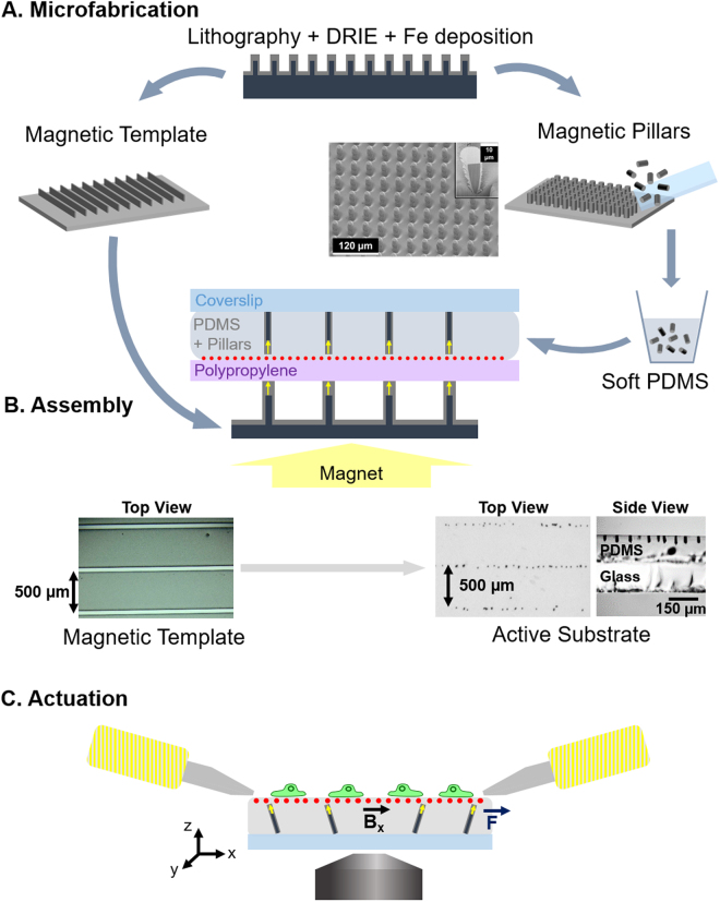
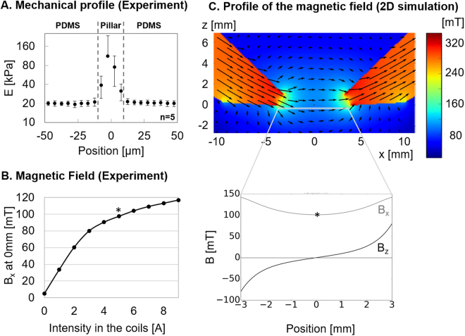
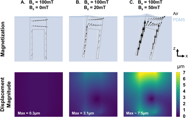
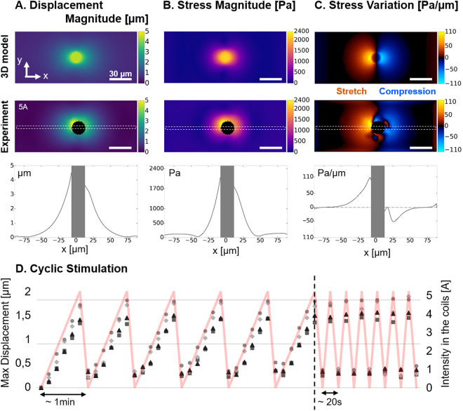
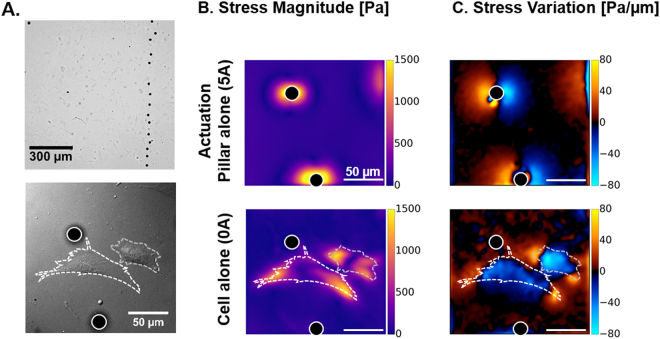
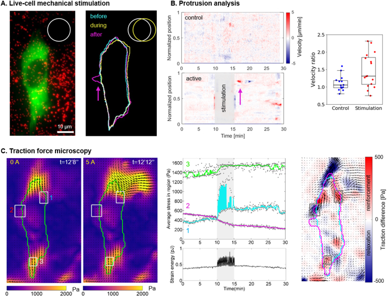
Cells are able to sense and react to their physical environment by translating a mechanical cue into an intracellular biochemical signal that triggers biological and mechanical responses. This process, called mechanotransduction, controls essential cellular functions such as proliferation and migration. The cellular response to an external mechanical stimulation has been investigated with various static and dynamic systems, so far limited to global deformations or to local stimulation through discrete substrates. To apply local and dynamic mechanical constraints at the single cell scale through a continuous surface, we have developed and modelled magneto-active substrates made of magnetic micro-pillars embedded in an elastomer. Constrained and unconstrained substrates are analysed to map surface stress resulting from the magnetic actuation of the micro-pillars and the adherent cells. These substrates have a rigidity in the range of cell matrices, and the magnetic micro-pillars generate local forces in the range of cellular forces, both in traction and compression. As an application, we followed the protrusive activity of cells subjected to dynamic stimulations. Our magneto-active substrates thus represent a new tool to study mechanotransduction in single cells, and complement existing techniques by exerting a local and dynamic stimulation, traction and compression, through a continuous soft substrate.


Conflict of interest statement

The authors declare that they have no competing interests.

Figures

Figure 1
Figure 1
Experimental workflow. (A) Magnetic micro-pillars and template are made by optical lithography followed by deep reactive ion etching (DRIE) and iron deposition by sputtering. The magnetic pillars are mechanically detached from the wafer and mixed with soft PDMS prior to casting between a sheet of polypropylene coated with fluorescent beads and a coverslip. (B) The resulting sandwich structure is positioned on a magnetic template laid on a large permanent magnet, so as to align the pillars vertically (side view) and organize them according to the pattern of the template (top view). (C) After peeling off the polypropylene sheet, coating the surface with proteins and plating cells on the top, the substrates are placed in the magnetic field generated by two electromagnets so as to actuate the pillars via a magnetic torque. The actuation setup is mounted on a microscope to quantify the local deformation of the surface by tracking the fluorescent beads and to follow the response of the cells to the mechanical stimulation.
Figure 2
Figure 2
Characterization. (A) Mechanical profiles of the magneto-active substrate measured around 5 pillars by atomic force microscopy reveal homogeneous PDMS substrates with local increases of the Young modulus above the pillars. (B) The horizontal component of the magnetic field Bx increases with the current input to the electromagnets and reaches 100mT for 5 A (*) at the mid-position between the two pole pieces. (C) A numerical model in 2D evaluates the distribution of the horizontal and vertical components of the magnetic field (Bx and Bz respectively) between the bevel shaped pole pieces of the electromagnets.
Figure 3
Figure 3
Contribution of Bz. The magnetization of the iron pillar induced by a purely horizontal magnetic field (A), a magnetic field with a slight (B) or large (C) vertical component Bz and the subsequent magnitude of displacement in the substrate have been predicted with the magneto-mechanical model. In each case, the undeformed positions of the PDMS free surface and the pillar are represented by blue and light grey lines, respectively.
Figure 4
Figure 4
Actuation of the substrate. (A) The magnitude of displacement induced by a pillar positioned 1 mm away from the electromagnet and experiencing Bx = 119mT and Bz = 27mT was estimated with a 3D model and compared to the magnitude of displacement measured experimentally around 5 pillars stimulated by an electromagnet supplied with 5 A. (B) Maps of stress magnitude were derived from the displacement fields by Fourier Transform Traction Cytometry, and (C) maps of stress variation were calculated to distinguish the regions under traction and compression. Scale bar: 30 µm. Profiles corresponding to the dashed region of each map are also displayed. (D) Maximum magnitude of displacement measured on 4 different pillars undergoing cyclic stimulations applied by manually adjusting the intensity of the current input (red curve).
Figure 5
Figure 5
Cells on magneto-active substrates. (A) Bright field images of NIH3T3 fibroblast cells spread on a magneto-active substrate. Stress magnitude (B) and variation (C) experienced by the surface were measured under the action of a pillar without cell (5 A) and in the presence of contractile cells in the vicinity of a pillar at rest. Scale bar: 50 µm.
Figure 6
Figure 6
Cell response to stimulation. (A) Fluorescent images of a NIH3T3 vinculin-eGFP fibroblast (green) and the beads spread under the surface (red) were used to draw the contours of the cell and the pillar, respectively. The cell was deformed (yellow) when the pillar was displaced, and a new protrusion appeared (magenta) a few minutes later. Corresponding movie as supplementary material (Movies 10) (B) Protrusion analysis. Velocity profiles of the boundary of a control cell and the stimulated cell (A) along a normalized perimeter as a function of time. Positive values (red) represent protrusions whereas negative values (blue) represent retractions. On the stimulated cell, a protrusion is visible (magenta arrow) shortly after the end of the stimulation and followed by a retraction. Quantification of the cellular response for 16 stimulated cells and 14 control cells: ratio of velocity after and before the stimulation were calculated as detailed in Fig. S8 and plotted for each cell. Approximately half of the cell population showed an increased protrusion activity after the mechanical stimulation. (C) Stress maps of the cell and the pillar at rest (0 A) and during actuation (5 A). Such maps were derived every 4 s during the entire protocol and the average stress in the 3 regions of interest (grey rectangles) and the strain energy of the cell (green contour) were plotted as a function of time. Map representing the local differences in averaged traction values before and after 5 min stimulations: blue indicates a local relaxation of the cell, and red indicates a reinforcement of the traction forces. The corresponding movie is given in the supplementary materials (Movies 10).

References

    1. Jaalouk DE, Lammerding J. Mechanotransduction gone awry. Nat. Rev. Mol. Cell Biol. 2009;10:63–73. doi: 10.1038/nrm2597. - DOI - PMC - PubMed
    1. Chin L, Xia Y, Discher DE, Janmey PA. Mechanotransduction in cancer. Curr. Opin. Chem. Eng. 2016;11:77–84. doi: 10.1016/j.coche.2016.01.011. - DOI - PMC - PubMed
    1. Lo CM, Wang HB, Dembo M, Wang YL. Cell movement is guided by the rigidity of the substrate. Biophys. J. 2000;79:144–152. doi: 10.1016/S0006-3495(00)76279-5. - DOI - PMC - PubMed
    1. Engler AJ, et al. Matrix elasticity directs stem cell lineage specification. Cell. 2006;126:677–89. doi: 10.1016/j.cell.2006.06.044. - DOI - PubMed
    1. Ruiz SA, Chen CS. Emergence of Patterned Stem Cell Differentiation Within Multicellular Structures. Stem Cells. 2008;26:2921–2927. doi: 10.1634/stemcells.2008-0432. - DOI - PMC - PubMed

Publication types