

Pituitary xanthogranulomas: clinical features, radiological appearances and post-operative outcomes
- PMID: 29363000
- PMCID: PMC5942345
- DOI: 10.1007/s11102-017-0859-x
Pituitary xanthogranulomas: clinical features, radiological appearances and post-operative outcomes
Abstract
Background: Xanthogranulomas are inflammatory masses most commonly found at peripheral sites such as the skin. Sellar and parasellar xanthogranulomas are rare and present a diagnostic challenge as they are difficult to differentiate from other sellar lesions such as craniopharyngiomas and Rathke's cleft cysts pre-operatively. Their radiological imaging features are yet to be clearly defined, and clinical outcomes after surgery are also uncertain. This study reviews clinical presentation, radiological appearances, and clinical outcomes in a cohort of patients with pituitary xanthogranulomas.
Methods: A prospectively maintained pituitary surgery database was screened for histologically confirmed pituitary xanthogranulomas between May 2011-December 2016. Retrospective case note assessments were then performed by three independent reviewers. Patient demographics, clinical presentations, imaging, and clinical outcomes were analysed.
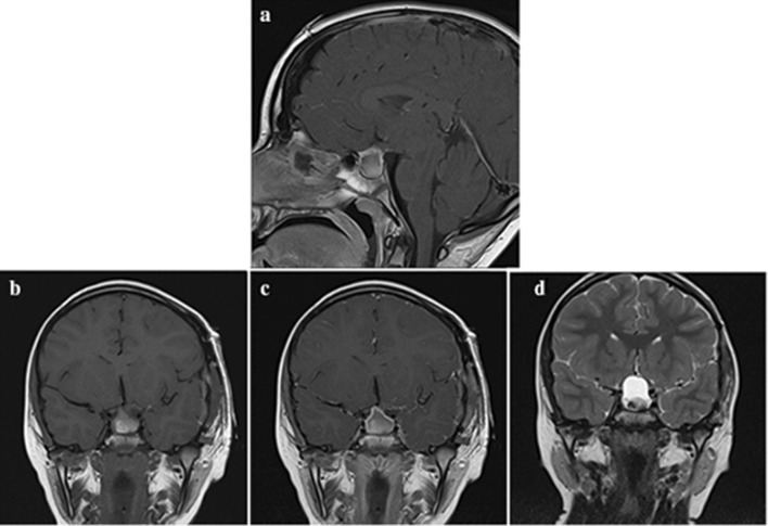
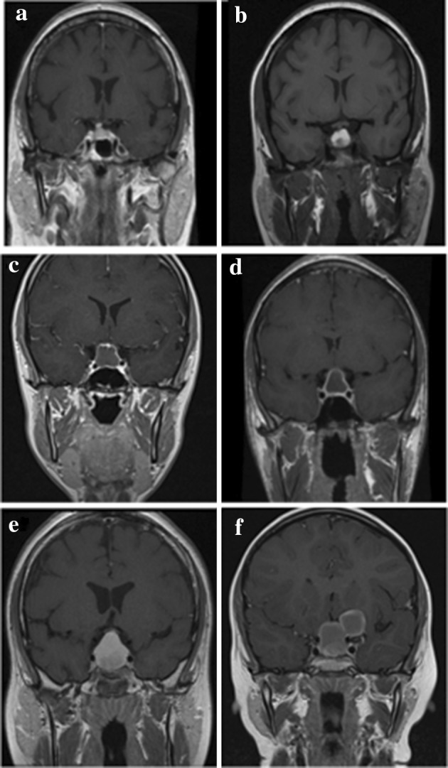
Results: During the study period 295 endoscopic endonasal pituitary surgeries were performed. Six patients had confirmed pituitary xanthogranulomas (2%). Patients most commonly presented with visual field deficits and/or endocrine dysfunction. Common imaging features included: a cystic consistency, hyperintensity on T1-weighted MR images, and contrast enhancement either peripherally (n = 3) or homogenously (n = 3). The most common pre-operative endocrine deficits were hyperprolactinaemia and hypoadrenalism (at least one of which was identified in 4/6 patients; 66%). Thirty-three percent (2/6) of patients presented with diabetes insipidus. The most common post-operative endocrinological deficits were adrenocortical dysfunction (66%) and gonadotropin deficiency (66%). Visual assessments normalised in all six patients post-operatively. Gross total resection was achieved in all patients, and at median follow up of 33.5 months there were no cases of tumour recurrence.
Conclusions: The prevalence of pituitary xanthogranulomas in our series is higher than that suggested in the literature. Surgery restored normal vision to all cases, however four patients (67%) required long-term hormonal replacement post-operatively. Imaging features such peripheral rim enhancement, a suprasellar tumour epicentre, and the absence of both calcification or cavernous sinus invasion were identified as potential indicators that together should alert clinicians to the possibility of pituitary xanthogranuloma when assessing patients with cystic sellar and parasellar tumours.
Keywords: Cystic; Pituitary; Sellar; Xanthogranuloma.
Conflict of interest statement
Conflict of interest
The authors declare that they have no conflicts of interest.
Informed consent
Informed consent was obtained were required from participants included in the study.
Ethical approval
All procedures performed in studies involving human participants were in accordance with the ethical standards of the institutional and/or national research committee and with the 1964 Helsinki declaration and its later amendments or comparable ethical standards.
Figures

References
-
- Petrakakis I, Pirayesh A, Krauss J, Raab P, Hartmann C, Nakamura M. The sellar and suprasellar region: a “hideaway” of rare lesions. Clinical aspects,imaging findings, surgical outcome and comparative analysis. Clin Neurol Neurosurg. 2016;149:154–165. doi: 10.1016/j.clineuro.2016.08.011. - DOI - PubMed
-
- Irataki K, Okada S, Matsumoto S. Histopathological study of the ‘‘cholesterol granuloma reaction. No To Shinkei. 1988;40(2):113–133. - PubMed
MeSH terms
Substances
LinkOut - more resources
Full Text Sources
Other Literature Sources
Medical
