. 2018 Mar;96(3):246-256.
doi: 10.1111/imcb.1033. Epub 2018 Jan 6.

Differential signaling pathways are initiated in macrophages during infection depending on the intracellular fate of Chlamydia spp

Affiliations

Differential signaling pathways are initiated in macrophages during infection depending on the intracellular fate of Chlamydia spp

Uma M Nagarajan et al. Immunol Cell Biol. 2018 Mar.

Abstract

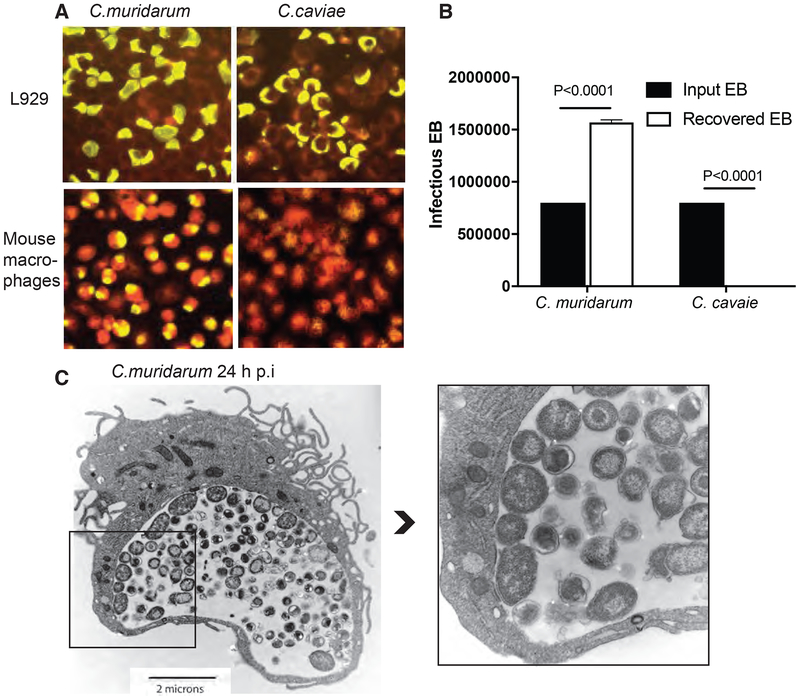
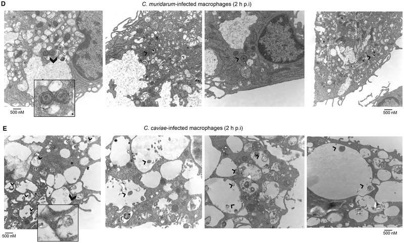
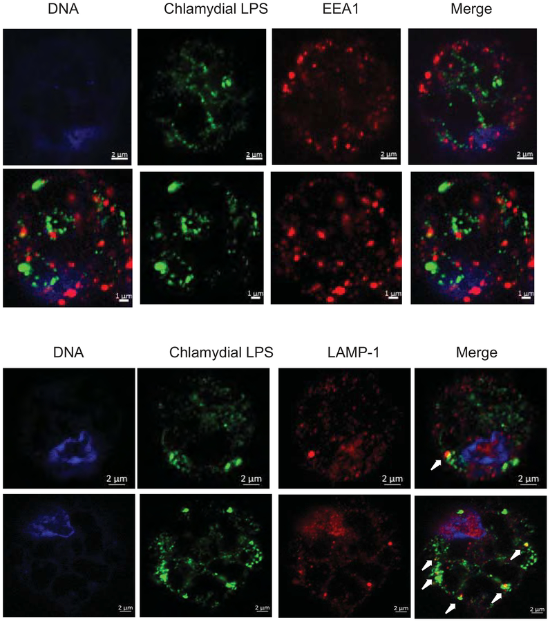
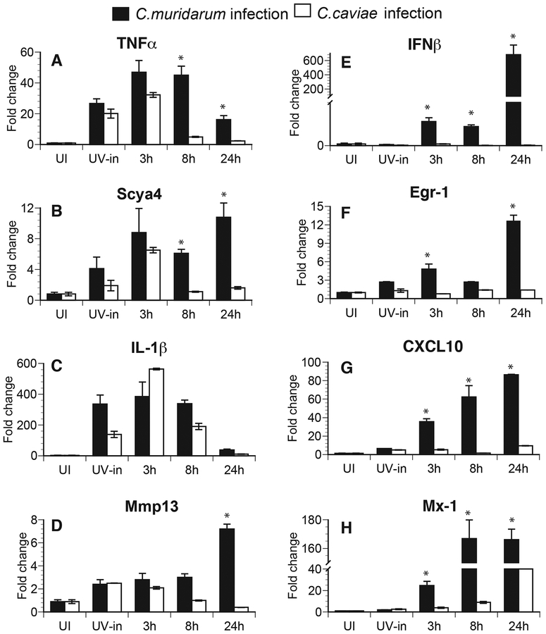
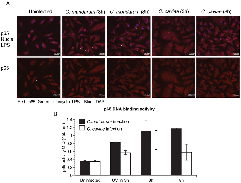
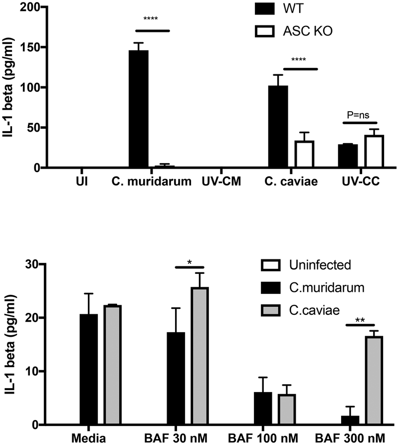
Chlamydia muridarum and Chlamydia caviae have equivalent growth rates in mouse epithelial cells but only C. muridarum replicates inside mouse macrophages, while C. caviae does not. Macrophages infected with C. muridarum or C. caviae were used to address the hypothesis that the early signaling pathways initiated during infection depend on the fate of chlamydiae in the host cell. Transmission electron microscopy of C. muridarum-infected macrophages showed intact chlamydial elementary bodies and reticulate bodies 2 h postinfection in compact vacuoles. Conversely, in macrophages infected with C. caviae, chlamydiae were observed in large phagocytic vacuoles. Furthermore, C. caviae infections failed to develop into inclusions or produce viable bacteria. Expression of proinflammatory cytokines TNFα, IL-1β and MMP13 was similar in C. caviae- or C. muridarum-infected macrophages at 3 h postinfection, indicating that chlamydial survival is not required for initiation of these responses. IL-1β secretion, dependent on inflammasome activation, occurred in C. caviae-infected macrophages despite no chlamydial growth. Conversely, IFNβ mRNA was observed only in C. muridarum- but not in C. caviae-infected macrophages. These data demonstrate that differential signaling events are initiated during a productive versus nonproductive chlamydial infection in a macrophage.

Keywords: Chlamydia; IFNβ; IL-1β; macrophages.


Conflict of interest statement

CONFLICT OF INTEREST

All authors have no conflict of interests

Figures

Fig 1:
Fig 1:. C. muridarum survives and forms mature inclusion in primary mouse macrophages while C. caviae does not.
L929 cells and primary mouse macrophages were infected with C. muridarum or C. caviae for 24 h at 1 MOI and stained using Pathfinder FITC-conjugated murine anti-chlamydial mAb (yellow) with evans blue counter stain (red) (A). Cell lysates from infected macrophages (done in triplicate) were collected and used to infect L929 cells for IFU counts. Data represent means plotted to show input and recovered EB from macrophages (B). Error bars represent SD and significance determined by one-way ANOVA. TEM image of macrophages infected with C. muridarum at 24 h post-infection (C and inset). Darker smaller forms inside the inclusions are chlamydial EBs and larger less dense forms are chlamydial RBs.
Fig 2:
Fig 2:. At 2 h post-infection, C. muridarum survives in small compact vacuoles, while C. caviae are in large vacuoles.
TEM images of macrophages infected at 10 MOI for 2 h with C. muridarum (A) or C. caviae (B). Inset in each panel shows chlamydiae in small compact vacuoles (C. muridarum) or in large vacuoles (C. caviae). Arrows point to chlamydiae inside vacuoles.
Fig 3:
Fig 3:. At 2 h post-infection, both C. muridarum and C. caviae do not co-localize with early endosomes, but more numbers of C. caviae co-localize with lysosomes.
Macrophages infected with C. muridarum or C. cavaie at 1 MOI infection for 3 h p.i were fixed and immuno-stained for an early endosomal marker EEA-1 (A) or lysosomal marker LAMP-1 (B) with chlamydial LPS staining. Arrows indicate co-localization of LAMP-1 and LPS. Image shows a single cell for clarity. Larger field is shown in Fig S3. A representative image from one of two experiments is shown.
Fig 4:
Fig 4:. Pro-inflammatory gene transcripts are induced following both C. muridarum and C. caviae infection, while IFNβ and IFN response gene transcripts are induced only during C. muridarum infection.
Quantitative RT-PCR data are expressed as fold changes compared to uninfected (UI) controls from RNA extracted from macrophages at 3, 8, and 24 h post-infection. RNA was extracted at 3 h post-infection in cells infected with UV inactivated (UV-in) Chlamydia. Individual gene expression was normalized to actin expression. TNFα (A), Scya4 (B), IL-1β (C) and Mmp13 (D) are shown on the left panel and data for IFNβ (E) and IFN-response genes, Egr-1 (F), CXL10 (G) and Mx-1 (H) are shown on the right panel. Samples were run in triplicates and data are representative of 3 independent experiments. Mean values are shown and error bars represent SD. * represents significant changes (P<0.05) by multiple t tests.
Fig 5:
Fig 5:. p65 nuclear translocation and its promoter binding activity were similar between C. muridarum- and C. caviae- infected macrophages.
Macrophages were infected with C. muridarum or C. caviae, and stained for p65, chlamydial inclusion (LPS) and DAPI for nuclear staining at 3 and 8 h post-infection (A). Cells were fixed and visualized using a Zeiss confocal microscope. Top panel shows staining for p65 (red), chlamydial inclusion (green) and DAPI (blue), while lower panel shows the same image for p65 staining only. Red signal inside the nucleus and similar area in the cytosol for each group was quantified using Image J software. Data represented as nuclei/cytoplasm ratio (B). Data shows mean and SEM from 8 cells. Significance determined by one way ANOVA with multiple comparison tests. Nuclear extracts were prepared from infected cells at 3 h and 8 h post-infection and p65 DNA-binding activity (C) was determined as described in Methods. Data represent mean of triplicate values from one of two experiments, error bars represent SD, and significance determined by one way ANOVA. * represents <0.05, ** represents<0.01, *** represents <0.001 and ns represents “not significant”.
Fig 6:
Fig 6:. C. caviae induces inflammasome activation in resting macrophages.
(A) Supernatants from WT and ASC KO-infected macrophages were analyzed for IL-1β protein by ELISA 24 h post-infection. Data represents averages from 3 experimental replicates (B) WT macrophages treated with three different concentrations of Bafilomycin were infected with C. muridarum or C. caviae and supernatants analyzed for IL-1β by ELISA. Mean values are shown and error bars represent SD. *** indicates p<0.001 and ** indicates p<0.01 by one way ANOVA. ND=Not detected.

References

    1. La Verda D, Byrne GI. Interactions between macrophages and chlamydiae. Immunol Ser 1994; 60: 381–99. - PubMed
    1. Gerard HC, Whittum-Hudson JA, Carter JD et al. The pathogenic role of Chlamydia in spondyloarthritis. Current Opinion in Rheumatology 2010; 22: 363–7. - PubMed
    1. Prantner D, Darville T, Nagarajan UM. Stimulator of IFN gene is critical for induction of IFN-beta during Chlamydia muridarum infection. J Immunol 2010; 184: 2551–60. - PMC - PubMed
    1. Zhang Y, Yeruva L, Marinov A et al. The DNA sensor, cyclic GMP-AMP synthase, is essential for induction of IFN-beta during Chlamydia trachomatis infection. J Immunol 2014; 193: 2394–404. - PMC - PubMed
    1. Prantner D, Darville T, Sikes JD et al. Critical role for interleukin-1beta (IL-1beta) during Chlamydia muridarum genital infection and bacterial replication-independent secretion of IL-1beta in mouse macrophages. Infection and immunity 2009; 77: 5334–46. - PMC - PubMed

Publication types

MeSH terms