. 2018 Apr 15;24(8):1891-1904.
doi: 10.1158/1078-0432.CCR-17-0691. Epub 2018 Jan 23.

Nitric Oxide Production by Myeloid-Derived Suppressor Cells Plays a Role in Impairing Fc Receptor-Mediated Natural Killer Cell Function

Affiliations

Nitric Oxide Production by Myeloid-Derived Suppressor Cells Plays a Role in Impairing Fc Receptor-Mediated Natural Killer Cell Function

Andrew Stiff et al. Clin Cancer Res. .

Abstract

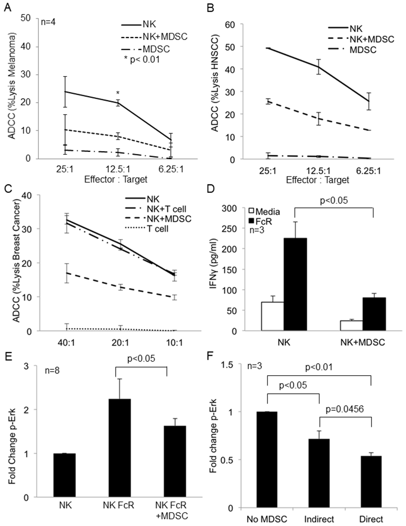
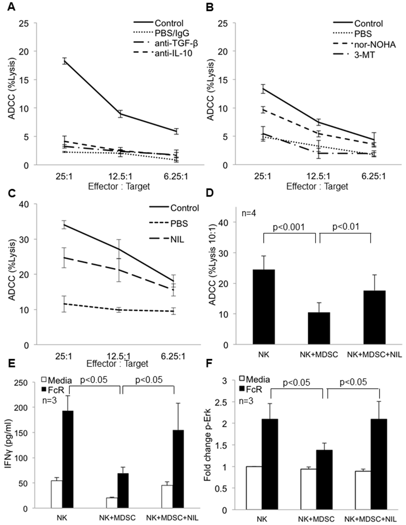
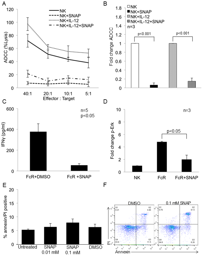
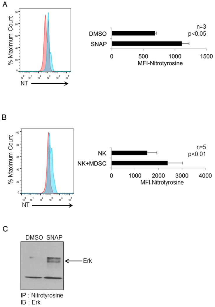
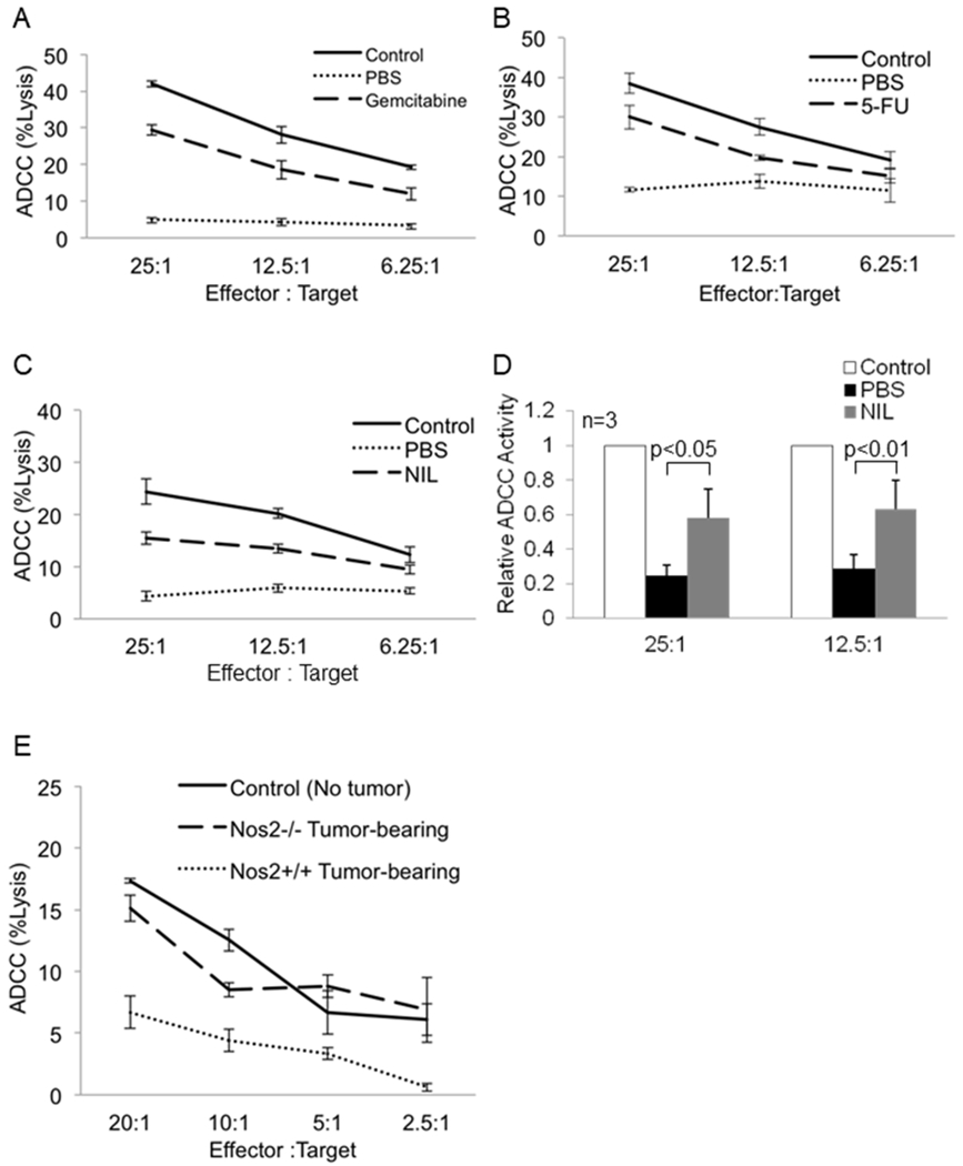
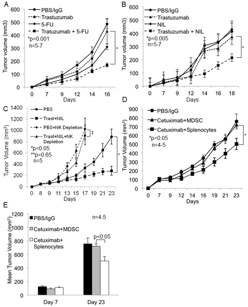
Purpose: mAbs are used to treat solid and hematologic malignancies and work in part through Fc receptors (FcRs) on natural killer cells (NK). However, FcR-mediated functions of NK cells from patients with cancer are significantly impaired. Identifying the mechanisms of this dysfunction and impaired response to mAb therapy could lead to combination therapies and enhance mAb therapy.Experimental Design: Cocultures of autologous NK cells and MDSC from patients with cancer were used to study the effect of myeloid-derived suppressor cells (MDSCs) on NK-cell FcR-mediated functions including antibody-dependent cellular cytotoxicity, cytokine production, and signal transduction in vitro Mouse breast cancer models were utilized to study the effect of MDSCs on antibody therapy in vivo and test the efficacy of combination therapies including a mAb and an MDSC-targeting agent.Results: MDSCs from patients with cancer were found to significantly inhibit NK-cell FcR-mediated functions including antibody-dependent cellular cytotoxicity, cytokine production, and signal transduction in a contact-independent manner. In addition, adoptive transfer of MDSCs abolished the efficacy of mAb therapy in a mouse model of pancreatic cancer. Inhibition of iNOS restored NK-cell functions and signal transduction. Finally, nonspecific elimination of MDSCs or inhibition of iNOS in vivo significantly improved the efficacy of mAb therapy in a mouse model of breast cancer.Conclusions: MDSCs antagonize NK-cell FcR-mediated function and signal transduction leading to impaired response to mAb therapy in part through nitric oxide production. Thus, elimination of MDSCs or inhibition of nitric oxide production offers a strategy to improve mAb therapy. Clin Cancer Res; 24(8); 1891-904. ©2018 AACR.


Conflict of interest statement

The authors have declared that no conflict of interest exists.

Conflict of interest

Authors declare no conflict of interest.

Figures

Figure 1:
Figure 1:. MDSC inhibit FcR mediated NK cell effector functions and signal transduction.
(a) NK cells from the peripheral blood of a melanoma patient were cultured alone or with autologous MDSC overnight and then used in a 51Cr release ADCC assay against cetuximab-coated HT-29 cells. Values represent the mean ± SD from four independent experiments, p<0.01. Significance was determined using the paired t-test. (b) Results from one ADCC assay as conducted in (a) using NK cells and autologous MDSC from a HNSCC patient. (c) Results from one ADCC assay as conducted in (a) using NK cells and autologous MDSC from a breast cancer patient. (d) Autologous NK cells and MDSC from the peripheral blood of melanoma patients were co-cultured at a 1:1 ratio in 96 well plates coated with human IgG (100 μg/ml) or media (control). Supernatants were collected after 48 hrs and cytokine levels measured by ELISA. Quantification of data from three independent experiments, values shown are the mean ± SE, p<0.05. Significance was determined using a paired t-test. (e) Quantification of changes in p-Erk levels in FcR activated CD56+ NK cells measured by flow cytometry in the presence or absence of MDSC. Values are mean ± SE from eight independent experiments, p<0.05. Significance was determined using a paired t-test. (f) NK cells were cultured alone, in direct contact with MDSC (Direct), or physically separated from MDSC by a permeable 0.4 μm Corning Transwell® membrane (Indirect) at a 1:1 ratio overnight. NK cells were then stimulated through the FcR using the anti-CD16 3G8 antibody and F(ab’)2 and levels of p-Erk determined as described above. Values are the mean ± SE from 3 independent experiments. Significance was determined using a paired t-test and Holms method. Representative flow cytometry dot plot for p-Erk is provided in Supplementary Figure 4.
Figure 2:
Figure 2:. iNOS inhibition restores FcR mediated NK cell functions and signal transduction in the presence of MDSC.
Female Balb/c mice were inoculated with 1x10⁵ 4T1 tumor cells or were left un-injected (no tumor-control). Following the establishment of tumors, mice were treated daily with (a) intraperitoneal injections of IgG (250 μg), anti-TGF-β antibody (200 μg), or anti-IL-10 antibody (250 μg) for four consecutive days prior to NK cell isolation for the ADCC assay. The mice were sacrificed 24 hrs after the last treatment. Each group consists of pooled samples from spleens of 4-5 mice. Values represent mean ± SD from one experiment. (b) Mice were treated with PBS (vehicle), arginase inhibitor nor-NOHA (20 mg/kg) i.p or the IDO inhibitor 3-methyltryptophan (MT, 400 mg/kg) via oral gavage prior to NK cell isolation for the ADCC assay. (c) Mice were given intraperitoneal injections of PBS (vehicle) or iNOS inhibitor, L-NIL (20 mg/kg) for one week. NK cells isolated from the spleen were employed in a standard ADCC assay using trastuzumab-coated CT26-HER2 positive tumor cells as targets. (d) NK cells and MDSC from the peripheral blood of melanoma patients were co-cultured overnight at a 1:1 ratio with or without the nitric oxide inhibitor L-NIL (2.5 mM). ADCC function of NK cells displayed as the mean percent lysis of cetuximab-coated HT-29 tumor cells at the 10:1 effector to target ratio. Means ± SE from four independent experiments are shown, p<0.05. Significance was determined using a linear mixed model. Treatment of NK cells with L-NIL alone did not enhance ADCC activity (not shown). (e) Autologous NK cells and MDSC isolated from peripheral blood of melanoma patients were co-cultured in 96 well plates coated with human IgG or media with or without the iNOS inhibitor L-NIL (2.5 mM). Supernatants were harvested after 48 hrs and analyzed for levels of IFNγ by ELISA. Values represent mean ± SE from three independent experiments, p<0.05. Significance was determined using a linear mixed model. (f) p-Erk expression in NK cells co-cultured overnight with MDSC in the presence or absence of L-NIL (2.5 mM). p-Erk levels are expressed as the average fold change ± SE from three independent experiments, p<0.05. Significance was determined using a linear mixed model.
Figure 3:
Figure 3:. The NO donor SNAP inhibits NK cell function and signal transduction.
The nitric oxide donor SNAP inhibits NK cell FcR-mediated function and signal transduction. (a) Purified NK cells from normal donors were treated with DMSO or SNAP (0.1 mM) in the presence or absence of IL-12 (10 ng/ml) and then used in a 51Cr release ADCC assay against cetuximab-coated HT-29 cells. Representative results shown are from one of three independent experiments. (b) Fold change in ADCC activity of NK cells (20:1 effector target ratio) treated in a similar fashion as in (a) against cetuximab-coated HT-29 cells. The mean ± SE from three independent experiments are shown, p<0.001. Significance was determined using a t-test. (c) Levels of IFN-γ production after 48 hrs measured by ELISA from healthy donor NK cells treated with DMSO or SNAP (0.1 mM) and activated with immobilized IgG. Values represent mean ± SE from five independent experiments, p<0.05. Significance was determined using a paired t-test. (d) Fold change in the expression of p-Erk in NK cells treated with DMSO or SNAP (0.1 mM) and then stimulated through the FcR via 3G8 antibody. Values represent mean ± SE from three independent experiments, p<0.05. Significance was determined using a paired t-test. (e) NK cells isolated from normal donors were treated with the indicated doses of SNAP or DMSO for 24 hrs. NK cells were then stained with annexin V/PI to determine the percentage of apoptotic cells. Values displayed are from three independent experiments. (f) NK cells isolated from normal donors were treated with the indicated doses of SNAP or DMSO for 24 hrs. NK cells were then stained with annexin V/PI to determine the percentage of apoptotic cells. A representative flow cytometric dot plot is provided for the quantification data in 3E.
Figure 4:
Figure 4:. Treatment of NK cells with the nitric oxide donor SNAP or co-culture with MDSC results in nitration of tyrosine residues in NK cells.
Purified NK cells from normal donors were treated with SNAP (0.01 mM) or DMSO (control). Following permeablization, cells were stained with anti-CD16 and anti-nitrotyrosine antibodies and analyzed by flow cytometry. (a) Representative flow cytometry profile and quantification of nitrotyrosine levels in CD16+ NK cells shown as mean fluorescence intensity (MFI) (right panel) from three independent experiments, p<0.05, significance was determined using a paired t-test. (b) Autologous NK cells and MDSC isolated from peripheral blood of melanoma patients were co-cultured overnight at a ratio of 1:1 and then stained with anti-CD16 and anti-nitrotyrosine antibodies. Representative flow cytometry profile (left) and quantification of nitrotyrosine in CD16+ NK cells as mean fluorescence intensity (MFI) (right) from five independent experiments, p<0.05, significance was determined using a paired t-test. (c) Immunoblot showing the nitration of Erk protein. Purified NK cells from normal donors were treated with SNAP or DMSO (control), immunoprecipitated with anti-nitrotyrosine beads and probed with anti-Erk antibody.
Figure 5:
Figure 5:. Depletion of MDSC or inhibition of nitric oxide production in mice augments NK cell-mediated ADCC activity.
Female Balb/c mice were inoculated with 1x10⁵ 4T1 tumor cells or were left un-injected (no tumor-control). The mice were sacrificed 24 hrs after treatment. NK cells isolated from the spleen were used in a standard ADCC assay using trastuzumab-coated CT26-HER2 positive tumor cells as targets. (a) Mice were treated with PBS (vehicle) or gemcitabine (80 mg/kg). Graph displays mean percent lysis by pooled NK cells from 4-5 mice per treatment group. One of two representative experiments is shown. (b) Mice were treated with PBS (vehicle) or 5-fluorouracil (50 mg/kg). Graph displays mean percent lysis by pooled NK cells from 4-5 mice per treatment group. One of two representative experiments is shown. (c) NK cells isolated from mice treated daily for one week with PBS (vehicle) or iNOS inhibitor, L-NIL (20 mg/kg) were used in a standard ADCC assay using trastuzumab-coated CT26-HER2 cells. Each group consists of pooled samples from spleens of 5 mice. (d) Fold change in NK cell ADCC activity from mice treated as in (c). Values displayed are the means ± SE from three independent experiments, p< 0.05 for the 25:1 and 12.5:1 effector target ratios. Data was log-transformed for testing group difference using a linear mixed effect model with random donor effect, and the p-value was calculated using Bonferroni method. (e) Female C57BL/6 (wild type) and Nos2−/− mice were inoculated with B16F10 tumor cells or left un-injected (control). NK cells were isolated from the spleen and used in a standard ADCC assay using trastuzumab coated CT26-HER2 tumor cells. Each group consists of pooled samples from spleens of 3-4 mice. Values represent mean ± SD from one experiment.
Figure 6:
Figure 6:. MDSC antagonize mAb therapy in vivo and MDSC depletion or inhibition of nitric oxide enhances the efficacy of mAb therapy.
Female Balb/c mice were inoculated with 1x10⁶ EMT6-HER2 tumor cells in the mammary fat pad. Following the establishment of tumors (day 7), mice were treated once a week i.p. with PBS or 5-FU and thrice weekly with IgG or trastuzumab and tumor growth was measured three times a week using digital calipers. (a) Tumor growth in mice treated with PBS and IgG (10 mg/kg), 5-FU (50 mg/kg), trastuzumab (10 mg/kg) or the combination of trastuzumab plus 5-FU. Differences in tumor volumes were tested using a linear mixed model and Student’s t-test. Values represent mean ± SE, p<0.001. (b) Tumor growth in mice treated with PBS and IgG (10 mg/kg), L-NIL (20 mg/kg), trastuzumab (10 mg/kg) or combination of trastuzumab plus L-NIL. Each group consisted of 5-7 mice. Values represent mean ± SE, p<0.005, significance was determined using a linear mixed model and Student’s t-test. (c) Depletion of NK cells with anti-asialo-GM1 abrogates the anti-tumor effect of trastuzumab plus NIL. Balb/c mice were inoculated with 1x10⁶ EMT6-HER2 tumor cells in the mammary fat pad. Three days prior to treatment, mice were administered PBS or 50 µg/mouse of anti asialo GM1 polyclonal antibody to deplete NK cells. On day 8, mice were randomized and treated thrice weekly with PBS and IgG or trastuzumab (10 mg/kg) plus L-NIL (20 mg/kg). NK depleted mice were administered the depleting antibody every 4 days until the end of the study. n=5 for each treatment group. (d) Athymic nude mice were inoculated with 1x10⁶ Panco2-EGFR tumor cells on day zero. After establishment of tumors mice were injected with PBS, splenocytes, or MDSC on day 7 prior to the initiation of treatment with IgG or cetuximab (0.5 mg/kg) on day 9. Mice were re-injected with PBS, splenocytes, or MDSC on day 14 and IgG or cetuximab treatment was continued. (d) Tumor growth curves of mice treated as described above. (e) Mean tumor volume in mice inoculated with Panco2-EGFR cells before (day 7) and after treatment (day 23). Each group consisted of 4-5 mice. Differences in tumor volumes were tested using a linear mixed model and Student’s t-test. Values represent mean ± SE, p<0.001.

Similar articles

Cited by

References

    1. Vivier E, Raulet DH, Moretta A, Caligiuri MA, Zitvogel L, Lanier LL, et al. Innate or adaptive immunity? The example of natural killer cells. Science 2011;331:44–9 - PMC - PubMed
    1. Parihar R, Dierksheide J, Hu Y, Carson WE. IL-12 enhances the natural killer cell cytokine response to Ab-coated tumor cells. J Clin Invest 2002;110:983–92 - PMC - PubMed
    1. Roda JM, Parihar R, Magro C, Nuovo GJ, Tridandapani S, Carson WE, 3rd. Natural killer cells produce T cell-recruiting chemokines in response to antibody-coated tumor cells. Cancer research 2006;66:517–26 - PubMed
    1. Scott AM, Wolchok JD, Old LJ. Antibody therapy of cancer. Nature reviews Cancer 2012;12:278–87 - PubMed
    1. Ishigami S, Natsugoe S, Tokuda K, Nakajo A, Che X, Iwashige H, et al. Prognostic value of intratumoral natural killer cells in gastric carcinoma. Cancer 2000;88:577–83 - PubMed

Publication types

MeSH terms

LinkOut - more resources