. 2018 Apr 1;78(7):1700-1712.
doi: 10.1158/0008-5472.CAN-17-1366. Epub 2018 Jan 23.

Tumor-Stroma IL1β-IRAK4 Feedforward Circuitry Drives Tumor Fibrosis, Chemoresistance, and Poor Prognosis in Pancreatic Cancer

Affiliations

Tumor-Stroma IL1β-IRAK4 Feedforward Circuitry Drives Tumor Fibrosis, Chemoresistance, and Poor Prognosis in Pancreatic Cancer

Daoxiang Zhang et al. Cancer Res. .

Abstract

Targeting the desmoplastic stroma of pancreatic ductal adenocarcinoma (PDAC) holds promise to augment the effect of chemotherapy, but success in the clinic has thus far been limited. Preclinical mouse models suggest that near-depletion of cancer-associated fibroblasts (CAF) carries a risk of accelerating PDAC progression, underscoring the need to concurrently target key signaling mechanisms that drive the malignant attributes of both CAF and PDAC cells. We previously reported that inhibition of IL1 receptor-associated kinase 4 (IRAK4) suppresses NFκB activity and promotes response to chemotherapy in PDAC cells. In this study, we report that CAF in PDAC tumors robustly express activated IRAK4 and NFκB. IRAK4 expression in CAF promoted NFκB activity, drove tumor fibrosis, and supported PDAC cell proliferation, survival, and chemoresistance. Cytokine array analysis of CAF and microarray analysis of PDAC cells identified IL1β as a key cytokine that activated IRAK4 in CAF. Targeting IRAK4 or IL1β rendered PDAC tumors less fibrotic and more sensitive to gemcitabine. In clinical specimens of human PDAC, high stromal IL1β expression associated strongly with poor overall survival. Together, our studies establish a tumor-stroma IL1β-IRAK4 feedforward signal that can be therapeutically disrupted to increase chemotherapeutic efficacy in PDAC.Significance: Targeting the IL1β-IRAK4 signaling pathway potentiates the effect of chemotherapy in pancreatic cancer. Cancer Res; 78(7); 1700-12. ©2018 AACR.


Conflict of interest statement

The authors declare no potential conflicts of interest

Figures
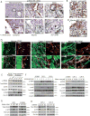
Figure 1
Figure 1. IRAK4 is constitutively phosphorylated and drives NF-κB activity in PDAC CAFs
A, Representative IHC images showing p-IRAK4 staining in CAFs in normal pancreas, pancreatitis and PDAC samples from a commercial TMA (US Biomax PA2081). Positive (+) or negative (−) staining was determined by GI pathologist. B, C, Representative IHC images showing presence of p-IRAK4 staining in CAFs of Capan-1 xenograft and murine KPC tumors. D, Confocal IF images of human PDAC, Capan-1 xenograft and KPC tumors showing p-IRAK4 staining in α-SMA+ in CAFs. (A–D, scale bars: 50µM). E, Western blots showing higher p-IRAK, p-p65 levels in four PDAC CAF lines specified by α-SMA expression, as opposed to normal dermal (hFB) and pancreatic (SC00A5) fibroblasts. F, Western blots showing dose-dependent suppression of p-IRAK4 and p-p65 levels in CAF-2 and CAF-8 following overnight treatment with AS2444697 or IRAK1/4 inhibitor at the indicated doses. G, H, Western blots showing changes in p-p65 levels following: (G) ectopic expression of empty vector, wild-type, or kinase-dead IRAK4 in two normal fibroblasts (hFB and SC00A5); (H) knockdown of IRAK4 using two shRNAs in CAF-2 and CAF-8.
Figure 2
Figure 2. CAF IRAK4 supports PDAC cell proliferation, migration and tumorigenesis
A, Co-culture assay showing abundance of MIA Paca-2 cells stably expressing luciferase/RFP, as determined by luciferase titer and RFP+ cells (Suppl. Fig. 2A), when cultured alone or at 1:1 ratio with GFP+ CAF-2 or CAF-8 cells expressing scramble shRNA or two different shIRAK4s. B, Wound-healing and C, transwell migration assay of MIA Paca-2 cells cultured in CM collected from SC00A5, CAF-2/8 cells expressing scramble shRNA or different shIRAK4. D, Final weights and picture of MIA Paca-2 tumors injected alone or in 1:2 ratio with the indicated CAFs (N=6 mice/group). E, Quantification and representative co-IF images of dual pan-cytokeratin+ and Ki-67+ cells in MIA Paca-2 tumors from experiment Fig. 2D. All in vitro experiments were conducted at least three times in triplicates and represented as mean ± SEM (*p<0.05, **p<0.01, ***p<0.001).
Figure 3
Figure 3. CAF IRAK4 contributes to PDAC tumor fibrosis
A, Quantification and representative images of gel contraction assay showing dose-dependent effect of IRAK4 inhibitor AS2444697 on the ability of CAF-2 and CAF-8 to contract type 1 collagen in vitro. B, Quantitative PCR of collagen 1 mRNA in the indicated fibroblast lines. Data (means ± SEM) represents one of three experiments done in biological duplicates and technical triplicates. C, Quantification and representative images (200X) of Sirius Red+ area in tumors arising from MIA Paca-2 injected alone or at 1:2 ratio with the indicated CAFs (N=6/group). D, Final weights of tumors arising from KPC2 cells injected subcutaneously in wild-type C57BL/6J mice, IRAK4-null littermates, alone or in combination with wild-type or IRAK4-null MEFs (N=6–10/group). E, F, Quantification and representative images of (E) dual pan-cytokeratin+ and Ki-67+ cells, and (F) Sirius Red+ area in the indicated KPC2 tumors. (6–8 tumors/group, ten 20X fields were analyzed per tumor). Data represents combined means ± SEM. (*p<0.05, **p<0.01, ***p<0.001).
Figure 4
Figure 4. Autocrine IL-1β activates IRAK4 in CAFs
A, Cytokine array showing altered abundance of chemokines/cytokines in serum-free CM collected from CAF-2 cells stably expressing scramble or IRAK4 shRNAs. B, Quantitative PCR showing fold changes in mRNA levels of the indicated genes in CAF-2 expressing scramble shRNA treated with DMSO, IRAK1/4 inhibitor or AS2444697 overnight, or stably expressing two different shIRAK4s. Data (means ± SEM) represents one of two experiments done in biological duplicates and technical triplicates. C, Consecutive IHC sections showing expression of IL-1β in α-SMA+ CAFs in two representative human PDAC samples from TMA (US Biomax PA 2081. Of all 117 PDAC samples, 96 showed positive, and 21 showed negative IL-1β IHC staining in α-SMA+ CAFs. D, Confocal IF images showing presence of IL-1β in α-SMA+ CAFs in two human surgical PDAC and murine KPC tumors. E, ELISA assay showing abundance of IL-1β protein in CM collected from the indicated cells treated with DMSO or AS2444697 overnight (left panel), or from CAF-2 cells stably expressing scramble shRNA or two difference shIRAK4s (right panel). Experiments were done twice in triplicates. Data presented as means ± SEM (*p<0.05, **p<0.01 by two-tailed t test). F, Western blots showing dose-dependent suppression of p-IRAK4 and p-p65 levels in CAF-2 treated overnight with nIL-1β antibody or Anakinra.
Figure 5
Figure 5. PDAC cells secrete IL-1β to activate CAF IRAK4
A, Western blots showing changes in p-IRAK4 and p-p65 levels of SC00A5 cells incubated overnight with serum-free DMEM alone or at 1:1 ratio with serum-free CM collected from the indicated PDAC lines. B, IL-1β ELISA of CM collected from the indicated PDAC lines used in Fig. 5A. Experiment was done twice in triplicates and data represented as means ± SEM. C. Quantitative PCR of IL-1β mRNA in the indicated fibroblast lines incubated with or without serum-free MIA Paca-2 CM overnight. D, Venn diagrams of genes expressed at ≥1.5-fold difference FDR <0.1 in DMSO vs. IRAK1/4 inhibitor treatment in Capan-1 and PANC-1 cells. Expression values used were gene-wise z-score normalized. E, Western blots showing changes in p-IRAK4 and p-p65 levels of SC00A5 cells incubated with serum-free DMEM, MIA Paca-2 or Capan-1 CM in the absence or presence of neutralizing anti-human IL-1β antibody. F, Gel contraction assay showing a role of IL-1β in MIA Paca-2 CM in inducing collagen contraction of SC00A5 cells. Experiment was done three time in triplicates and presented as means ± SEM. G, Western blots confirming IL-1R protein knockdown in SC00A5 cells stably expressing two different shIL-1R (left panel). These three SC00A5 lines were then treated with serum-free DMEM or MIA Paca-2 CM overnight, followed by western blots showing changes in p-IRAK4 and p-p65 levels. (*p<0.05, **p<0.01, ***p<0.001).
Figure 6
Figure 6. CAF IRAK4 drives IL-1β secretion to promote PDAC cell NF-κB activity and survival
A, Alamar blue assay showing viability of the indicated PDAC lines cultured in serum-free medium alone or at 1:1 ratio with different CAF CM for 72 hours. B, NF-κB luciferase reporter assay of MIA Paca-2 cells incubated without or with different CAF CM overnight. C, Alamar blue assay showing gemcitabine dose-response inhibition of MIA Paca-2 and CFPAC-1 cells cultured with CM collected from SC00A5, or CAF-2 cells expressing scramble shRNA or two different shIRAK4s for 72 hours. Changes in IC50 of gemcitabine in each condition were shown. D, Caspase 3/7 luciferase reporter activity in MIA Paca-2 cells cultured in the indicated CAF CM, treated with vehicle (PBS) or gemcitabine for 24 hours. E, Alamar blue assay showing viability of two PDAC lines cultured in serum free medium alone or CAF-2 CM for 72 hours in the absence or presence of neutralizing anti-human IL-1β antibody. F, NF-κB luciferase reporter assay of MIA Paca-2 cells treated with anti-IL-1β neutralizing antibody without or with CAF-2 CM overnight. All experiments were done three times in triplicates and data presented as means ± SEM. (*p<0.05, **p<0.01, ***p<0.001).
Figure 7
Figure 7. Disruption of tumor-stroma IL-1β-IRAK4 circuitry attenuates tumor fibrosis and augments the effect of gemcitabine in vivo
A, Final weights and representative pictures of Capan-1 tumors injected subcutaneously alone, or in 1:2 ratio with CAF-2 expressing scramble shRNA, shIRAK4 or shIL-1R. (N=8/group). B, C, Quantification of (B) proliferating (dual pan-cytokeratin+ and Ki-67+ cells) or (C) α-SMA+ cells in the indicated Capan-1 tumors. D, Final weights and representative pictures of KPC2 tumors grown in mice treated with vehicle (V), anti-mouse IL-1β neutralizing antibody (nIL-1β), gemcitabine (GEM), AS2444697, or in dual combinations. (N=10–14/group combined from two independent experiments). E, F, G, Quantification of proliferating (dual pan-cytokeratin+ and Ki-67+ cells), apoptotic cells (cleaved caspase-3+), or degree of fibrosis (Sirius Red+ area) of KPC2 tumors treated as indicated (N=10/group). H, (Left) Representative IHC images showing assigned intensity scores (0=absence/faint, 1=weak, 2=moderate, 3=strong) of stromal IL-1β staining in a PDAC tissue microarray (N=125). (Right) Kaplan-Meier survival analysis and median survival of 125 patients based on low (0–1) or high (2–3) stromal IL-1β IHC score. I, Schematics depicting the proposed model in which PDAC cells and CAFs conspire to establish a feedforward IL-1β–IRAK4 circuitry that leads to tumor fibrosis, chemoresistance and poor prognosis.

Similar articles

Cited by

References

    1. Feig C, Gopinathan A, Neesse A, Chan DS, Cook N, Tuveson DA. The pancreas cancer microenvironment. Clin Cancer Res. 2012;18(16):4266–76. - PMC - PubMed
    1. Mitchem JB, Brennan DJ, Knolhoff BL, Belt BA, Zhu Y, Sanford DE, et al. Targeting Tumor-Infiltrating Macrophages Decreases Tumor-Initiating Cells, Relieves Immunosuppression, and Improves Chemotherapeutic Responses. Cancer Res. 2013 - PMC - PubMed
    1. Goedegebuure P, Mitchem JB, Porembka MR, Tan MC, Belt BA, Wang-Gillam A, et al. Myeloid-derived suppressor cells: general characteristics and relevance to clinical management of pancreatic cancer. Curr Cancer Drug Targets. 2011;11(6):734–51. - PMC - PubMed
    1. Bayne LJ, Beatty GL, Jhala N, Clark CE, Rhim AD, Stanger BZ, et al. Tumor-derived granulocyte-macrophage colony-stimulating factor regulates myeloid inflammation and T cell immunity in pancreatic cancer. Cancer cell. 2012;21(6):822–35. - PMC - PubMed
    1. Provenzano PP, Cuevas C, Chang AE, Goel VK, Von Hoff DD, Hingorani SR. Enzymatic targeting of the stroma ablates physical barriers to treatment of pancreatic ductal adenocarcinoma. Cancer Cell. 2012;21(3):418–29. - PMC - PubMed

Publication types

MeSH terms