. 2018 Apr 1;78(7):1820-1832.
doi: 10.1158/0008-5472.CAN-17-1958. Epub 2018 Jan 23.

Fc-Mediated Anomalous Biodistribution of Therapeutic Antibodies in Immunodeficient Mouse Models

Affiliations

Fc-Mediated Anomalous Biodistribution of Therapeutic Antibodies in Immunodeficient Mouse Models

Sai Kiran Sharma et al. Cancer Res. .

Abstract

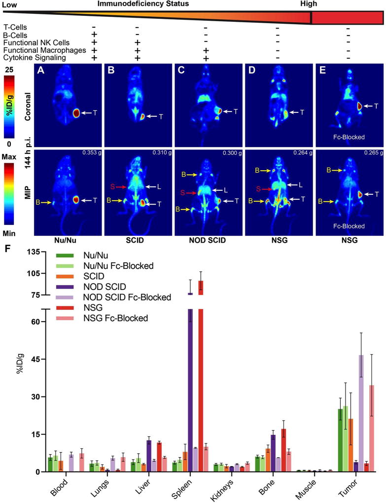
A critical benchmark in the development of antibody-based therapeutics is demonstration of efficacy in preclinical mouse models of human disease, many of which rely on immunodeficient mice. However, relatively little is known about how the biology of various immunodeficient strains impacts the in vivo fate of these drugs. Here we used immunoPET radiotracers prepared from humanized, chimeric, and murine mAbs against four therapeutic oncologic targets to interrogate their biodistribution in four different strains of immunodeficient mice bearing lung, prostate, and ovarian cancer xenografts. The immunodeficiency status of the mouse host as well as both the biological origin and glycosylation of the antibody contributed significantly to the anomalous biodistribution of therapeutic monoclonal antibodies in an Fc receptor-dependent manner. These findings may have important implications for the preclinical evaluation of Fc-containing therapeutics and highlight a clear need for biodistribution studies in the early stages of antibody drug development.Significance: Fc/FcγR-mediated immunobiology of the experimental host is a key determinant to preclinical in vivo tumor targeting and efficacy of therapeutic antibodies. Cancer Res; 78(7); 1820-32. ©2018 AACR.


Conflict of interest statement

Conflicts of Interest: The authors declare no potential conflicts of interest.

Figures

Figure 1
Figure 1. Degree of immunodeficiency of the preclinical host is associated with increased anomalous biodistribution and inefficient in vivo tumor targeting of humanized therapeutic antibodies
(A–D) Coronal slices and maximum intensity projection PET images (MIPs, 0–100%) acquired between 140 h – 148 h after the injection of 89Zr-labeled hSC16 in subcutaneous H82 xenografts developed in mice with different degrees of immunodeficiency (increasing from left to right): Nu/Nu < SCID << NOD SCID <<< NSG. Arrows indicate the tumor (T), spleen (S), bone (B), and liver (L); (E) Restoration of 89Zr-labeled hSC16-associated activity concentration in H82 tumor and a concomitant decrease in activity in non-target organs by pre-injection of a 25-fold excess of a humanized isotype control. (F) Ex vivo biodistribution analysis of 89Zr-labeled hSC16 at 144 h after the injection of the tracer with and without 25-fold excess of humanized isotype control that serves as an Fc-block in xenograft mice with high immunodeficiency status (n=4 mice per group). Tumor weights are mentioned at the top right hand corner of the MIP images. %ID/g values are shown in Table S2.
Figure 2
Figure 2. Deglycosylated and Fc-silent variants of humanized antibodies can escape Fc-mediated uptake in non-target organs of highly immunodeficient mice
(A) Gel electrophoresis of various hSC16 constructs showing a downward shift in the mobility of antibody heavy chains from the chemoenzymatically deglycosylated DFO-hSC16 (purple arrow) and the DFO-conjugated Fc-silent variant of hSC16 (green arrow) (B) Ponceau S-stained nitrocellulose membrane showing successful transfer of protein from the gel; (C) Lens Culinaris Agglutinin (LCA) blot showing absence of glycans on the heavy chains of the Fc-engineered hSC16 antibody immunoconjugates; (D–E) PET images and ex vivo biodistribution analysis of 89Zr-labeled deglycosylated and Fc-silent hSC16 radioimmunoconjugates in Nu/Nu (Nu/Nu) versus NSG mice bearing H82 tumors showing highly specific uptake of the radiotracer in the tumors in both strains. The tumor weights are mentioned at the top right hand corner of the MIP images. %ID/g values are shown in Tables S2 and S3.
Figure 3
Figure 3. A recurring theme of Fc-mediated reversible off-target in vivo biodistribution of humanized therapeutic antibodies in highly immunodeficient mice
(A) PET images acquired at 142 h – 146 h after the injection of 89Zr-labeled huJ591 in PSMA-positive PC3-PIP xenografts developed in a spectrum of immunodeficient background (increasing from left to right): Nu/Nu < SCID << NOD SCID <<< NSG. PET images showing the delineation of similarly sized subcutaneous PC3-PIP tumors with different activity concentration and signal intensities in the various strains. MIP images of the highly immunodeficient NODSCID and NSG mice showed higher signal and activity concentrations in the spleen (S), bone (B) and liver (L). (B) PET images acquired at 148 h – 150 h after the injection of 89Zr-labeled Trastuzumab in Her2-positive SKOV3 xenografts in Nu/Nu mice (left) versus NSG mice (right). PET images reveal strongly-delineated PET-avid SKOV3 tumors in both strains; however, NSG mice showed relatively higher background signal in the bone (B) and liver (L); (C) Ex vivo biodistribution analysis of 89Zr-labeled huJ591 antibody at 144 h after the injection of the tracer, showing activity concentrations in PC3-PIP tumors and selected non-target organs from xenograft mice with varying immunodeficient backgrounds. (D) Ex vivo biodistribution of 89Zr-labeled Trastuzumab at 144 h after the injection of the tracer, showing the concentration of radioactivity in SKOV3 tumors and select non-target organs from Nu/Nu versus NSG xenograft mice. The tumor weights are mentioned at the top right hand corner of the MIP images. %ID/g values are shown in Tables S4 and S5.
Figure 4
Figure 4. Chimeric antibodies are subject to Fc-mediated anomalous in vivo biodistribution in NSG mice, but murine antibodies are indifferent to the immunodeficiency status of the preclinical host
(A) PET images of 89Zr-labeled Cetuximab in EGFR-positive A431 xenografts in Nu/Nu versus NSG mice showing relatively higher specific uptake of the radiotracer in the tumors of Nu/Nu mice. (B) PET images of 89Zr-labeled mSC16 antibody acquired at 144 h – 146 h after the injection of the tracer in H82 xenografts in Nu/Nu (left) versus NSG mice (right). PET images revealed well-delineated H82 tumors with high uptake, while also highlighting the liver (L) in the background of mice from both strains; (C) Ex vivo biodistribution analysis of 89Zr-labeled Cetuximab revealing the anomalous pattern for high splenic concentration of activity and concomitantly low uptake in the tumors of NSG mice xenografts. A reversal of this pattern and decreased non-target organ accumulation was obtained by co-injection of a 50-fold excess of the humanized isotype control antibody in NSG mice xenografts. %ID/g values are shown in Table S6; (D) Ex vivo biodistribution of 89Zr-labeled mSC16 antibody at 144 h post injection showed no differences in the biodistribution of the tracer between Nu/Nu versus NSG xenograft mice. A slightly higher concentration of activity was found in the blood, spleen and bones of NSG mice. However, co-injection of a 42–50-fold excess of humanized anti-hapten antibody did not significantly alter the in vivo radiopharmacologic profile of the murine antibody-based tracer in this NSG mice. The tumor weights are mentioned at the top right hand corner of the MIP images. %ID/g values are shown in Table S7.
Figure 5
Figure 5. Absence of endogenous IgG leads to the anomalous biodistribution of humanized antibodies in NSG mice
(A) PET images and (B) ex vivo biodistribution analysis of NSG H82 xenograft mice with (w/) and without (w/o) spleen, showing no significant difference in the in vivo radiopharmacologic profile of the 89Zr-labeled hSC16 antibody. The tumor weights are mentioned at the top right hand corner of the MIP images. %ID/g values are shown in Table S8; (C) Ex vivo biodistribution of 89Zr-labeled hSC16 antibody in NSG H82 xenograft mice versus those pre-injected with 1 mg of murine IgG showing a reversal of the anomalous biodistribution by reconstituting the antibody titers in NSG mice prior to injection of the of 89Zr-labeled hSC16 antibody tracer. %ID/g values are shown in Table S9; (D) A plot showing the percent injected dose (% ID) on the Y axis from the various experiments using NSG H82 xenografts and the humanized SC16 antibody tracer to identify the major non-target tissue sinks bereft of the contribution of the weight (g) of the tissue. %ID values are shown in Table S10; (E) A time course ex vivo biodistribution analysis demonstrating the in vivo pharmacokinetics of 89Zr-labeled hSC16 and the rapidly increasing accretion of activity (% ID) in the liver, spleen and bone within the first 24 h after the injection of the tracer.
Figure 6
Figure 6. Fc-mediated anomalous in vivo biodistribution of radiolabeled humanized antibodies in highly immunodeficient mice causes hematopoietic aplasia in the spleen and bone marrow
H&E- and Myeloperoxidase (MPO)-stained images of cross sections of the spleens harvested from (A) NSG mice showing normal splenic histology, characterized by absence of white pulp (lymphocytes) and a highly cellular red pulp (hematopoietic cells) in experimentally naïve and NSG H82 mice injected with unlabeled hSC16. The spleens of NSG H82 xenograft mice injected with the 89Zr-labeled hSC16 show a dramatic loss of cellularity and shrinkage in size regardless of being co-injected with a 25-fold excess of the isotype control antibody used for FcR-blockade; (B) Nude mice showing a characteristic anatomical architecture of white pulp (B-cell follicles) and red pulp (hematopoietic cells) in experimentally naïve mice as well as nude H82 xenografts mice injected with the 89Zr-labeled hSC16; (C) H&E- and MPO-stained images of cross sections of the femur/sternum harvested from NSG mice showing densely packed marrow with high cellularity in the experimentally naïve mice and those injected with unlabeled hSC16. A marked depletion of cells in this compartment is seen in NSG H82 xenografts injected with the 89Zr-labeled hSC16 regardless of the co-injection with a 25-fold excess of isotype control antibody; (D) H&E- and MPO-stained images of cross sections of the femur/sternum harvested from nude mice showing densely packed marrow in the experimentally naïve mice and no recognizable loss in cellularity in mice injected with 89Zr-labeled hSC16.
Figure 7
Figure 7. Ex vivo multicolor flow cytometry characterizes Fc receptor expression in splenic and hepatic myeloid cells and tracks the in vivo fate of hSC16
(A) FcR expression measured by mean fluorescence intensity (MFI) minus the negative signal in the fluorescence minus one (FMO) control in 4 immune cell populations of the spleen in Nu/Nu and NSG mice. (n=3). (B) Total hSC16 sink in spleen, liver and bone marrow as a product of MFI and tissue cellularity (N=5). Significance testing was performed by Student’s t-test (1–sided). ns = < 0.05; *** = p ≤ 0.001; **** = p ≤ 0.0001. (C) Heatmap of hSC16 localization measured in 5 immune cell populations from spleen, liver, and bone marrow in Nu/Nu or NSG mice as a product of MFI and tissue cellularity (n=5).

References

    1. Reichert JM. Antibodies to watch in 2017. MAbs. 2017;9:167–81. - PMC - PubMed
    1. Geng X, Kong X, Hu H, Chen J, Yang F, Liang H, et al. Research and development of therapeutic mAbs: An analysis based on pipeline projects. Hum Vaccin Immunother. 2015;11:2769–76. - PMC - PubMed
    1. Ecker DM, Jones SD, Levine HL. The therapeutic monoclonal antibody market. MAbs. 2015;7:9–14. - PMC - PubMed
    1. Hay M, Thomas DW, Craighead JL, Economides C, Rosenthal J. Clinical development success rates for investigational drugs. Nat Biotechnol. 2014;32:40–51. - PubMed
    1. Kohler G, Milstein C. Continuous cultures of fused cells secreting antibody of predefined specificity. Nature. 1975;256:495–7. - PubMed

Publication types

MeSH terms