. 2018 Apr;93(4):355-367.
doi: 10.1124/mol.117.110916. Epub 2018 Jan 23.

Probing the Mechanism of Receptor Activity-Modifying Protein Modulation of GPCR Ligand Selectivity through Rational Design of Potent Adrenomedullin and Calcitonin Gene-Related Peptide Antagonists

Affiliations

Probing the Mechanism of Receptor Activity-Modifying Protein Modulation of GPCR Ligand Selectivity through Rational Design of Potent Adrenomedullin and Calcitonin Gene-Related Peptide Antagonists

Jason M Booe et al. Mol Pharmacol. 2018 Apr.

Abstract

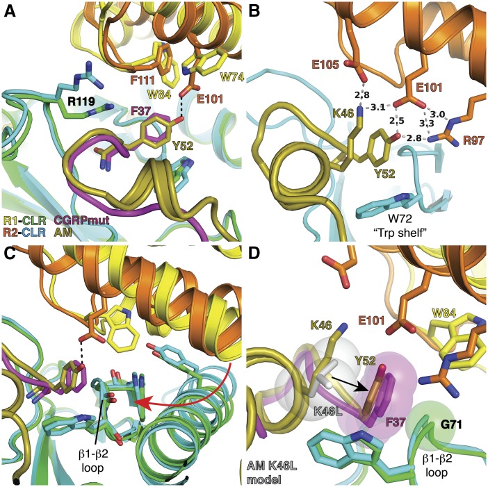
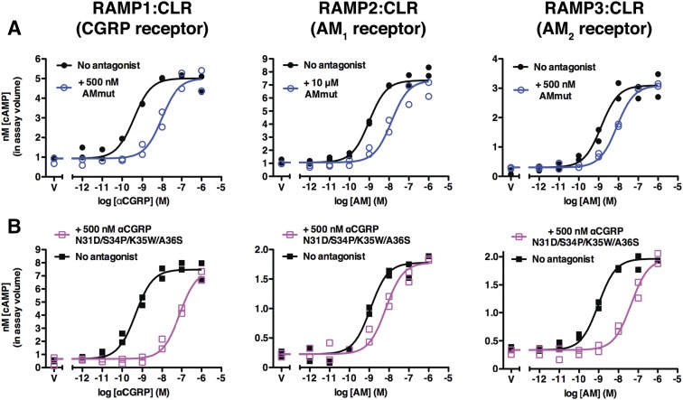
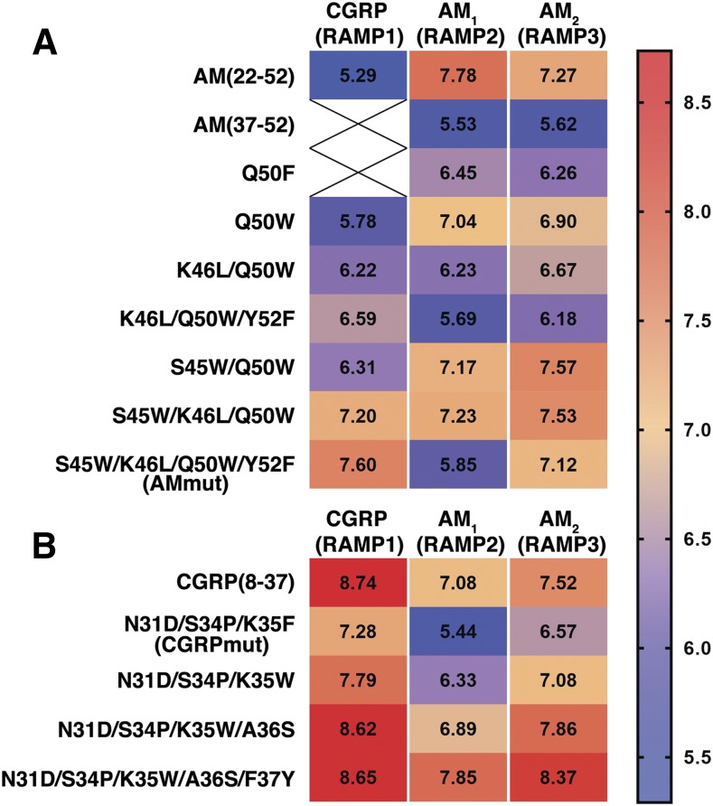
Binding of the vasodilator peptides adrenomedullin (AM) and calcitonin gene-related peptide (CGRP) to the class B G protein-coupled receptor calcitonin receptor-like receptor (CLR) is modulated by receptor activity-modifying proteins (RAMPs). RAMP1 favors CGRP, whereas RAMP2 and RAMP3 favor AM. Crystal structures of peptide-bound RAMP1/2-CLR extracellular domain (ECD) heterodimers suggested RAMPs alter ligand preference through direct peptide contacts and allosteric modulation of CLR. Here, we probed this dual mechanism through rational structure-guided design of AM and CGRP antagonist variants. Variants were characterized for binding to purified RAMP1/2-CLR ECD and for antagonism of the full-length CGRP (RAMP1:CLR), AM1 (RAMP2:CLR), and AM2 (RAMP3:CLR) receptors. Short nanomolar affinity AM(37-52) and CGRP(27-37) variants were obtained through substitutions including AM S45W/Q50W and CGRP K35W/A36S designed to stabilize their β-turn. K46L and Y52F substitutions designed to exploit RAMP allosteric effects and direct peptide contacts, respectively, yielded AM variants with selectivity for the CGRP receptor over the AM1 receptor. AM(37-52) S45W/K46L/Q50W/Y52F exhibited nanomolar potency at the CGRP receptor and micromolar potency at AM1 A 2.8-Å resolution crystal structure of this variant bound to the RAMP1-CLR ECD confirmed that it bound as designed. CGRP(27-37) N31D/S34P/K35W/A36S exhibited potency and selectivity comparable to the traditional antagonist CGRP(8-37). Giving this variant the ability to contact RAMP2 through the F37Y substitution increased affinity for AM1, but it still preferred the CGRP receptor. These potent peptide antagonists with altered selectivity inform the development of AM/CGRP-based pharmacological tools and support the hypothesis that RAMPs alter CLR ligand selectivity through allosteric effects and direct peptide contacts.


Figures

Fig. 1.
Fig. 1.
Enhancing AM affinity through rational structure-based design. (A) “Two-domain” peptide-binding model for RAMP:CLR complexes. (B) Crystal structures of AM-bound MBP-RAMP2-CLR ECD (Protein Data Bank 4RWF) and CGRPmut-bound MBP-RAMP1-CLR ECD (PDB 4RWG) in cartoon representation. MBP is omitted for clarity. Dotted lines represent disordered linkers connecting the ECDs. (C) Detailed view centered on the pocket occupied by the C-terminal AM Y52 and CGRPmut F37. (D) Model of AM S45W/Q50W bound to RAMP2-CLR ECD. Two conformations are modeled for Q50W. (E and F) Competition AlphaLISA peptide-binding assays with purified MBP-RAMP1-(GS)5-CLR ECD (E) or MBP-RAMP2[L160R]-(GS)5-CLR ECD fusion proteins (F) and indicated competitor peptides. Each panel is representative of two to three independent experiments performed in duplicate. Individual data points for each technical replicate are shown. (G) Amino acid sequence alignment of human AM(13–52), αCGRP(1–37), and CGRPmut. Numbers above the sequences correspond to AM amino acid position, whereas numbers below the sequences correspond to CGRP amino acid position. Dark-blue line represents the N-terminal disulfide linkage.
Fig. 2.
Fig. 2.
FP peptide-binding assay utilizing an AM(37–52) S45W/Q50W probe. (A and B) Saturation binding assays with 7 nM FITC-AM(37–52) S45W/Q50W and indicated concentrations of purified MBP-RAMP1-(GS)5-CLR ECD (A) or MBP-RAMP2[L106R]-(GS)5-CLR ECD (B) tethered fusion proteins. (C and D) Competition binding assays using 7 nM FITC-AM(37–52) S45W/Q50W and 15 nM MBP-RAMP1-(GS)5-CLR ECD (C) or 110 nM MBP-RAMP2[L106R]-(GS)5-CLR ECD complexes (D) and indicated concentration of unlabeled competitor AM(37–52) S45W/Q50W. Each panel is representative of at least three independent experiments performed in duplicate. Individual data points for each technical replicate are shown.
Fig. 3.
Fig. 3.
Enhancing CGRP peptide affinity through rational structure-based design. (A) Modeled CGRP K35W showing two possible conformations. (B) Modeled CGRP A36S showing hydrogen bonds in two possible conformations. (C and D) Representative competition binding FP assays using 7 nM FITC-AM(37–52) S45W/Q50W and 15 nM MBP-RAMP1-(GS)5-CLR ECD (C) or 110 nM MBP-RAMP2[L106R]-(GS)5-CLR ECD (D) with competitor peptides CGRPmut or CGRP(27–37) N31D/S34P/K35W/A36S. Each panel is representative of at least three independent experiments performed in duplicate. Individual data points for each technical replicate are shown.
Fig. 4.
Fig. 4.
Designing peptide substitutions to exploit RAMP-peptide contacts and putative RAMP allosteric modulation of CLR ECD conformation. (A) Detailed view of AM and CGRPmut C-terminal residue contacts with the RAMP subunits (Protein Data Bank 4RWF and 4RWG). Dotted line represents a hydrogen bond. (B) Hydrogen bond network involving AM K46, Y52, and RAMP2 R97, E101, and E105 (Protein Data Bank 4RWF). Hydrogen bond distances in angstroms are in black text. (C) Differences in the CLR β1-β2 loop position in the RAMP1- and RAMP2-CLR ECD complexes (Protein Data Bank 4RWF and 4RWG). The red arrow indicates the putative allosteric pathway propagating changes from the RAMP:CLR interface to the CLR β1-β2 loop (see also Supplemental Movie S1). (D) Modeling AM K46L suggests that it would push Y52 to a position similar to that of CGRPmut F37, allowing contact with G71 in RAMP1-CLR while clashing with G71 in RAMP2-CLR. Semitransparent space-filling spheres are shown for the modeled Leu at AM position 46, CGRPmut F37, and G71 in RAMP1-CLR ECD.
Fig. 5.
Fig. 5.
Binding of AM variants substituted at position 46 and the AMmut quadruple mutant to purified MBP-RAMP1/2-CLR ECD complexes. (A and B) Single-point competition FP assays using 7 nM FITC-AM S45W/Q50W and 15 nM MBP-RAMP1-(GS)5-CLR ECD (A) or 110 nM MBP-RAMP2[L106R]-(GS)5-CLR ECD (B) with indicated concentration of competitor peptides. Each of the variants is in the AM(37–52) scaffold. (C and D) Competition binding FP assays using 7 nM FITC-AM(37–52) S45W/Q50W and 15 nM MBP-RAMP1-(GS)5-CLR ECD (C) or 110 nM MBP-RAMP2[L106R]-(GS)5-CLR ECD (D) with competitor peptides AM(37–52) and the S45W/K46L/Q50W/Y52F variant (AMmut). (A) and (B) are representative of at least two independent experiments performed in duplicate with error bars shown as S.D., and (C) and (D) are representative of three independent experiments performed in duplicate with individual data points for each technical replicate shown.
Fig. 6.
Fig. 6.
Summary of AM and CGRP pKI values obtained in the FP assay with purified ECD complexes. Plot of the mean pKI values for AM variants (A) or CGRP variants (B) at either MBP-RAMP1-CLR ECD or MBP-RAMP2-CLR ECD purified fusion proteins. AM variants are in the context of AM(37–52), whereas CGRP variants are in the context of CGRP(27–37). Open circles indicate that pKI values were only obtainable at one of the purified RAMP1/2-CLR ECD complexes, whereas binding the other receptor had an estimated pKI of <4.3. Error bars indicate 95% confidence intervals. See Supplemental Fig. S4, Supplemental Table S2, and Table 1 for statistical analysis.
Fig. 7.
Fig. 7.
Antagonism of AM and CGRP variants at intact RAMP:CLR complexes in COS-7 cells. Concentration-response curves for CGRP, AM1, and AM2 receptors transiently expressed in COS-7 cells stimulated with αCGRP (CGRP receptor) or AM (AM receptors) agonists in the presence or absence of the indicated concentrations of AMmut (A) or CGRP(27–37) N31D/S34P/K35W/A36S (B). V, represents vehicle control with no agonist. Each panel is representative of three independent experiments performed in duplicate. Individual data points for each technical replicate are shown.
Fig. 8.
Fig. 8.
Heat map summary of AM and CGRP variant apparent pKB values obtained in the cell-based signaling assay in COS-7 cells. Apparent pKB values obtained for transiently expressed CGRP receptor (RAMP1), AM1 receptor (RAMP2), or AM2 receptor (RAMP3) stimulated with αCGRP (CGRP receptor) or AM (AM receptors) in the presence or absence of the indicated AM antagonist variants (A) or CGRP antagonist variants (B). AM variants are in the context of AM(37–52), whereas CGRP variants are in the context of CGRP(27–37). The mean apparent pKB values for AM(37–52) and AM Q50F at the CGRP receptor were estimated to be less than 5.3 (indicated by X symbol). See Table 2 for apparent pKB values with associated error and Supplemental Fig. S7 and Supplemental Table S3 for statistical analysis.
Fig. 9.
Fig. 9.
Structural basis for AMmut binding to the RAMP1-CLR ECD complex. (A) Cartoon representation of the 2.8-Å resolution crystal structure of AMmut-bound MBP-RAMP1-CLR ECD with AMmut in gray, CLR ECD in blue, and RAMP1 ECD in red. Two alternate conformations were modeled for S45W. The (GSA)3 linker sequence connecting the two ECDs was disordered. MBP is omitted for clarity. (B) Superimposition of AMmut-bound RAMP1-CLR ECD [colored as in (A)] and AM (gold) bound to RAMP2 ECD (orange)-CLR ECD (cyan) (Protein Data Bank 4RWF). (C) Superimposition of AMmut-bound RAMP1-CLR ECD [colored as in (A)] and CGRPmut (violet) bound to RAMP1 ECD (yellow)-CLR ECD (green) (Protein Data Bank 4RWG). (D) Detailed view comparing peptide-binding interactions for the three peptide-bound RAMP ECD-CLR ECD heterodimer crystal structures (current structure and Protein Data Bank 4RWF and 4RWG). Key peptide and RAMP residues are labeled and colored as in (A)–(C).

Similar articles

Cited by

References

    1. Adams PD, Afonine PV, Bunkóczi G, Chen VB, Davis IW, Echols N, Headd JJ, Hung LW, Kapral GJ, Grosse-Kunstleve RW, et al. (2010) PHENIX: a comprehensive Python-based system for macromolecular structure solution. Acta Crystallogr D Biol Crystallogr 66:213–221. - PMC - PubMed
    1. Aubdool AA, Thakore P, Argunhan F, Smillie SJ, Schnelle M, Srivastava S, Alawi KM, Wilde E, Mitchell J, Farrell-Dillon K, et al. (2017) A novel α-calcitonin gene-related peptide analogue protects against end-organ damage in experimental hypertension, cardiac hypertrophy, and heart failure. Circulation 136:367–383. - PMC - PubMed
    1. Bailey RJ, Hay DL. (2006) Pharmacology of the human CGRP1 receptor in Cos 7 cells. Peptides 27:1367–1375. - PubMed
    1. Barwell J, Gingell JJ, Watkins HA, Archbold JK, Poyner DR, Hay DL. (2012) Calcitonin and calcitonin receptor-like receptors: common themes with family B GPCRs? Br J Pharmacol 166:51–65. - PMC - PubMed
    1. Booe JM, Walker CS, Barwell J, Kuteyi G, Simms J, Jamaluddin MA, Warner ML, Bill RM, Harris PW, Brimble MA, et al. (2015) Structural basis for receptor activity-modifying protein-dependent selective peptide recognition by a G protein-coupled receptor. Mol Cell 58:1040–1052. - PMC - PubMed

Publication types

MeSH terms

LinkOut - more resources