Comparative Study
. 2018 Mar 9;293(10):3758-3769.
doi: 10.1074/jbc.RA117.000295. Epub 2018 Jan 23.

Fibroblast growth factor 2 induces proliferation and fibrosis via SNAI1-mediated activation of CDK2 and ZEB1 in corneal endothelium

Affiliations
Comparative Study

Fibroblast growth factor 2 induces proliferation and fibrosis via SNAI1-mediated activation of CDK2 and ZEB1 in corneal endothelium

Jeong Goo Lee et al. J Biol Chem. .

Abstract

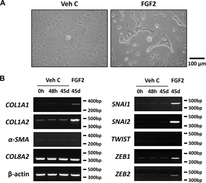
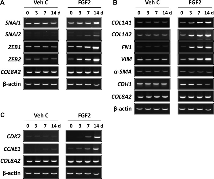
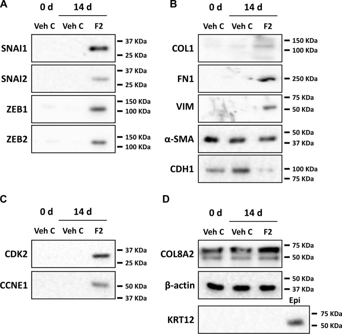
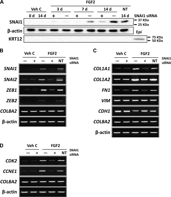
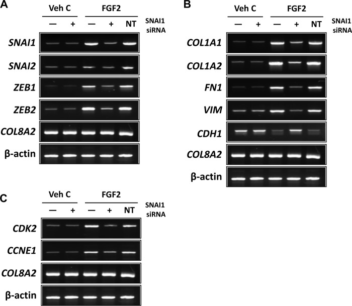
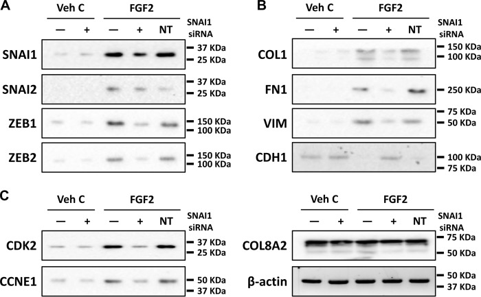
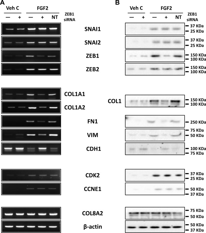
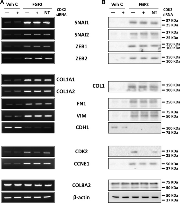
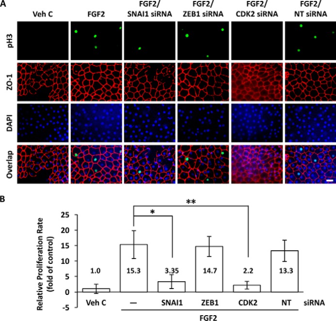
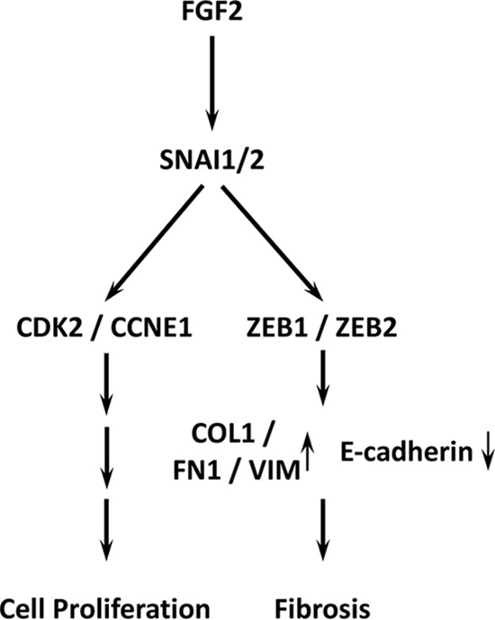
Investigating stimulation of endogenous wound healing in corneal endothelial cells (CECs) may help address the global shortage of donor corneas by decreasing the number of transplants performed for blindness because of endothelial dysfunction. We previously reported that IL-1β stimulation leads to fibroblast growth factor (FGF2) expression, enhancing migration and proliferation of mammalian CECs. However, FGF2 also promotes the endothelial-mesenchymal transition, which can lead to retrocorneal membrane formation and blindness. This prompted us to investigate downstream FGF2 signaling targets that could be manipulated to prevent retrocorneal membrane formation. FGF2 stimulation altered cell morphology and induced expression of mesenchymal transition marker genes such as snail family transcriptional repressor 1 (SNAI1), SNAI2, zinc finger E-box-binding homeobox 1 (ZEB1), and ZEB2 This, in turn, induced expression of fibronectin, vimentin, and type I collagen, and suppressed E-cadherin in CECs in vitro and ex vivo siRNA-mediated SNAI1 knockdown revealed that SNAI1 induces ZEB1 expression, in turn inducing expression of type I collagen, the major component of retrocorneal membranes, and of cyclin-dependent kinase 2 (CDK2) and cyclin E1, promoting cell proliferation. siRNA-mediated knockdown of SNAI1 or ZEB1, but not of CDK2, inhibited FGF2-dependent expression of fibronectin, vimentin, and type I collagen and of suppression of E-cadherin expression. We conclude that SNAI1 is a key regulator of FGF2-dependent mesenchymal transition in human ex vivo corneal endothelium, with ZEB1 regulating type I collagen expression and CDK2 regulating cell proliferation. These results suggest that SNAI1 promotes fibrosis and cell proliferation in human corneal endothelium through ZEB1 and CDK2.

Keywords: FGF2; SNAI1; ZEB1; cornea; corneal endothelium; endothelial-mesenchymal transition; endothelium; fibroblast growth factor (FGF); fibrosis; mesenchymal transition; zinc finger.


Conflict of interest statement

The authors declare that they have no conflicts of interest with the contents of this article

Figures

Figure 1.
Figure 1.
FGF2 induces mesenchymal transition-related gene expression in human primary corneal endothelial cells in vitro. A, after 45 days of FGF2 stimulation, the polygonal cell morphology of corneal endothelial cells (CEC) is altered to the elongated and spindle shape, whereas CEC treated with vehicle control showed normal morphology. B, following treatment for designated times (d, day) and culture conditions, total RNA from CEC were purified and RT-PCR was performed. Marked induction of COL1A1, COL1A2, SNAI1, SNAI2, ZEB1, and ZEB2 were noted in FGF2 but not in vehicle control treated human primary CEC. Expression of αSMA and TWIST was not altered by FGF2 treatment. COL8A2 and β-actin were used as corneal endothelial marker and loading control respectively. Veh C, vehicle control.
Figure 2.
Figure 2.
FGF2 regulates mesenchymal transition- and proliferation-related gene expression at the transcriptional level in human ex vivo corneal endothelium. Following treatment for designated times, total RNA from human ex vivo corneal endothelium was purified and RT-PCR performed. A, expression of SNAI1, SNAI2, ZEB1, and ZEB2 was increased by FGF2 treatment in a time-dependent manner. Treatment with vehicle control had no effect. B, FGF2 treatment led to increased expression of COL1A1, COL1A2, fibronectin, and vimentin, while it suppressed expression of E-cadherin. Similar to human CECs in vitro, FGF2 treatment had no effect on expression of αSMA. C, increased expression of CDK2 and Cyclin E1 were also noted in FGF2-treated, but not in vehicle control–treated, human ex vivo corneal endothelium. COL8A2 and β-actin were used as corneal endothelial marker and loading control, respectively. Veh C, vehicle control.
Figure 3.
Figure 3.
FGF2 regulates mesenchymal transition- and proliferation-related gene expression at the translational level in human ex vivo corneal endothelium. After treatment of human ex vivo corneas for indicated times (0 or 14 days), total protein was isolated from the endothelium-Descemet's membrane complex. A, increased protein levels of SNAI1, SNAI2, ZEB1, and ZEB2 were noted in FGF2-treated, but not in vehicle control–treated, human ex vivo corneal endothelium. B, increased expression of COL1, fibronectin, and vimentin and suppression of E-cadherin were observed in FGF2-treated, but not in vehicle control–treated, human ex vivo corneal endothelium. FGF2 treatment also had no effect on expression of αSMA protein. C, CDK2 and cyclin E1 protein levels were also increased in FGF2-treated, but not in vehicle control–treated, human ex vivo corneal endothelium. D, COL8A2 and keratin 12 were used as markers of corneal endothelium and epithelium, respectively, and β-actin was used as loading control. Veh C, vehicle control.
Figure 4.
Figure 4.
FGF2 regulates mesenchymal transition- and proliferation-related gene expression at the transcriptional level through SNAI1 in human primary corneal endothelial cells in vitro. A, to determine application time point of SNAI1 siRNA, human primary corneal endothelial cells were transfected with SNAI1 siRNA and total protein was isolated at indicated times (3, 7, or 14 days) post transfection. siRNA knockdown of FGF2-dependent SNAI1 protein expression was observed at up to 14 days post transfection. Keratin 12 was used as control for corneal epithelial cell contamination. B, SNAI1 siRNA knocked down FGF2-dependent SNAI1, SNAI2, ZEB1, and ZEB2 expression in human primary CEC. C, SNAI1 siRNA knockdown led to inhibition of FGF2-dependent expression of COL1A1, COL1A2, and fibronectin; however, there was no change in expression of vimentin. FGF2-dependent suppression of E-cadherin was also reversed by SNAI1 siRNA. D, SNAI1 siRNA transfection inhibited FGF2-dependent expression of CDK2 and cyclin E1 in primary human CEC. COL8A2 and β-actin were used as corneal endothelial marker and loading control, respectively. Transfection with non-targeting control siRNA did not have any effect on gene expression in all experiments. Veh C, vehicle control; Epi, corneal epithelium; NT, non-targeting control.
Figure 5.
Figure 5.
FGF2 regulates mesenchymal transition- and proliferation-related gene expression at the transcriptional level through SNAI1 in human ex vivo corneal endothelium. A, SNAI1 siRNA knockdown attenuated FGF2-dependent expression of SNAI1, SNAI2, ZEB1, and ZEB2 in human ex vivo corneal endothelium. B, SNAI1 siRNA knockdown decreased FGF2-dependent expression of COL1A1, COL1A2, fibronectin, and vimentin. There also was an inhibition of FGF2-dependent suppression of E-cadherin expression by SNAI1 siRNA knockdown. C, SNAI1 siRNA knockdown attenuated FGF2-dependent expression of CDK2 and cyclin E1. There was no effect on gene expression with non-targeting siRNA transfection in all experiments. COL8A2 and β-actin were used as corneal endothelial marker and loading control, respectively. Veh C, vehicle control; NT, non-targeting control.
Figure 6.
Figure 6.
FGF2 regulates mesenchymal transition- and proliferation-related gene expression at the translational level through SNAI1 in human ex vivo corneal endothelium. A, SNAI1 siRNA knockdown inhibited FGF2-dependent expression of SNAI1, SNAI2, ZEB1, and ZEB2 proteins. B, FGF2-dependent expression of COL1, fibronectin and vimentin was inhibited SNAI1 siRNA knockdown. FGF2-dependent suppression of E-cadherin was also inhibited by SNAI1 siRNA knockdown. C, SNAI1 siRNA knockdown inhibited FGF2-dependent expression of CDK2 and CCNE1. Transfection with non-targeting control siRNA did not alter gene expression in all experiments. COL8A2 and β-actin were used as corneal endothelial marker and loading control, respectively. Veh C, vehicle control; NT, non-targeting control.
Figure 7.
Figure 7.
ZEB1 regulates fibrosis but not proliferation in human ex vivo corneal endothelium. A and B, RT-PCR (A) and Western blotting (B) showed ZEB1 siRNA knockdown inhibited FGF2-dependent expression of COL1, fibronectin, and vimentin, and inhibited FGF2-dependent suppression of E-cadherin. SNAI1, SNAI2, and ZEB2 levels were not affected by ZEB1 siRNA knockdown. ZEB1 siRNA transfection also did not affect FGF2-dependent expression of CDK2 and cyclin E1. Transfection with non-targeting control siRNA did not alter gene expression in all experiments. COL8A2 and β-actin were used as corneal endothelial marker and loading control, respectively. Veh C, vehicle control; NT, non-targeting control.
Figure 8.
Figure 8.
CDK2 regulates proliferation but not fibrosis in human ex vivo corneal endothelium. A and B, RT-PCR (A) and immunoblotting (B) showed CDK2 siRNA knockdown inhibited FGF2-dependent expression of CDK but not cyclin E1. Moreover, CDK2 siRNA knockdown did not alter FGF2-dependent expression of SNAI1, SNAI2, ZEB1, ZEB2, fibronectin, vimentin, and type I collagen. There also was effect on FGF2-depedent suppression of E-cadherin by CDK2 siRNA knockdown. Transfection with non-targeting control siRNA did not alter gene expression in all experiments. COL8A2 and β-actin were used as corneal endothelial marker and loading control respectively. Veh C, vehicle control; NT, non-targeting control.
Figure 9.
Figure 9.
FGF2 promotes proliferation through SNAI1 but not ZEB1 in human ex vivo corneal endothelium. A, FGF2 treatment following injury of human ex vivo corneal endothelium induced phosphorylation of histone H3 (pH3) in the nuclei (green) of endothelial cells immediately adjacent to the injury site. FGF2-induced histone H3 phosphorylation was severely attenuated by SNAI1 or CDK2 siRNA knockdown, but not by ZEB1 siRNA knockdown. Transfection with non-targeting control siRNA had no effect on FGF2-induced histone H3 phosphorylation. Corneal endothelial cell membranes were visualized using anti-ZO-1 (red) antibody and nuclei were stained with DAPI (blue). Scale bar = 50 μm. B, FGF2 treatment increased the relative proliferation rate compared with vehicle control in human ex vivo corneal endothelium, 15.3 ± 4.5 versus 1.0 ± 1.52, p < 0.05. SNAI1 and CDK2 siRNA knockdown reduced relative proliferation rates compared with FGF2 treatment alone, 3.35 ± 2.26 versus 15.3 ± 4.5, *, p < 0.05 and 2.2 ± 1.24 versus 15.3 ± 4.5, **, p < 0.05, respectively. There was no significant change in relative proliferation rate for ZEB1 siRNA knockdown versus FGF2 treatment alone, 14.7 ± 3.23 versus 15.3 ± 4.5. One-way ANOVA, F(5,9) = 9.0, p < 0.003, n = 9 per sample. Tukey's post hoc test, HSD[0.05] = 11.4 and HSD[0.01] = 15.0. Veh C, vehicle control; NT, non-targeting control.
Figure 10.
Figure 10.
FGF2 signaling enhances mesenchymal transition through SNAI1 and ZEB1 in human corneal endothelium. FGF2 induced SNAI1 activates parallel and independent ZEB1 and CDK2 pathways, leading to regulation of fibrosis and proliferation.

References

    1. Geroski D. H., and Edelhauser H. F. (1984) Quantitation of Na/K ATPase pump sites in the rabbit corneal endothelium. Invest. Ophthalmol. Vis. Sci. 25, 1056–1060 - PubMed
    1. Kreutziger G. O. (1976) Lateral membrane morphology and gap junction structure in rabbit corneal endothelium. Exp. Eye Res. 23, 285–293 10.1016/0014-4835(76)90129-9 - DOI - PubMed
    1. Joyce N. C., Navon S. E., Roy S., and Zieske J. D. (1996) Expression of cell cycle-associated proteins in human and rabbit corneal endothelium in situ. Invest. Ophthalmol. Vis. Sci. 37, 1566–1575 - PubMed
    1. Senoo T., and Joyce N. C. (2000) Cell cycle kinetics in corneal endothelium from old and young donors. Invest. Ophthalmol. Vis. Sci. 41, 660–667 - PubMed
    1. Kay E. P., Gu X., Ninomiya Y., and Smith R. E. (1993) Corneal endothelial modulation: A factor released by leukocytes induces basic fibroblast growth factor that modulates cell shape and collagen. Invest. Ophthalmol. Vis. Sci. 34, 663–672 - PubMed

Publication types

MeSH terms