. 2018 Mar 23;293(12):4403-4410.
doi: 10.1074/jbc.M117.817890. Epub 2018 Jan 23.

The arrestin-1 finger loop interacts with two distinct conformations of active rhodopsin

Affiliations

The arrestin-1 finger loop interacts with two distinct conformations of active rhodopsin

Matthias Elgeti et al. J Biol Chem. .

Abstract

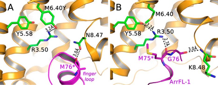
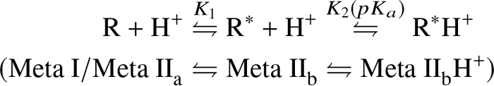
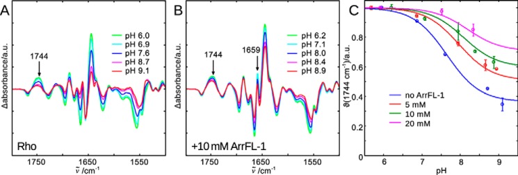
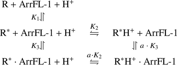
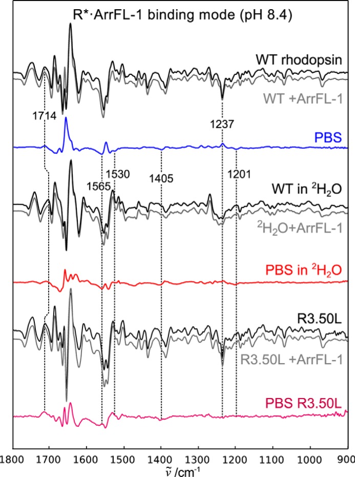
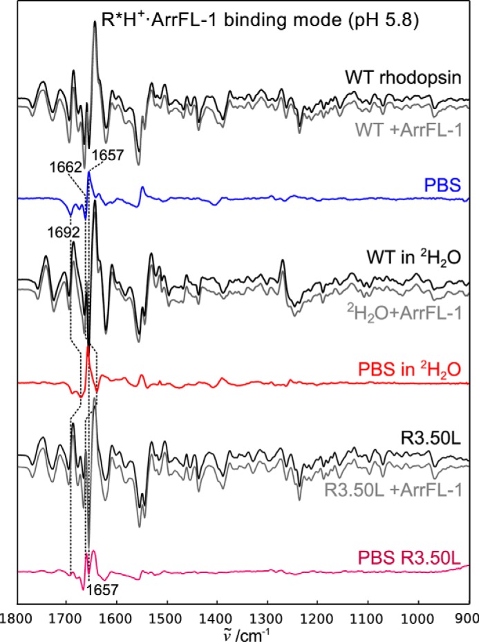
Signaling of the prototypical G protein-coupled receptor (GPCR) rhodopsin through its cognate G protein transducin (Gt) is quenched when arrestin binds to the activated receptor. Although the overall architecture of the rhodopsin/arrestin complex is known, many questions regarding its specificity remain unresolved. Here, using FTIR difference spectroscopy and a dual pH/peptide titration assay, we show that rhodopsin maintains certain flexibility upon binding the "finger loop" of visual arrestin (prepared as synthetic peptide ArrFL-1). We found that two distinct complexes can be stabilized depending on the protonation state of E3.49 in the conserved (D)ERY motif. Both complexes exhibit different interaction modes and affinities of ArrFL-1 binding. The plasticity of the receptor within the rhodopsin/ArrFL-1 complex stands in contrast to the complex with the C terminus of the Gt α-subunit (GαCT), which stabilizes only one specific substate out of the conformational ensemble. However, Gt α-subunit binding and both ArrFL-1-binding modes involve a direct interaction to conserved R3.50, as determined by site-directed mutagenesis. Our findings highlight the importance of receptor conformational flexibility and cytoplasmic proton uptake for modulation of rhodopsin signaling and thereby extend the picture provided by crystal structures of the rhodopsin/arrestin and rhodopsin/ArrFL-1 complexes. Furthermore, the two binding modes of ArrFL-1 identified here involve motifs of conserved amino acids, which indicates that our results may have elucidated a common modulation mechanism of class A GPCR-G protein/-arrestin signaling.

Keywords: Fourier transform IR (FTIR); G protein; G protein-coupled receptor (GPCR); arrestin; biased signaling; functional selectivity; rhodopsin.


Conflict of interest statement

The authors declare that they have no conflicts of interest with the contents of this article

Figures

Figure 1.
Figure 1.
Crystal structures presenting the rhodopsin/arrestin-1 finger loop binding interface. A, crystal structure of a fusion protein of constitutively active, thermostable human opsin (orange, substitutions: E3.28Q/M6.40Y and N2C/N282C) with preactivated mouse visual arrestin (purple). B, crystal structure of light-activated native bovine rhodopsin (orange) and ArrFL-1 (purple), an 11-mer peptide derived from the arrestin-1 finger loop (67YGQEDIDVMGL77). PDB IDs are 4ZWJ and 4PXF for A and B, respectively. Parts of TM6 were omitted for clarity (asterisk).
Scheme 1
Scheme 1
Figure 2.
Figure 2.
FTIR difference spectra of rhodopsin light activation in the native membrane and at different pHs are recorded to illuminate the different agonist-bound receptor conformations in equilibrium (Scheme 1). A, lowering the bulk pH causes E3.49 protonation and disruption of the E3.49-R3.50 ionic lock (23), which leads to stabilization of the active R*H+ conformation and an intensity increase of difference bands indicating activating structural changes. The band at 1744 cm−1 is a suitable monitor of the active conformations R* and R*H+ and it is isolated from other changing absorbance bands. B, addition of 10 mm ArrFL-1 leads to several additional difference bands (e.g. 1659 cm−1). It also stabilizes the active conformation, because even at high pH the intensity of the 1744 cm−1 marker band reflects predominantly active conformation formed. C, evaluation of 1744 cm−1 intensity changes as a function of pH and ArrFL-1. With increasing ArrFL-1 concentration an upshift of the apparent pKa of proton uptake and an increase of the alkaline end point level are observed, which indicates stabilization of R* and R*H+ because of ArrFL-1 binding. For each pH value two datasets have been acquired independently and averaged, the deviation is shown as error bar.
Scheme 2
Scheme 2
Figure 3.
Figure 3.
Peptide-binding spectra of the deprotonated binding mode R*·ArrFL-1. Difference spectra of wildtype rhodopsin (WT) in native disk membranes were recorded in the absence (black) and presence (gray) of 20 mm ArrFL-1 peptide. The resulting double difference (PBS) is shown in blue and reflects structural changes because of ArrFL-1 binding to the deprotonated receptor. Identical experiments were performed after H2O/2H2O buffer exchange and R3.50L mutation, the resulting PBS are shown in red and purple, respectively.
Figure 4.
Figure 4.
The R*H+·ArrFL-1 binding mode stabilized at low pH. Difference spectra recorded in the absence of ArrFL-1 peptide are shown in black, in the presence of 20 mm ArrFL-1 in gray. The resulting PBS are shown in blue (WT), red (in 2H2O), or purple (R3.50L mutant). Note the difference in band pattern compared with the deprotonated complex. The small effect of 2H2O on the positive 1657 cm−1 indicates that this band is a structurally sensitive amide I band.

References

    1. Farrens D. L., Altenbach C., Yang K., Hubbell W. L., and Khorana H. G. (1996) Requirement of rigid-body motion of transmembrane helices for light activation of rhodopsin., Science 274, 768–770 10.1126/science.274.5288.768 - DOI - PubMed
    1. Scheerer P., Park J. H., Hildebrand P. W., Kim Y. J., Krauss N., Choe H.-W. W., Hofmann K. P., and Ernst O. P. (2008) Crystal structure of opsin in its G-protein-interacting conformation, Nature 455, 497–502 10.1038/nature07330 - DOI - PubMed
    1. Hofmann K. P., Scheerer P., Hildebrand P. W., Choe H.-W., Park J. H., Heck M., and Ernst O. P. (2009) A G protein-coupled receptor at work: the rhodopsin model., Trends Biochem. Sci. 34, 540–552 10.1016/j.tibs.2009.07.005 - DOI - PubMed
    1. Shenoy S. K., and Lefkowitz R. J. (2011) β-Arrestin-mediated receptor trafficking and signal transduction., Trends Pharmacol. Sci. 32, 521–533 10.1016/j.tips.2011.05.002 - DOI - PMC - PubMed
    1. Zhou X. E., He Y., de Waal P. W., Gao X., Kang Y., Van Eps N., Yin Y., Pal K., Goswami D., White T. A., Barty A., Latorraca N. R., Chapman H. N., Hubbell W. L., Dror R. O., Stevens R. C., Cherezov V., Gurevich V. V., Griffin P. R., Ernst O. P., Melcher K., and Xu H. E. (2017) Identification of phosphorylation codes for arrestin recruitment by G protein-coupled receptors. Cell 170, 457–469.e13 10.1016/j.cell.2017.07.002 - DOI - PMC - PubMed

Publication types

LinkOut - more resources