. 2018 Feb 21;38(8):2043-2056.
doi: 10.1523/JNEUROSCI.2257-17.2018. Epub 2018 Jan 23.

Ephexin1 Is Required for Eph-Mediated Limb Trajectory of Spinal Motor Axons

Affiliations

Ephexin1 Is Required for Eph-Mediated Limb Trajectory of Spinal Motor Axons

Chih-Ju Chang et al. J Neurosci. .

Abstract

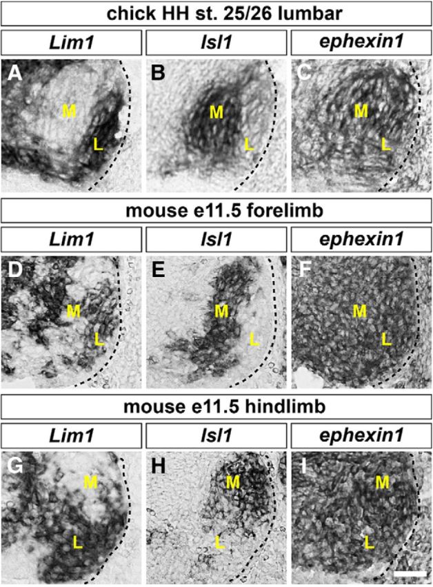
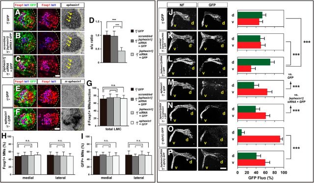
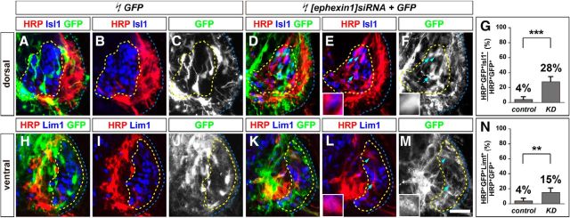
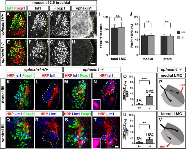
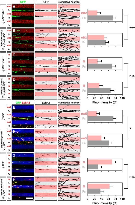
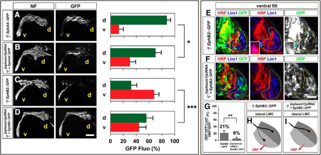
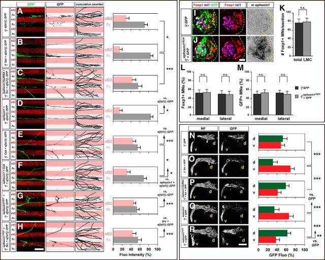
The precise assembly of a functional nervous system relies on the guided migration of axonal growth cones, which is made possible by signals transmitted to the cytoskeleton by cell surface-expressed guidance receptors. We investigated the function of ephexin1, a Rho guanine nucleotide exchange factor, as an essential growth-cone guidance intermediary in the context of spinal lateral motor column (LMC) motor axon trajectory selection in the limb mesenchyme. Using in situ mRNA detection, we first show that ephexin1 is expressed in LMC neurons of chick and mouse embryos at the time of spinal motor axon extension into the limb. Ephexin1 loss of function and gain of function using in ovo electroporation in chick LMC neurons, of either sex, perturbed LMC axon trajectory selection, demonstrating an essential role of ephexin1 in motor axon guidance. In addition, ephexin1 loss in mice of either sex led to LMC axon trajectory selection errors. We also show that ephexin1 knockdown attenuates the growth preference of LMC neurites against ephrins in vitro and Eph receptor-mediated retargeting of LMC axons in vivo, suggesting that ephexin1 is required in Eph-mediated LMC motor axon guidance. Finally, both ephexin1 knockdown and ectopic expression of nonphosphorylatable ephexin1 mutant attenuated the retargeting of LMC axons caused by Src overexpression, implicating ephexin1 as an Src target in Eph signal relay in this context. In summary, our findings demonstrate that ephexin1 is essential for motor axon guidance and suggest an important role in relaying ephrin:Eph signals that mediate motor axon trajectory selection.SIGNIFICANCE STATEMENT The proper development of functioning neural circuits requires precise nerve connections among neurons or between neurons and their muscle targets. The Eph tyrosine kinase receptors expressed in neurons are important in many contexts during neural-circuit formation, such as axon outgrowth, axon guidance, and synaptic formation, and have been suggested to be involved in neurodegenerative disorders, including amyotrophic lateral sclerosis and Alzheimer's disease. To dissect the mechanism of Eph signal relay, we studied ephexin1 gain of function and loss of function and found ephexin1 essential for the development of limb nerves toward their muscle targets, concluding that it functions as an intermediary to relay Eph signaling in this context. Our work could thus shed new light on the molecular mechanisms controlling neuromuscular connectivity during embryonic development.

Keywords: axon guidance; motor neuron; spinal cord.


Figures

Figure 1.
Figure 1.
Expression of ephexin1 in chick and mouse LMC motor neurons. A–I, Detection of mRNA in consecutive sections of spinal cords: all chick sections are HH stage 25/26 lumbar spinal cords; all mouse sections are E11.5 lumbar spinal cords. A, B, Detection of Lim1 (A) and Isl1 (B) mRNA in the chick spinal cord, highlighting lateral and medial LMC neurons, respectively. C, Detection of ephexin1 mRNA in both medial and lateral LMC neurons. D, E, G, H, Detection of Lim1 (D, G) and Isl1 (E, H) mRNA in mouse LMC neurons. F, I, Detection of ephexin1 mRNA in both medial and lateral mouse LMC neurons. M, Medial; L, lateral. Scale bar: (in I) A–I, 30 μm.
Figure 2.
Figure 2.
Ephexin1 function is required for the selection of limb axon trajectory. All images are from chick HH stage 28/29 lumbar levels. A–C, Detection of Foxp1, Isl1, GFP, and ephexin1 in LMC neurons of chick embryos electroporated with GFP (A), scrambled [ephexin1]siRNA, and GFP (B), or with [ephexin1]siRNA and GFP (C). D, Quantification of effects of GFP electroporation, of scrambled [ephexin1]siRNA and GFP electroporation, or of [ephexin1]siRNA and GFP electroporation on ephexin1 mRNA levels. The ratio of immunoreactivity signal levels in LMC neurons of the electroporated to the unelectroporated contralateral side (e/u ratio) was obtained in ≥20 sections of six embryos. E, F, Detection of Isl1, Foxp1, and GFP protein and mouse ephexin1 mRNA in the LMC of chick HH stage 28/29 electroporated with GFP (E) or ephexin1 and GFP (F) expression plasmids. G, Number of LMC motor neurons expressed as the average number of total (Foxp1+) LMC neurons per section (# Foxp1+ MNs/section). Number of embryos: n = 5 for all groups. H, I, Number of total or electroporated medial (Foxp1+ Isl1+) and lateral (Foxp1+ Isl1) LMC motor neurons in lumbar spinal cord expressed as the percentage of total motor neurons [Foxp1+ MNs (%); H] or electroporated motor neurons [GFP+ MNs (%); I]. Number of embryos: n = 5 for all groups. J–P, Neurofilament and GFP detection in the limb nerve branches in the crural plexus of chick embryos electroporated with the following expression plasmids and siRNAs: GFP (J); scrambled [ephexin1]siRNA and GFP (K); [ephexin1]siRNA and GFP (L); ephexin1 and GFP (M); [ephexin1]siRNA, mouse ephexin1, and GFP (N); medial LMC axonal marker e[Isl1]::GFP (O); or [ephexin1]siRNA and e[Isl1]::GFP (P). Quantification of GFP signals in all electroporation experiments expressed as, respectively, percentage in dorsal and ventral limb nerves [GFP Fluo (%)]. Number of embryos: n = 5 for all groups. d, Dorsal; v, ventral; error bars, SD; n.s., not significant; ***p < 0.001; statistical significance computed using Mann–Whitney U test; all values are mean ± SD. Scale bar: (in F) A–C, E, F, 45 μm; (in P) J–O, 150 μm.
Figure 3.
Figure 3.
Ephexin1 function is required for the fidelity of LMC motor axon limb trajectories. Retrograde labeling of LMC neurons by HRP injections into the dorsal or ventral limb muscles of chick HH stage 28/29 embryos. A–F, Detection of HRP, Lim1, and GFP in the LMC regions of GFP (A–C) or of [ephexin1]siRNA and GFP (D–F) electroporated embryos injected with HRP into dorsal hindlimb shank muscles. Examples of HRP+ GFP+ neurons are indicated by arrows and arrowheads (E, F). E, F, Insets, Examples indicated by arrowheads are shown at higher magnification. G, Proportions of electroporated medial LMC motor neurons labeled with HRP in dorsally filled embryos. Number of embryos: n = 5 for all groups. H–M, Detection of HRP, Lim1, and GFP in the LMC regions of GFP (H–J) or of [ephexin1]siRNA and GFP (K–M) electroporated embryos injected with HRP into ventral hindlimb shank muscles. Examples of HRP+ GFP+ neurons are indicated by arrows and arrowheads (L, M). L, M, Insets, Examples indicated by arrowheads are shown at higher magnification. N, Proportions of electroporated lateral LMC motor neurons labeled with HRP in ventrally filled embryos. Number of embryos: n = 5 for all groups. control, GFP electroporated groups; KD, [ephexin1]siRNA and GFP electroporated groups; error bars, SD; ***p < 0.001; **p < 0.01; statistical significance computed using Fisher's exact test; all values are mean ± SD. Scale bars: (in M) A–F, H–M, 45 μm; E, F, L, M, insets, 8 μm.
Figure 4.
Figure 4.
Ephexin1 is required for the fidelity of LMC motor axon trajectory selection. Retrograde labeling of LMC neurons by HRP injections into the dorsal or ventral limb muscles of mouse E12.5 embryos. A–H, Detection of Isl1 (green), Foxp1 (red), and ephexin1 protein in the LMC region at the brachial level of mouse e12.5 ephexin1+/+ (A–D) or ephexin1−/− (E–H) embryos. I, Number of LMC motor neurons expressed as the average number of total (Foxp1+) LMC neurons per section (# FoxP1+/section). Number of embryos: n = 6 for all groups. J, Number of total medial (FoxP1+ Isl1+) and lateral (FoxP1+ Isl1) LMC motor neurons in brachial spinal cord expressed as the percentage of total motor neurons [FoxP1+ MNs (%)]. Number of embryos: n = 6 for all groups. K–N, Detection of HRP (red), Isl1 (blue), and Foxp1 (green), which marks all LMC neurons, in the LMC regions of ephexin1+/+ (K, L) and ephexin1−/− (M, N) embryos injected with HRP into dorsal forelimb muscles. Examples of medial LMC motor neurons labeled with HRP are indicated by arrows and arrowheads. Examples indicated by arrowheads are shown at a higher magnification (N, inset). O, Quantification of retrogradely labeled medial LMC axon projections. The graph depicts the mean percentage ±SD of HRP+ motor neurons that express the medial LMC marker Isl1 following a dorsal limb injection. Numbers of embryos: n = 4 for all groups. P, Summary scheme of medial LMC projections in ephexin1−/− mice, depicting a significant misrouting of medial LMC axons into the dorsal limb. Q–T, Detection of HRP (red), Lim1 (blue), and Foxp1 (green) in the LMC regions of ephexin1+/+ (Q, R) and ephexin1−/− (S, T) embryos injected with HRP into ventral forelimb muscles. Examples of lateral LMC motor neurons labeled with HRP are indicated by arrows and arrowheads. Examples indicated by arrowheads are shown at a higher magnification (T, inset). U, Quantification of retrogradely labeled lateral LMC axon projections. The graph depicts the mean percentage ±SD of HRP+ motor neurons that express the lateral LMC marker Lim1 following a ventral limb injection. Number of embryos: n = 4 for all groups. V, Summary scheme of lateral LMC projections in ephexin1−/− mice, depicting a significant misrouting of lateral LMC axons into the ventral limb. Error bars, SD; n.s., not significant; ***p < 0.001; **p < 0.01; statistical significance computed using Fisher's exact test; all values are mean ± SD. Scale bars: (in H) A–H, 20 μm; (in T) K–N, Q–T, 20 μm; N, T, insets, 7 μm.
Figure 5.
Figure 5.
Ephexin1 function is required for ephrin-mediated but not netrin-mediated guidance of cultured LMC neurites. Growth preference on protein stripes exhibited by medial or lateral LMC axons. Each experiment is composed of three panels (left, middle, and right) and one quantification. A–D, Left, Detection of medial (GFP+) LMC neurites of explants on eB2/Fc (A) or N/Fc (C) stripes, and of [ephexin1]siRNA coelectroporated explants on eB2/Fc (B) or N/Fc (D) stripes. Middle, Inverted images where GFP signals are dark pixels overlaid on substrate stripes. Right, Superimposed images of five representative explants from each experimental group, highlighting the distribution of medial LMC neurites. Quantification of medial (GFP+) LMC neurites on first (pink) and second (pale) stripes expressed as a percentage of total GFP signals. Neurites, n = ≥85; explants, n = ≥12. E–H, Left, Detection of lateral (GFP+ EphA4+) LMC neurites of explants on eA5/Fc (E) or N/Fc (G) stripes, and [ephexin1]siRNA coelectroporated explants on eA5/Fc (F) or N/Fc (H) stripes. Middle, Inverted images where EphA4 signals are dark pixels overlaid on substrate stripes. Right, Superimposed images of five representative explants from each experimental group, highlighting the distribution of lateral LMC neurites. Quantification of lateral (EphA4+) LMC neurites on first (pink) and second (pale) stripes expressed as a percentage of total EphA4 signals. Neurites, n = ≥82; explants, ≥11. eB2, Ephrin-B2-Fc; eA5, ephrin-A5-Fc; N, netrin-1; error bars, SD; ***p < 0.001; *p < 0.05; n.s., not significant; statistical significance computed using Mann–Whitney U test; all values are mean ± SD. Scale bar: (in H) A–H, 150 μm.
Figure 6.
Figure 6.
Ephexin1 loss attenuates both EphA4-induced and EphB2-induced LMC motor axon redirection. All images are from chick HH stage 28/29 lumbar levels. A–D, Neurofilament and GFP detection in the limb nerve branches in the crural plexus of chick embryos electroporated with the following expression plasmids and siRNAs: EphA4::GFP (A), [ephexin1]siRNA and EphA4::GFP (B), EphB2::GFP (C), or [ephexin1]siRNA and EphB2::GFP (D). Quantification of GFP signals in all electroporation experiments expressed as, respectively, percentage in dorsal and ventral limb nerves [GFP Fluo (%)]. Number of embryos: n = 5 for all groups. E, F, Detection of HRP, Lim1, and GFP in the LMC regions of EphB2::GFP-electroporated embryos (E) or of embryos electroporated with [ephexin1]siRNA and EphB2::GFP (F) injected with HRP into ventral hindlimb shank muscles. Examples of HRP+ GFP+ neurons are indicated by arrows and arrowheads (E). Examples indicated by arrowheads are shown at higher magnification (E, insets). G, Proportions of electroporated lateral LMC motor neurons labeled with HRP in ventrally filled embryos. Number of embryos: n = 5 for all groups. H, I, Summary scheme of lateral LMC projections in EphB2::GFP-electroporated embryos (H) or in embryos electroporated with [ephexin1]siRNA and EphB2::GFP (I), depicting the attenuation of EphB2-induced lateral LMC axon misrouting by ephexin1 knockdown. d, Dorsal; v, ventral; error bars, SD; ***p < 0.001; **p < 0.01; *p < 0.05; statistical significance computed using Mann–Whitney U test (A–D) or Fisher's exact test (G); all values are mean ± SD. Scale bars: (in D) A–D, 150 μm; (in F) E, F, 45 μm; E, insets, 8 μm.
Figure 7.
Figure 7.
Ephexin1 functions downstream of Src in Eph-mediated LMC axon guidance. A–H, Growth preference on protein stripes exhibited by medial LMC axons. Each experiment is composed of three panels (left, middle, and right) and one quantification. Left, Detection on eB2/Fc stripes of medial (GFP) LMC neurites of explants expressing the following: e[Isl1]::GFP (A); Src and e[Isl1]::GFP (B); [ephexin1]siRNA, Src, and e[Isl1]::GFP (C); ephexin1 and e[Isl1]::GFP (D); Csk and e[Isl1]::GFP (E); ephexin1, Csk, and e[Isl1]::GFP (F); ephexin1Y87F and e[Isl1]::GFP (G); ephexin1Y87F, Src, and e[Isl1]::GFP (H). Middle, Inverted images where GFP signals are dark pixels overlaid on substrate stripes. Right, Superimposed images of five representative explants from each experimental group, highlighting the distribution of medial LMC neurites. Quantification of medial (GFP+) LMC neurites on first (pink) and second (pale) stripes expressed as a percentage of total GFP signals. Neurites, n ≥ 95; explants, n ≥ 15. I, J, Detection of Isl1, Foxp1, and GFP protein and mouse ephexin1 mRNA in the LMC of chick HH stage 28/29 electroporated with GFP (I) or ephexin1Y87F and GFP (J) expression plasmids. K, Number of LMC motor neurons expressed as the average number of total (Foxp1+) LMC neurons per section (# Foxp1+ MNs/section). Number of embryos: n = 6 for all groups. L, M, Number of total or electroporated medial (Foxp1+ Isl1+) and lateral (Foxp1+ Isl1) LMC motor neurons in lumbar spinal cord expressed as the percentage of total motor neurons [Foxp1+ MNs (%)] (L) or electroporated motor neurons [GFP+ MNs (%)] (M). Number of embryos: n = 6 for all groups. N–R, All images are from chick HH stage 28/29 lumbar levels. Neurofilament and GFP detection in the limb nerve branches in the crural plexus of chick embryos electroporated with the following expression plasmids and siRNAs: GFP (N); Src and GFP (O); [ephexin1]siRNA, Src, and GFP (P); ephexin1 and GFP (Q); or ephexin1Y87F and GFP (R). Quantification of GFP signals in all electroporation experiments expressed as, respectively, percentage in dorsal and ventral limb nerves [GFP Fluo (%)]. Number of embryos: n = 6 for all groups. d, Dorsal; v, ventral; error bars, SD; ***p < 0.001; **p < 0.01; *p < 0.05; n.s., not significant; statistical significance computed using Mann–Whitney U test; all values are mean ± SD. Scale bars: (in A) A–H, 150 μm; (in J) I, J, 45 μm; (in R) N–R, 150 μm.

Similar articles

Cited by

References

    1. Bladt F, Aippersbach E, Gelkop S, Strasser GA, Nash P, Tafuri A, Gertler FB, Pawson T (2003) The murine nck SH2/SH3 adaptors are important for the development of mesoderm-derived embryonic structures and for regulating the cellular actin network. Mol Cell Biol 23:4586–4597. 10.1128/MCB.23.13.4586-4597.2003 - DOI - PMC - PubMed
    1. Bonanomi D, Chivatakarn O, Bai G, Abdesselem H, Lettieri K, Marquardt T, Pierchala BA, Pfaff SL (2012) Ret is a multifunctional coreceptor that integrates diffusible- and contact-axon guidance signals. Cell 148:568–582. 10.1016/j.cell.2012.01.024 - DOI - PMC - PubMed
    1. Bülow HE, Boulin T, Hobert O (2004) Differential functions of the C. elegans FGF receptor in axon outgrowth and maintenance of axon position. Neuron 42:367–374. 10.1016/S0896-6273(04)00246-6 - DOI - PubMed
    1. Defourny J, Poirrier AL, Lallemend F, Mateo Sánchez S, Neef J, Vanderhaeghen P, Soriano E, Peuckert C, Kullander K, Fritzsch B, Nguyen L, Moonen G, Moser T, Malgrange B (2013) Ephrin-A5/EphA4 signalling controls specific afferent targeting to cochlear hair cells. Nat Commun 4:1438. 10.1038/ncomms2445 - DOI - PubMed
    1. Dudanova I, Gatto G, Klein R (2010) GDNF acts as a chemoattractant to support ephrinA-induced repulsion of limb motor axons. Curr Biol 20:2150–2156. 10.1016/j.cub.2010.11.021 - DOI - PubMed

Publication types

Substances