. 2018 Feb 6;115(6):E1309-E1318.
doi: 10.1073/pnas.1717948115. Epub 2018 Jan 23.

The eardrums move when the eyes move: A multisensory effect on the mechanics of hearing

Affiliations

The eardrums move when the eyes move: A multisensory effect on the mechanics of hearing

Kurtis G Gruters et al. Proc Natl Acad Sci U S A. .

Abstract

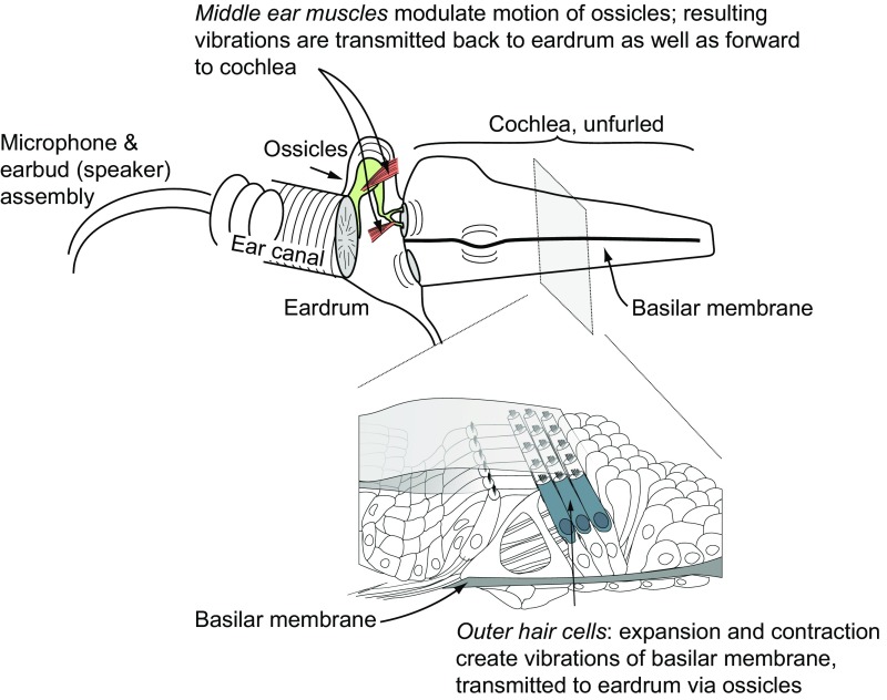
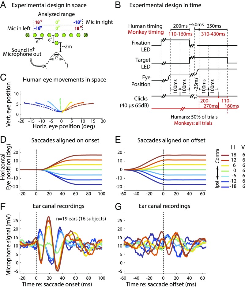
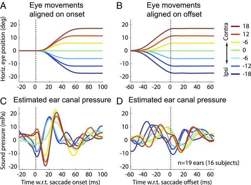
Interactions between sensory pathways such as the visual and auditory systems are known to occur in the brain, but where they first occur is uncertain. Here, we show a multimodal interaction evident at the eardrum. Ear canal microphone measurements in humans (n = 19 ears in 16 subjects) and monkeys (n = 5 ears in three subjects) performing a saccadic eye movement task to visual targets indicated that the eardrum moves in conjunction with the eye movement. The eardrum motion was oscillatory and began as early as 10 ms before saccade onset in humans or with saccade onset in monkeys. These eardrum movements, which we dub eye movement-related eardrum oscillations (EMREOs), occurred in the absence of a sound stimulus. The amplitude and phase of the EMREOs depended on the direction and horizontal amplitude of the saccade. They lasted throughout the saccade and well into subsequent periods of steady fixation. We discuss the possibility that the mechanisms underlying EMREOs create eye movement-related binaural cues that may aid the brain in evaluating the relationship between visual and auditory stimulus locations as the eyes move.

Keywords: EMREO; middle ear muscles; otoacoustic emissions; reference frame; saccade.


Conflict of interest statement

The authors declare no conflict of interest.

Figures

Fig. 1.
Fig. 1.
Motile cochlear OHCs expand and contract in a way that depends both on the incoming sound and on the descending input received from the superior olivary complex in the brain. OHC motion moves the basilar membrane, and subsequently the eardrum, via fluid/mechanical coupling of these membranes through the ossicular chain. The MEMs also pull on the ossicles, directly moving the eardrum. These muscles are innervated by motor neurons near the facial and trigeminal nerve nuclei, which receive input from the superior olive bilaterally. In either case, eardrum motion can be measured with a microphone in the ear canal.
Fig. 2.
Fig. 2.
Experimental design and results. (A) Recordings for all subjects were made via a microphone (Mic) in the ear canal set into a custom-fit ear bud. On each trial, a subject fixated on a central LED and then made a saccade to a target LED (−24° to +24° horizontally in 6° increments and 6° above the fixation point) without moving his/her head. The ±24° locations were included on only 4.5% of trials and were excluded from analysis (Methods); other target locations were equally likely (∼13% frequency). (B) Humans (black text) received randomly interleaved silent and click trials (50% each). Clicks were played via a sound transducer coupled with the microphone at four times during these trials: during the initial fixation and saccade and at 100 ms and 200 ms after target fixation. Monkeys’ trials had minor timing differences (red text), and all trials had one click at 200–270 ms after target fixation (red click trace). (C) Average eye trajectories for one human subject and session for each of the included targets are shown; colors indicate saccade target locations from ipsilateral (blue) to contralateral (red). deg, degrees; Horiz., horizontal; Vert., vertical. Mean eye position is shown as a function of time, aligned on saccade onset (D) and offset (E). H, horizontal; V, vertical. Mean microphone recordings of air pressure in the ear canal, aligned on saccade onset (F) and offset (G), indicate that the eardrum oscillates in conjunction with eye movements. The phase and amplitude of the oscillation varied with saccade direction and amplitude, respectively. The oscillations were, on average, larger when aligned on saccade onset than when aligned on saccade offset.
Fig. 3.
Fig. 3.
Regression results for data aligned to saccade onset (A, C, and E) and offset (B, D, and F). (A and B) Mean ± SEM slope of regression of microphone voltage vs. saccade target location (conducted separately for each subject and then averaged across the group) at each time point for real (red) vs. scrambled (gray) data. In the scrambled Monte Carlo analysis, the true saccade target locations were shuffled and arbitrarily assigned to the microphone traces for individual trials. (C and D) Proportion of variance (Var.) accounted for by regression fit (R2). (E and F) Percentage of subject ears showing P < 0.05 for the corresponding time point. Additional details are provided in Methods.
Fig. 4.
Fig. 4.
Recordings in left (A) and right (B) ears in an individual human subject. The eye movement-related signals were similar in the two ears when saccade direction is defined with respect to the recorded ear. The remaining individual subjects’ data can be found in Fig. S2. Contra, contralateral; Ipsi, ipsilateral; Mic, microphone.
Fig. 5.
Fig. 5.
Estimated EMREO pressure for human subjects at saccade onset (A and C) and saccade offset (B and D). Pressures were obtained from the measured microphone voltage using the microphone’s complex frequency response measured at low frequencies as described by Christensen et al. (29). Contra, contralateral; deg, degrees; Horiz., horizontal; Ipsi, ipsilateral.
Fig. 6.
Fig. 6.
Eye position (A), microphone signal of ear canal pressure (B), and results of point-by-point regression (CE) for monkey subjects (n = 5 ears). All analyses were calculated in the same way as for the human data (Figs. 2 and 3). Contra, contralateral; deg, degrees; Ipsi, ipsilateral.
Fig. 7.
Fig. 7.
EMREOs recorded normally (A) are not observed when the microphone input port is plugged to eliminate acoustic but not electrical (B) contributions to the microphone signal. The plugged microphone sessions were run as normal sessions except that after calibration, the microphone was placed in a closed earbud before continuing with the session (n = 4 subjects). (C) Similarly, EMREOs were not evident when the microphone was placed in a test tube behind a human subject’s pinna while he/she performed the experiment as normal. Contra, contralateral; Ipsi, ipsilateral.
Fig. 8.
Fig. 8.
Clicks do not appear to alter EMREOs. (Upper Row) Mean microphone signals for human subjects (AD) and monkeys (E) for click presentations at various time points are shown: during the initial fixation (A), during the saccade (B, ∼20 ms after saccade onset), and 100 ms (C) and 200 ms (D) after target fixation was obtained in humans, and 200–270 ms after target fixation in monkeys (E). (Insets) Zoomed-in views of the periclick timing for a more detailed view (gray backgrounds). (Middle, B–D) Residual microphone signal after the first click in each trial was subtracted (human subjects) is shown. There were no obvious distortions in the EMREOs at the time of the (removed) click, suggesting that the effects of EMREOs interact linearly with incoming sounds. (Lower, A–E) Mean peak-to-peak amplitude of the clicks (mean ± SE) is shown. There were no clear differences in the peak click amplitudes for any epochs, indicating that the acoustic impedance did not change as a function of saccade target location. Contra, contralateral; Ipsi, ipsilateral.

Similar articles

Cited by

References

    1. Groh JM, Sparks DL. Two models for transforming auditory signals from head-centered to eye-centered coordinates. Biol Cybern. 1992;67:291–302. - PubMed
    1. Groh JM, Trause AS, Underhill AM, Clark KR, Inati S. Eye position influences auditory responses in primate inferior colliculus. Neuron. 2001;29:509–518. - PubMed
    1. Zwiers MP, Versnel H, Van Opstal AJ. Involvement of monkey inferior colliculus in spatial hearing. J Neurosci. 2004;24:4145–4156. - PMC - PubMed
    1. Porter KK, Metzger RR, Groh JM. Representation of eye position in primate inferior colliculus. J Neurophysiol. 2006;95:1826–1842. - PubMed
    1. Bulkin DA, Groh JM. Distribution of eye position information in the monkey inferior colliculus. J Neurophysiol. 2012;107:785–795. - PMC - PubMed