Review
. 2018 May;19(5):297-312.
doi: 10.1038/nrm.2017.127. Epub 2018 Jan 24.

Once and only once: mechanisms of centriole duplication and their deregulation in disease

Affiliations
Review

Once and only once: mechanisms of centriole duplication and their deregulation in disease

Erich A Nigg et al. Nat Rev Mol Cell Biol. 2018 May.

Abstract

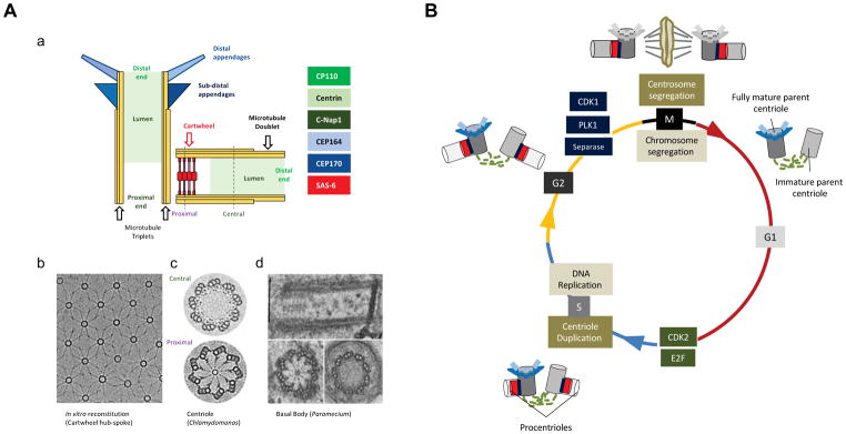
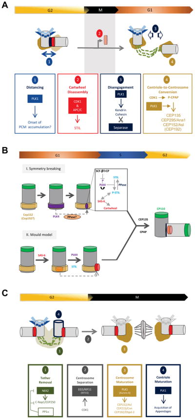
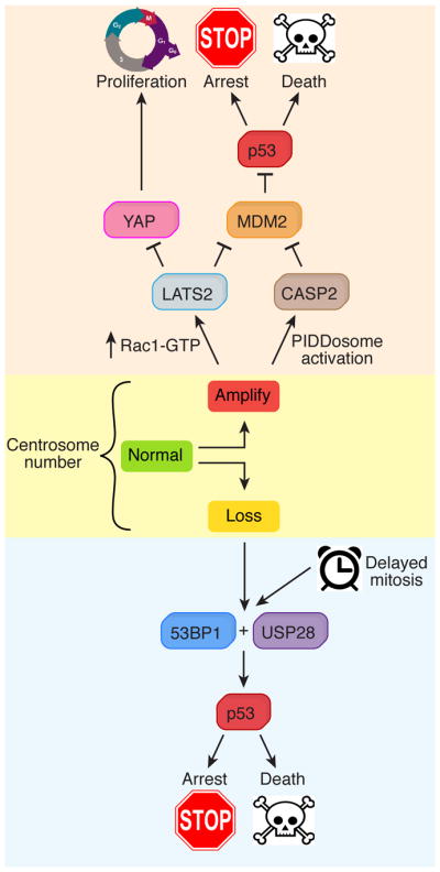
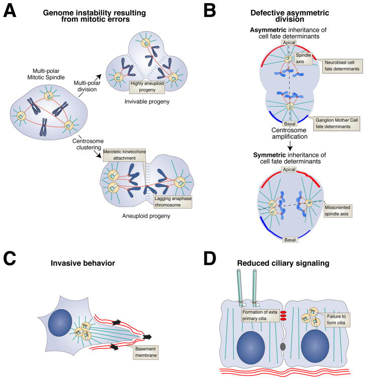
Centrioles are conserved microtubule-based organelles that form the core of the centrosome and act as templates for the formation of cilia and flagella. Centrioles have important roles in most microtubule-related processes, including motility, cell division and cell signalling. To coordinate these diverse cellular processes, centriole number must be tightly controlled. In cycling cells, one new centriole is formed next to each pre-existing centriole in every cell cycle. Advances in imaging, proteomics, structural biology and genome editing have revealed new insights into centriole biogenesis, how centriole numbers are controlled and how alterations in these processes contribute to diseases such as cancer and neurodevelopmental disorders. Moreover, recent work has uncovered the existence of surveillance pathways that limit the proliferation of cells with numerical centriole aberrations. Owing to this progress, we now have a better understanding of the molecular mechanisms governing centriole biogenesis, opening up new possibilities for targeting these pathways in the context of human disease.


Figures

Figure 1
Figure 1. Centriole architecture and the centrosome duplication-segregation cycle
(A) (a) Schematic showing fully mature parent centriole (upright) and tightly associated procentriole. Prominent markers representative for the different structures are indicated to the right. (b) Micrograph shows lattice of in vitro reconstituted cartwheel hub and spoke structures visualized by cryo-electron microscopy. Adapted with permission from. (c) Image derived from cryotomogram sections of Chlamydomonas procentriole emphasizes cartwheel and triplet microtubules. Adapted with permission from. (d) Transmission electron microscopy shows longitudinal section (top) and cross sections at proximal (lower left) and distal parts (lower right) of Paramecium basal body (Anne-Marie Tassin, unpublished). (B) Shared pathways ensure coordination of centrosome duplication-segregation and chromosome replication-segregation cycles. At the G1/S transition both centriole duplication and DNA replication depend on CDK2 as well as phosphorylation of the retinoblastoma protein pRb and liberation of E2F transcription factors. Similarly, overlapping sets of enzymes, including the kinases CDK1 and PLK1 and the protease Separase govern entry into mitosis, chromosome segregation, and licensing of DNA and centrioles for a new round of duplication. Lastly, several proteins with well-established functions in DNA transactions have been proposed to play additional roles in the centrosome cycle, but indirect effects on centrosomes remain difficult to exclude. Centrioles are depicted in different shades of grey to indicate different states of maturity. A procentriole (light grey) is a newly created centriole that is not yet duplication competent. A procentriole converts into an immature parent centriole (middle grey) following disengagement in mitosis. An immature parent centriole becomes a mature parent centriole (dark grey) following the acquisition of appendages. Appendage structures undergo a transient modification/disassembly during mitosis. Cartwheels are shown in red; loose tethers connecting parent centrioles in dashed green lines; tight linkers connecting procentrioles to their parents in dark blue; subdistal and distal appendages are shown in light and dark blue respectively.
Figure 2
Figure 2. Key aspects of the centrosome duplication cycle
(A) Mitotic events licensing a new round of centriole duplication. Schematic describing four major events that occur during the progression from late G2 through M and into early G1. All four events, distancing, removal of the cartwheel, centriole disengagement and centriole-to-centrosome conversion, are considered necessary for the licensing of centrioles for a new round of duplication. Although the four events are conceptually distinct, they are expected to be integrated at a molecular and structural level. (B) The birth of a new centriole. The master regulator PLK4 is initially recruited to a ring of CEP152 and CEP192 at the proximal end of the parent centriole. According to one model (I), a symmetry breaking event triggers accumulation of active PLK4 at one single site (dot) on the ring. The mechanism underlying symmetry breaking remains to be understood, but presumably involves self-enforcing feedback loops centered on PLK4, STIL, proteases and yet unidentified phosphatases. An alternative model (II) attributes an important role to the lumen of the parent centriole in assisting SAS-6 self-assembly into a cartwheel structure. PLK4 and STIL subsequently cooperate to remove the pre-formed cartwheel scaffold from the mould and position it laterally on the parent centriole. (C) Coming of age: centriole and centrosome maturation. A G2 cell typically comprises 2 pairs of centrioles. The two parent centrioles are initially connected by a loose tether and form a single microtubule-organizing center. This tether is removed by a shift in the balance of activities of the NEK2 kinase and an opposing type 1 phosphatase (PP1α) acting on C-Nap1/CEP250 and other substrates,,. Subsequently, the two centrosomes are separated by the microtubule-dependent motor EG5 (and the partially redundant motor KIF15), with EG5 being recruited to centrosomes in response to CDK1 phosphorylation. Entry into mitosis requires expansion of PCM, termed centrosome maturation, in preparation for mitotic spindle formation. This step is triggered by PLK1 and Aurora A and results in the sequential recruitment of CEP152/Asl, CEP215/Cnn and CEP192/DSpd-2. Finally, only one parent centriole is fully mature (i.e. carries appendages) in a G2 cell, but during G2 and/or M phase the second parent centriole matures and acquires appendages in an event triggered by PLK1. Centrioles are depicted in different shades of grey and PCM in different shades of brown, to indicate different states of maturity. Cartwheels are shown in red; loose tethers connecting parent centrioles as dashed green lines; tight linkers connecting procentrioles to their parents in dark blue; subdistal and distal appendages in light and dark blue respectively.
Figure 3
Figure 3. Responding to centrosome defects
Pathways activated by centrosome loss (bottom) and centrosome amplification (top). Centrosome loss leads to 53BP1 and USP28-dependent stabilization of p53, which in turn promotes either cell death or cell cycle arrest,–. An increased duration of mitosis also activates p53 through the same pathway. Centrosome amplification leads to hyper-activation of Rac1 and a corresponding decline in RhoA-GTP. RhoA-GTP activates the LATS2 kinase, which stabilizes p53 through inhibition of MDM2. In addition, LATS2 phosphorylates and inactivates the transcription factor YAP to inhibit proliferation. In an alternative pathway, supernumerary centrosomes promote activation of the PIDDosome, which leads to activation of Caspase-2. Active Caspase-2 cleaves MDM2 and thereby stabilizes p53.
Figure 4
Figure 4. Mechanisms through which centrosome amplification can contribute to tumorigenesis
(A) Genome instability. Cells with supernumerary centrosomes form multi-polar mitotic spindles. Multipolar divisions lead to the production of highly aneuploid daughter cells that are typically inviable. To avoid multipolar divisions, cells cluster their centrosomes prior to anaphase. Centrosome clustering enriches for incorrect merotelic attachments of chromosomes to the mitotic spindle, resulting in chromosome segregation errors,,. In addition to creating whole chromosome aneuploidy, mitotic errors caused by extra centrosomes can promote the acquisition of DNA double strand breaks that result in chromosomal rearrangements. (B) Defective asymmetric divisions. Drosophila neuroblasts undergo asymmetric cell division to self-renew and produce a differentiated Ganglion Mother Cell. Centrosome amplification can lead to a failure to correctly align the spinde resulting in the equal partioning of cell fate determinates (red and green crescents) into the daughter cells. This leads to an expansion of the stem cell pool and tissue overgrowth. However, centrosome amplification did not produce spindle orientation defects in mouse neuronal cells, indicating this defect is likely to species or cell type specific. (C) Invasive behavior. Increased microtubule nucleation promotes Rac1 hyper-activation that drives invasive behavior. (D) Reduced ciliary signaling. Ciliary signaling can be disrupted in response to centrosome amplification by either dilution of cilia signaling components or a failure to form cilia,.

References

    1. Nigg EA, Raff JW. Centrioles, centrosomes, and cilia in health and disease. Cell. 2009;139:663–678. - PubMed
    1. Bornens M. The centrosome in cells and organisms. Science. 2012;335:422–426. - PubMed
    1. Conduit PT, Wainman A, Raff JW. Centrosome function and assembly in animal cells. Nat Rev Mol Cell Biol. 2015;16:611–624. - PubMed
    1. Fu J, Hagan IM, Glover DM. The centrosome and its duplication cycle. Cold Spring Harb Perspect Biol. 2015;7:a015800. - PMC - PubMed
    1. Woodruff JB, Wueseke O, Hyman AA. Pericentriolar material structure and dynamics. Philos Trans R Soc Lond B Biol Sci. 2014;369 - PMC - PubMed

Publication types

LinkOut - more resources