. 2018 Jan 24;19(2):344.
doi: 10.3390/ijms19020344.

Lycopene Attenuates Tulathromycin and Diclofenac Sodium-Induced Cardiotoxicity in Mice

Affiliations

Lycopene Attenuates Tulathromycin and Diclofenac Sodium-Induced Cardiotoxicity in Mice

Mohamed M Abdel-Daim et al. Int J Mol Sci. .

Abstract

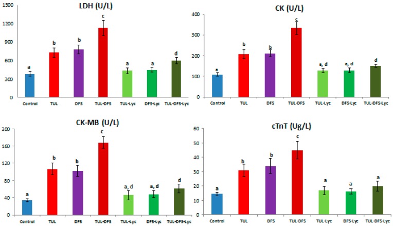
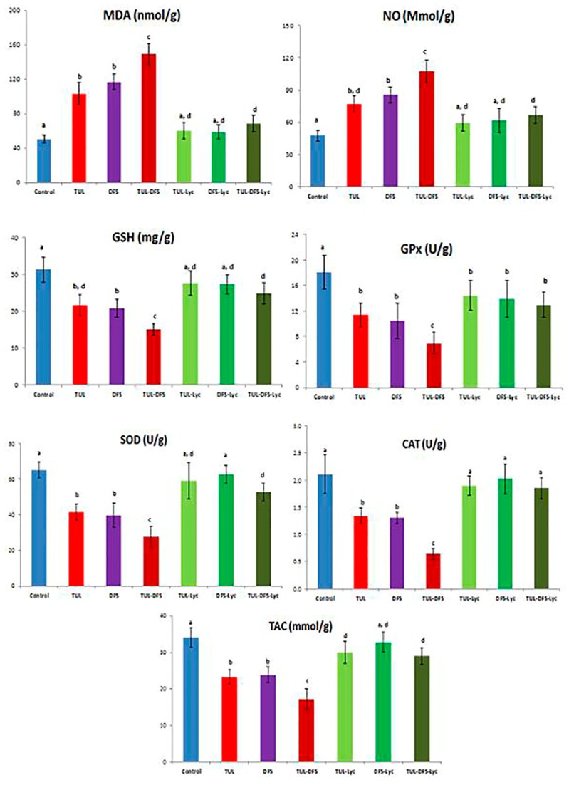
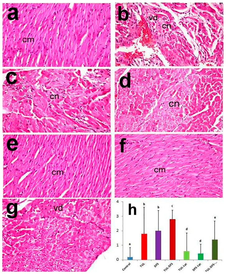
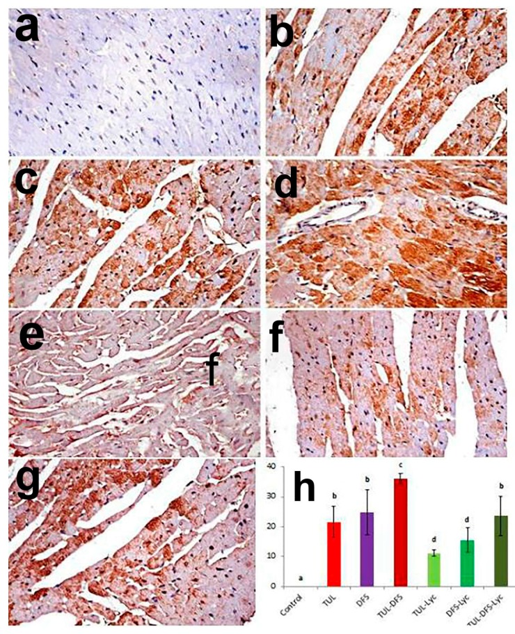
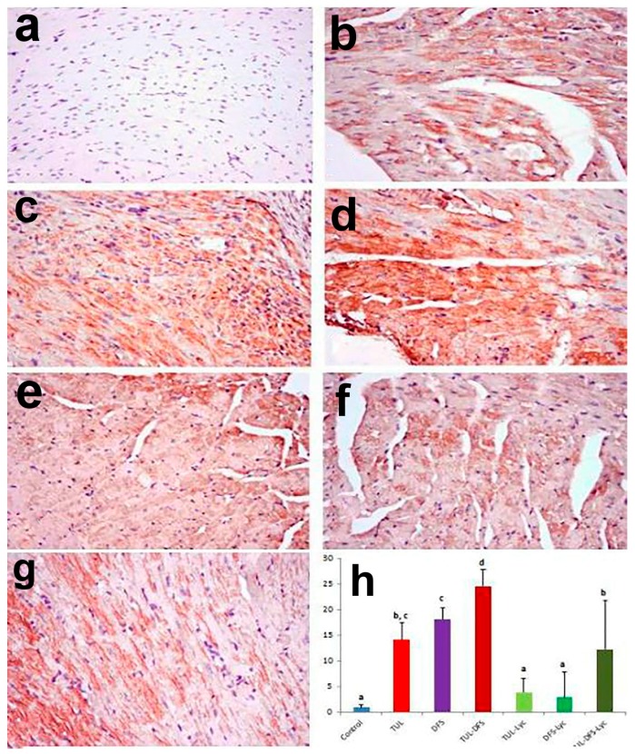
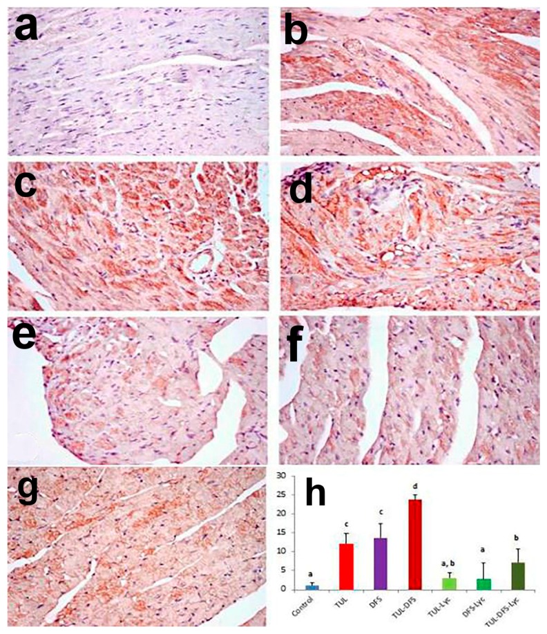
Recent experiments showed a potential cardiotoxic effect of the macrolide antibiotic (tulathromycin). This study was performed to investigate whether diclofenac sodium (DFS) potentiates the cardiotoxicity of tulathromycin and increases the cardioprotective effects of lycopene against DFS and tulathromycin. Seven groups (eight per group) of adult Swiss albino mice received saline (control), tulathromycin (a single subcutaneous dose of 28 mg/kg/bw on day 14), DFS (a single oral dose of 100 mg/kg/bw on day 14), tulathromycin plus DFS, or lycopene (oral, 10 mg/kg/bw daily for 15 d) combined with tulathromycin, DFS, or both. Compared to the control group, the administration of tulathromycin or DFS (individually or in combination) caused significantly elevated (p < 0.05) serum levels of Creatine kinase-myocardial B fraction (CK-MB), lactate dehydrogenase, and cardiac-specific troponin-T and tissue levels of nitric oxide and malondialdehyde that were accompanied by significantly decreased tissue reduced glutathione content and glutathione peroxidase, superoxide dismutase, and catalase antioxidant enzyme activity. Upon histopathological and immunohistochemical examination, the mean pathology scores and the percentages of caspase-3-, Bax-, and CK-positive regions were significantly higher in the tulathromycin- and/or DFS-treated groups than in control mice. For all these parameters, the pathological changes were more significant in the tulathromycin-DFS combination group than in mice treated with either drug individually. Interestingly, co-administration of lycopene with tulathromycin and/or DFS significantly ameliorated the changes described above. In conclusion, DFS could potentiate the cardiotoxic effects of tulathromycin, whereas lycopene can serve as a cardioprotective agent against DFS and tulathromycin.

Keywords: cardiotoxicity; diclofenac sodium; lycopene; mice; tulathromycin.


Conflict of interest statement

The authors declare no conflict of interest.

Figures

Figure 1
Figure 1
The effect of lycopene treatment on serum levels of cardiac injury biomarkers in tulathromycin and diclofenac sodium intoxicated rats. Data are means ± SD. Means carrying different superscripts (a,b,c,d) are significantly different at (p < 0.05). Abbreviations: CK: Creatine kinase, cTnT: Cardiac-specific troponin-T, DFS: Diclofenac sodium, LDH: Lactate Dehydrogenase, Lyc: Lycopene, TLR: Tulathromycin.
Figure 2
Figure 2
The effect of lycopene treatment on tissue lipid peroxidation and activities of antioxidant enzymes in tulathromycin and diclofenac sodium intoxicated rats. Data are means ± Standard deviation (SD). Means carrying different superscripts (a,b,c,d) are significantly different at (p < 0.05). Abbreviations: CAT: Catalase, DFS: Diclofenac Sodium, GSH: Reduced glutathione, GPx: Glutathione peroxidase, Lyc: Lycopene, MDA: Malondialdehyde, NO: Nitric oxide, SOD: Superoxide dismutase, TAC: Total antioxidant capacity, TLR: Tulathromycin.
Figure 3
Figure 3
Light photomicrographs of heart tissue in (a) control mice showing normal architecture of cardiac muscle fibers (cm); (b) tulathromycin-treated mice showing extensive vacuolar degeneration (vd) associated with coagulative necrosis (cn) of cardiac myocytes in addition to apoptotic changes; (c) DFS-treated mice showing coagulative necrosis of cardiomyocytes (cn) with loss of cross-striation in cardiac muscle fibers; (d) tulathromycin–DFS-treated mice showing cardiomyocyte necrosis (cn), apoptosis and myocytolysis; (e) tulathromycin–lycopene-treated mice showing normal cardiac myocytes (cm); (f) DFS–lycopene-treated mice showing marked restoration of cardiomyocytes (cm); (g) tulathromycin–DFS–lycopene-treated mice showing mild vacuolar degeneration of cardiomyocytes (vd) (H&E, ×400); and (h) pathologic scoring of cardiotoxicity in the heart tissue of control and treated mice. Means carrying different superscripts (a,b,c,d,e) are significantly different at (p < 0.05).
Figure 4
Figure 4
Staining for caspase-3 protein in a cardiac tissue section of (a) control mice showing no caspase-3 immune-reactive cells; (b) tulathromycin-treated mice showing multiple caspase-3 immune-reactive cells; (c) DFS-treated mice showing abundant caspase-3 immune-reactive cells; (d) tulathromycin–DFS-treated mice showing diffuse, intensely stained caspase-3 immune-reactive cells; (e) tulathromycin–lycopene-treated mice showing fewer caspase-3 immune-reactive cells; (f) DFD–lycopene-treated mice showing sparse caspase-3 immune-reactive cells; (g) tulathromycin–DFS–lycopene-treated mice showing sporadic caspase-3 immune-reactive cells (caspase-3 immunohistochemical staining, ×400); and (h) immunohistochemical scoring of caspase-3 in the heart tissue of control and treated mice. Means carrying different superscripts (a,b,c,d) are significantly different at (p < 0.05).
Figure 5
Figure 5
Staining for Bax protein in a section of heart tissue in (a) control mice showing no Bax immune-reactive cells; (b) tulathromycin-treated mice showing an increased number of Bax immune-reactive cells; (c) DFS-treated mice showing numerous Bax immune-reactive cells; (d) tulathromycin–DFS-treated mice showing intense staining of Bax immune-reactive cells; (e) tulathromycin–lycopene-treated mice showing fewer Bax immune-reactive cells; (f) DFS–lycopene-treated mice showing sparse Bax immune-reactive cells; (g) tulathromycin–DFS–lycopene-treated mice showing a reduced number of Bax immune-reactive cells (Bax immunohistochemical staining, ×400); and (h) immunohistochemical scoring of Bax protein in the heart tissue of control and treated mice. Means carrying different superscripts (a,b,c,d) are significantly different at (p < 0.05).
Figure 6
Figure 6
Staining for CK protein in a section of heart tissue in (a) control mice showing no CK immune-reactive cells; (b) tulathromycin-treated mice showing an increased number of CK immune-reactive cells; (c) DFS-treated mice showing abundant CK immune-reactive cells; (d) tulathromycin–DFS-treated mice showing numerous CK immune-reactive cells; (e) tulathromycin–lycopene-treated mice showing few individual CK immune-reactive cells; (f) DFS–lycopene-treated mice showing sparse CK immune-reactive cells; (g) tulathromycin–DFS–lycopene-treated mice showing sporadic CK immune-reactive cells (CK immunohistochemical staining, ×400); and (h) immunohistochemical scoring of Bax protein in the heart tissue of control and treated mice. Means carrying different superscripts (a,b,c,d) are significantly different at (p < 0.05).

References

    1. Abu-Gharbieh E., Vasina V., Poluzzi E., de Ponti F. Antibacterial macrolides: A drug class with a complex pharmacological profile. Pharmacol. Res. 2004;50:211–222. doi: 10.1016/j.phrs.2004.01.008. - DOI - PubMed
    1. Anadon A., Reeve-Johnson L. Macrolide antibiotics, drug interactions and microsomal enzymes: Implications for veterinary medicine. Res. Vet. Sci. 1999;66:197–203. doi: 10.1053/rvsc.1998.0244. - DOI - PubMed
    1. Ibrahim N., Burke J., Beattie D. The sensitivity of rat liver and yeast mitochondrial ribosomes to inhibitors of protein synthesis. J. Biol. Chem. 1974;249:6806–6811. - PubMed
    1. Villa P., Sassella D., Corada M., Bartosek I. Toxicity, uptake, and subcellular distribution in rat hepatocytes of roxithromycin, a new semisynthetic macrolide, and erythromycin base. Antimicrob. Agents Chemother. 1988;32:1541–1546. doi: 10.1128/AAC.32.10.1541. - DOI - PMC - PubMed
    1. De Vries H., Arendzen A., Kroon A. The interference of the macrolide antibiotics with mitochondrial protein synthesis. Biochim. Biophys. Acta. 1973;331:264–275. doi: 10.1016/0005-2787(73)90439-5. - DOI - PubMed

LinkOut - more resources