Review
. 2018 Jan 24;553(7689):427-436.
doi: 10.1038/nature25177.

Skin microbiota-host interactions

Affiliations
Review

Skin microbiota-host interactions

Y Erin Chen et al. Nature. .

Erratum in

Abstract

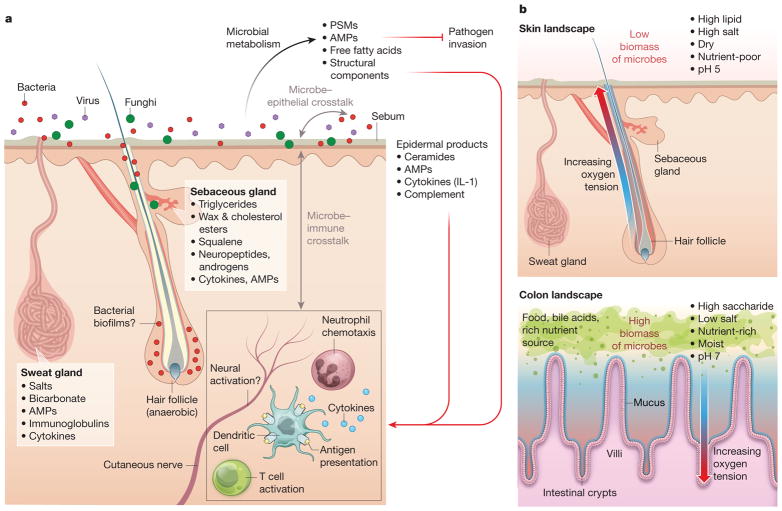
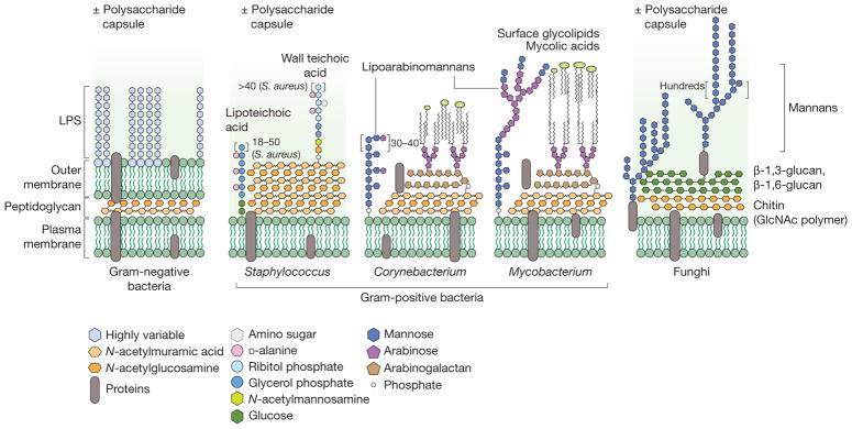
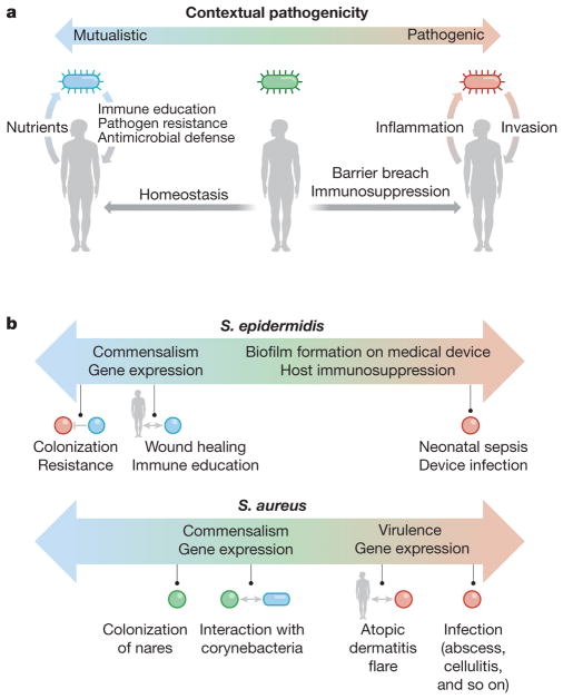
The skin is a complex and dynamic ecosystem that is inhabited by bacteria, archaea, fungi and viruses. These microbes-collectively referred to as the skin microbiota-are fundamental to skin physiology and immunity. Interactions between skin microbes and the host can fall anywhere along the continuum between mutualism and pathogenicity. In this Review, we highlight how host-microbe interactions depend heavily on context, including the state of immune activation, host genetic predisposition, barrier status, microbe localization, and microbe-microbe interactions. We focus on how context shapes the complex dialogue between skin microbes and the host, and the consequences of this dialogue for health and disease.


Conflict of interest statement

The authors declare no competing financial interests.

Figures

Figure 1
Figure 1. Crosstalk between skin microbiota and the host
a, Diverse microbes (viruses, fungi and bacteria) cover the skin surface and associated structures (hair follicles, sebaceous glands and sweat glands), possibly forming biofilms at some sites. These microbes metabolize host proteins and lipids and produce bioactive molecules, such as free fatty acids, AMPs, phenol-soluble modulins (PSMs), cell wall components, and antibiotics,. These products act on other microbes to inhibit pathogen invasion, on the host epithelium to stimulate keratinocyte-derived immune mediators such as complement and IL-1, and on immune cells in the epidermis and dermis. In turn, host products and immune cell activity influence microbial composition on the skin. b, The skin differs from the gut in its physical and chemical properties. The skin is a dry, acidic, lipid-rich, high-salt environment without exogenous nutrient sources, and therefore has low microbial biomass. By contrast, the gut is moist and has abundant nutrients and a thick layer of mucin,, enabling it to support much greater microbial biomass. While hair follicles become more anaerobic deeper into the follicle, crypts become more aerobic closer to the epithelium,. In addition, material within crypts regularly exchanges with material in the gut lumen, owing to peristalsis, whereas hair follicles have narrow openings filled with sebum and keratinocytic debris, making them more isolated.
Figure 2
Figure 2. Chemistry of the skin
The skin surface consists of a highly organized basketweave structure of keratinocytic proteins and lipids, which are produced by keratinocytes (epidermal lipids) and sebaceous glands (sebaceous lipids). Ceramides are unique to epidermal origin, whereas squalene and wax esters are unique to sebaceous origin, with variable composition under endocrine control. Other dominant skin lipids are cholesterol, triglycerides and free fatty acids (which are often microbial products). Some lipids, such as sphingosine and free fatty acids, demonstrate antimicrobial activity against bacteria, fungi, and viruses and may have immunomodulatory effects. Of keratinocytic proteins, more than 70% of the dry protein weight consists of loricrin, a glycine-rich protein that is thought to have important barrier properties.
Figure 3
Figure 3. Chemistry of microbial surfaces
Bacteria and fungi have diverse cell envelopes loaded with immunogenic molecules. Gram-negative bacteria (left) have two lipid bilayers separated by a peptidoglycan cell wall. The outer leaflet of the outer membrane is studded with an immunogenic lipoglycan called lipopolysaccharide (LPS), which has a lipid anchor and highly variable polysaccharide region called the O-antigen. In Escherichia coli, for example, 184 different O-antigen structures are known. Gram-positive bacteria (middle) lack an outer lipid bilayer but have a thicker peptidoglycan cell wall. Staphylococcus species have wall-bound and membrane-anchored teichoic acids, which are analogous to Gram-negative LPS, with a host-facing strain-specific branched polysaccharide. Corynebacterium and Mycobacterium species also have complex lipoglycans, called lipoarabinomannans. They also have a unique lipid outer layer made up of cell-wall-bound mycolic acids and other noncovalently bound glycolipids. Like Gram-positive bacteria, fungi (right) have only one lipid bilayer membrane. This is covered by a cell wall usually consisting of chitin and a β-D-glucan mesh. The outer layer of the fungal cell wall often contains heavily mannosylated proteins and sometimes a capsule of various polysaccharides. GlcNAc, N-acetylglucosamine.
Figure 4
Figure 4. Contextual pathogenicity
a, Microbes exhibit contextual pathogenicity along a spectrum. Host factors such as barrier breaches and immunosuppression bias microbes towards pathogenic behaviour, whereas homeostatic conditions bias them towards mutualistic behaviour. On the microbial side, virulence gene expression and microbe–microbe interactions can also push microbial behaviour to be mutualistic or pathogenic. In a mutualistic host–microbe relationship, the host provides nutrients, while the microbe promotes epithelial and immune homeostasis as well as pathogen resistance through microbial products and occupation of metabolic niches. In a pathogenic relationship, the microbe invades past the epithelium, causing inflammation, and sometimes also benefiting from a host inflammatory response. b, Both S. epidermidis and S. aureus are examples of contextual pathogenicity; S. epidermidis is biased towards mutualistic behaviour whereas S. aureus displays more pathogenic character.

References

    1. De Luca C, Valacchi G. Surface lipids as multifunctional mediators of skin responses to environmental stimuli. Mediators Inflamm. 2010;2010:321494. - PMC - PubMed
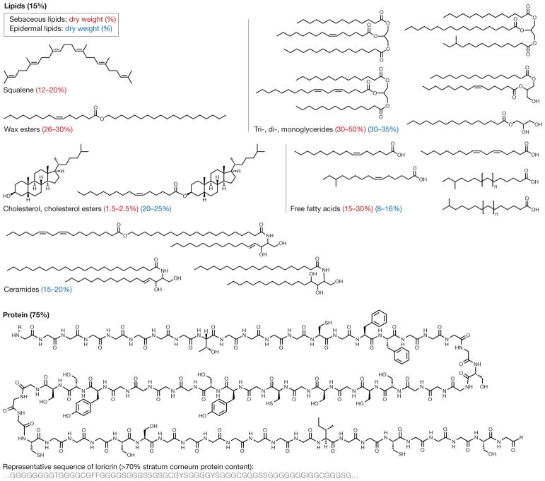
    1. van Smeden J, Bouwstra JA. Stratum corneum lipids: their role for the skin barrier function in healthy subjects and atopic dermatitis patients. Curr Probl Dermatol. 2016;49:8–26. - PubMed
    1. Niyonsaba F, Kiatsurayanon C, Chieosilapatham P, Ogawa H. Friends or foes? Host defense (antimicrobial) peptides and proteins in human skin diseases. Exp Dermatol. 2017;26:989–998. - PubMed
    1. Bek-Thomsen M, Lomholt HB, Scavenius C, Enghild JJ, Brüggemann H. Proteome analysis of human sebaceous follicle infundibula extracted from healthy and acne-affected skin. PLoS One. 2014;9:e107908. - PMC - PubMed
    1. Lee DY, et al. Sebocytes express functional cathelicidin antimicrobial peptides and can act to kill propionibacterium acnes. J Invest Dermatol. 2008;128:1863–1866. - PMC - PubMed

Publication types