. 2018 Apr 17;207(0):307-327.
doi: 10.1039/c7fd00190h.

Engineered photoproteins that give rise to photosynthetically-incompetent bacteria are effective as photovoltaic materials for biohybrid photoelectrochemical cells

Affiliations

Engineered photoproteins that give rise to photosynthetically-incompetent bacteria are effective as photovoltaic materials for biohybrid photoelectrochemical cells

Juntai Liu et al. Faraday Discuss. .

Abstract

Reaction centre/light harvesting proteins such as the RCLH1X complex from Rhodobacter sphaeroides carry out highly quantum-efficient conversion of solar energy through ultrafast energy transfer and charge separation, and these pigment-proteins have been incorporated into biohybrid photoelectrochemical cells for a variety of applications. In this work we demonstrate that, despite not being able to support normal photosynthetic growth of Rhodobacter sphaeroides, an engineered variant of this RCLH1X complex lacking the PufX protein and with an enlarged light harvesting antenna is unimpaired in its capacity for photocurrent generation in two types of bio-photoelectrochemical cells. Removal of PufX also did not impair the ability of the RCLH1 complex to act as an acceptor of energy from synthetic light harvesting quantum dots. Unexpectedly, the removal of PufX led to a marked improvement in the overall stability of the RCLH1 complex under heat stress. We conclude that PufX-deficient RCLH1 complexes are fully functional in solar energy conversion in a device setting and that their enhanced structural stability could make them a preferred choice over their native PufX-containing counterpart. Our findings on the competence of RCLH1 complexes for light energy conversion in vitro are discussed with reference to the reason why these PufX-deficient proteins are not capable of light energy conversion in vivo.


Figures
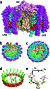
Fig. 1
Fig. 1. Structure and mechanism of the Rba. sphaeroides RCLH1X and RCLH1 complexes. (a) A view approximately in the plane of the photosynthetic membrane of a RCLH1X monomer. The colour/representation coding is: pink surface – RC H-polypeptide, lime-green surface – RC L-polypeptide, beige surface – RC M-polypeptide, yellow ribbon – PufX, cyan spheres – LH1 α-polypeptides, magenta spheres – LH1 β-polypeptides, red/orange spheres – alternating LH1 BChls with the central Mg shown in magenta, black spheres – side chain of the RC QB ubiquinone-10. (b) A view from the periplasmic side of the membrane of the Rba. sphaeroides RCLH1X complex. (c) The same view of the T. tepidum RCLH1 complex. (d) The 32 BChls and 16 carotenoids of T. tepidum LH1 (spheres) arranged around the cofactors of the central RC (sticks). The colour coding for the LH1 cofactors is: red/orange spheres – alternating LH1 BChls with the central Mg shown in magenta, green spheres – carotenoids. The colour coding for the RC cofactors is: yellow carbons – P BChls, green carbons – monomeric BChls, pink carbons – bacteriopheophytins, cyan carbons – ubiquinones, brown spheres – iron atoms, magenta spheres – BChl Mg atoms. (e) Cofactors of the Rba. sphaeroides RC and the route of electron transfer. The cofactor representation is the same as for (d). For clarity, cofactor hydrocarbon side chains are not shown.
Fig. 2
Fig. 2. A variety of RCLH1 complexes from Rba. sphaeroides. (a) Bands formed by pigment-proteins separated on sucrose density gradients, (b) visible region absorbance spectra for the RCLH1Xr and RCLH1Xg complexes, (c) molar absorption coefficients for the purified RCLH1(X) or RC complexes and near-IR absorbance spectra for 1 μM solutions.
Fig. 3
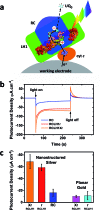
Fig. 3. Photocurrents from the RCLH1Xr, RCLH1r and RC complexes interfaced with metal electrodes. (a) Photoexcitation of the BChls (alternating red/orange) and carotenoids (not shown) of the LH1 antenna (green domain) produces charge separation in the RC (blue domain). The RC is re-reduced by electrons from the working electrode in a process mediated by cyt c (orange protein). The transfer of electrons to the counter electrode is mediated by 1 mM Q0. For a representation of the RCLH1 components, see Fig. 1. The haem of cyt c is shown as slate-blue spheres with a brown Fe sphere. (b) Photocurrent transients in response to 180 seconds of illumination of the RCLH1Xr, RCLH1r or RC complexes adhered to nanostructured silver working electrodes. (c) A comparison of the steady state photocurrent densities for proteins adhered to nanostructured silver or planar gold working electrodes.
Fig. 4
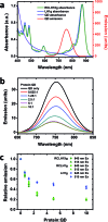
Fig. 4. Quenching of QD emission by the RCLH1Xg or RCLH1g complexes. (a) Absorbance spectra of the RCLH1Xg and LH1g complexes, and absorbance and emission spectra of 6.5 nm-diameter water soluble CdTe QDs, (b) quenching of emission of 50 nM QDs by 31.25–500 nM RCLH1Xg complexes, with excitation at 515 nm, (c) decay of relative QD emission as a function of protein : QD ratio, using data collected at two excitation wavelengths. The data points are means with standard deviation (n = 3).
Fig. 5
Fig. 5. Binding of RCLH1(X) complexes to 6.5 nm CdTe QDs. (a) Distribution of acidic and basic residues on the surface of the Rba. sphaeroides RCLH1X complex exposed at the periplasmic (top) and cytoplasmic (bottom) side of the membrane. Glu and Asp residues are shown in red and Lys and Arg residues are shown in blue. PufX is shown in yellow. (b) Schematic of three 12 nm diameter RCLH1X complexes (view parallel to the membrane) positioned around a 6.5 nm diameter QD (blue sphere). (c) Separation of the unbound RCLH1Xg protein (upper band, green arrow) from the RCLH1Xg protein bound to the QDs (lower band, red arrow) on three step sucrose density gradients. (d) Variation with QD : protein ratio of unbound protein as a fraction of the total.
Fig. 6
Fig. 6. Thermal stabilities of the RCLH1Xr and RCLH1r proteins. (a) Near-IR absorbance spectra of the pigment-protein solutions before and after heating at 60 °C for six hours, (b) decay of the amplitude of the Qy absorbance band of the LH1 BChls over a 10 hour incubation at 60 °C. The data were fitted with two exponential terms and an offset to illustrate their biphasic nature.
Fig. 7
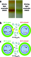
Fig. 7. Proteins from cells grown in DMSO and the cause of the phenotype of PufX-minus strains. (a) Monomeric RCLH1 complexes from cells grown under light/anaerobic conditions in the presence of DMSO. (b) In native RCLH1X complexes, a 75% reduction of the intramembrane quinone pool results in 10% of the RCs being closed due to QA being reduced (the electrons are represented as grey spheres). (c) In engineered RCLH1 complexes, 62% of the RCs are closed under the same conditions, but quinone exchange with the intramembrane pool is not prevented.

References

    1. LaVan D. A., Cha J. N. Proc. Natl. Acad. Sci. U. S. A. 2006;103:5251–5255. - PMC - PubMed
    1. Badura A., Kothe T., Schuhmann W., Rogner M. Energy Environ. Sci. 2011;4:3263–3274.
    1. Boghossian A. A., Ham M.-H., Choi J. H., Strano M. S. Energy Environ. Sci. 2011;4:3834–3843.
    1. Wang F., Liu X., Willner I. Adv. Mater. 2013;25:349–377. - PubMed
    1. Kim Y., Shin S. A., Lee J., Yang K. D., Nam K. T. Nanotechnology. 2014;25:342001. - PubMed

Publication types

MeSH terms

Substances

LinkOut - more resources