. 2018 Mar 1;141(3):698-712.
doi: 10.1093/brain/awx358.

De novo mutations in GRIN1 cause extensive bilateral polymicrogyria

Affiliations

De novo mutations in GRIN1 cause extensive bilateral polymicrogyria

Andrew E Fry et al. Brain. .

Abstract

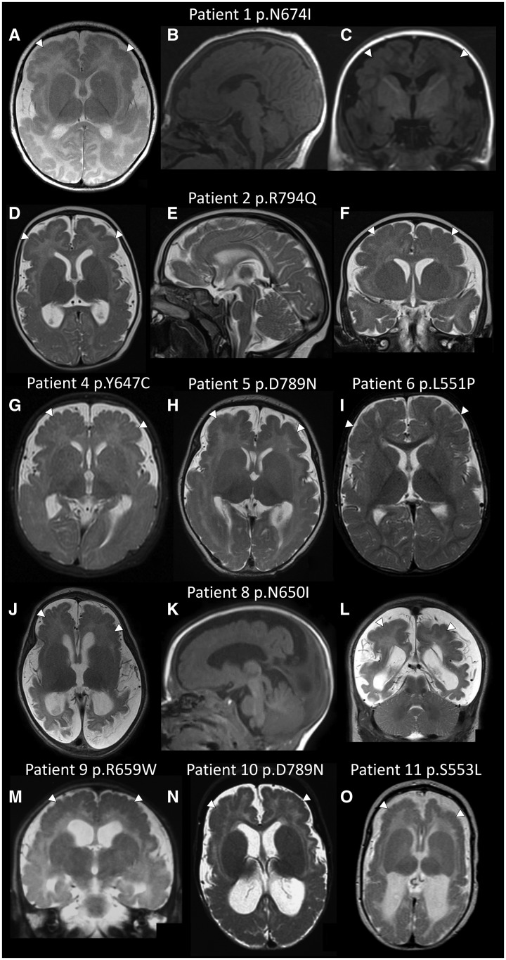
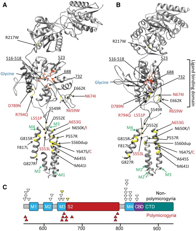
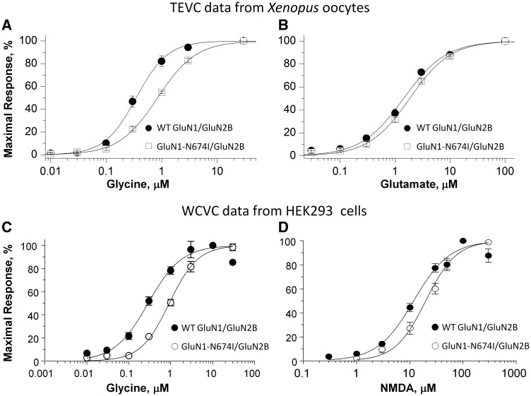
Polymicrogyria is a malformation of cortical development. The aetiology of polymicrogyria remains poorly understood. Using whole-exome sequencing we found de novo heterozygous missense GRIN1 mutations in 2 of 57 parent-offspring trios with polymicrogyria. We found nine further de novo missense GRIN1 mutations in additional cortical malformation patients. Shared features in the patients were extensive bilateral polymicrogyria associated with severe developmental delay, postnatal microcephaly, cortical visual impairment and intractable epilepsy. GRIN1 encodes GluN1, the essential subunit of the N-methyl-d-aspartate receptor. The polymicrogyria-associated GRIN1 mutations tended to cluster in the S2 region (part of the ligand-binding domain of GluN1) or the adjacent M3 helix. These regions are rarely mutated in the normal population or in GRIN1 patients without polymicrogyria. Using two-electrode and whole-cell voltage-clamp analysis, we showed that the polymicrogyria-associated GRIN1 mutations significantly alter the in vitro activity of the receptor. Three of the mutations increased agonist potency while one reduced proton inhibition of the receptor. These results are striking because previous GRIN1 mutations have generally caused loss of function, and because N-methyl-d-aspartate receptor agonists have been used for many years to generate animal models of polymicrogyria. Overall, our results expand the phenotypic spectrum associated with GRIN1 mutations and highlight the important role of N-methyl-d-aspartate receptor signalling in the pathogenesis of polymicrogyria.


Figures

Figure 1
Figure 1
Polymicrogyria in patients with GRIN1 mutations. Axial, midline sagittal and coronal brain magnetic resonance images for Patient 1 at age 2 months (A–C) and Patient 2 at age 5 months (D–F); axial magnetic resonance images for Patient 4 at age 3 months (G), Patient 5 at age 6 weeks (H) and Patient 6 at age 8 months (I); axial, sagittal and coronal images for Patient 8 at age 3 months (J–L); a coronal image for Patient 9 at age 4 months (M); axial images from Patient 10 at age 8 months (N) and Patient 11 at age 2 months (O). Images B, C and K are T1-weighted. All other images are T2-weighted. The images demonstrate bilateral extensive polymicrogyria (white arrows) more severe anteriorly. Note the increased extra-axial spaces and enlarged lateral ventricles (in most images apart from I) suggesting cerebral volume loss.
Figure 2
Figure 2
Position of the GRIN1 mutations. (A) This ribbon diagram of the GluN1 subunit of the NMDA receptor demonstrates the location of the binding site of glycine in the ligand binding domain, transmembrane helices (M1-M4 in green) and glycine binding residues of GluN1 (orange residues and blue underlined numbers). Mutated residues are in yellow. Polymicrogyria-associated mutations are in red text and previous GRIN1 mutations are in black text. (B) The same ribbon model rotated 90 degrees in the axial plane. (C) A model of the C-terminal end of GRIN1. In addition to showing M1-M4, the model shows the position of the second ligand binding domain (S2), calmodulin biding domain (CBD) and the C-terminal domain (CTD). Codon position is listed below the model. Heterozygous GRIN1 variants reported or reviewed by Lemke et al. (2016) (non-polymicrogyria, white triangles) are shown above the model. The two yellow triangles represent subjects with CT brain imaging only. Polymicrogyria-associated GRIN1 mutations (red triangles) cluster in the S2 domain (6/11) or in the adjacent Lurcher motif of M3 (3/11).
Figure 3
Figure 3
The p.Asn674Ile mutation changes the response of the NMDA receptor to agonists. The top graphs display concentration-response curves for (A) glycine (in the presence of 100 µM glutamate) and (B) glutamate (in the presence of 100 µM glycine) determined by two-electrode voltage-clamp (TEVC) recordings from Xenopus oocytes expressing either wild-type (WT)-GluN1/GluN2B or GluN1-N674I /GluN2B. The bottom graphs display concentration-response curves for (C) glycine (in the presence of 100 µM NMDA) and (D) NMDA (in the presence of 100 µM glycine) determined by whole-cell voltage-clamp (WCVC) recordings from transfected HEK 293 cells expressing either WT-GluN1/GluN2B or GluN1-N674I /GluN2B. Error bars represent SEM.

Comment in

References

    1. Adams DR, Yuan H, Holyoak T, Arajs KH, Hakimi P, Markello TC, et al.Three rare diseases in one Sib pair: RAI1, PCK1, GRIN2B mutations associated with Smith-Magenis Syndrome, cytosolic PEPCK deficiency and NMDA receptor glutamate insensitivity. Mol Genet Metab 2014; 113: 161–70. - PMC - PubMed
    1. Adzhubei IA, Schmidt S, Peshkin L, Ramensky VE, Gerasimova A, Bork P, et al.A method and server for predicting damaging missense mutations. Nat Methods 2010; 7: 248–9. - PMC - PubMed
    1. Behar TN, Scott CA, Greene CL, Wen X, Smith SV, Maric D, et al.Glutamate acting at NMDA receptors stimulates embryonic cortical neuronal migration. J Neurosci 1999; 19: 4449–61. - PMC - PubMed
    1. Bosch DG, Boonstra FN, de Leeuw N, Pfundt R, Nillesen WM, de Ligt J, et al.Novel genetic causes for cerebral visual impairment. Eur J Hum Genet 2016; 24: 660–5. - PMC - PubMed
    1. Carvill GL, Regan BM, Yendle SC, O’Roak BJ, Lozovaya N, Bruneau N, et al.GRIN2A mutations cause epilepsy-aphasia spectrum disorders. Nat Genet 2013; 45: 1073–6. - PMC - PubMed

Publication types

MeSH terms