

Microparticulate Caspase 1 Regulates Gasdermin D and Pulmonary Vascular Endothelial Cell Injury
- PMID: 29365280
- PMCID: PMC6039876
- DOI: 10.1165/rcmb.2017-0393OC
Microparticulate Caspase 1 Regulates Gasdermin D and Pulmonary Vascular Endothelial Cell Injury
Abstract
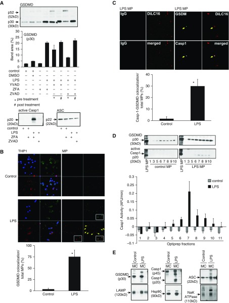
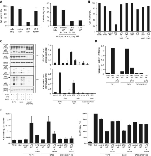
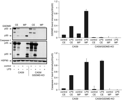
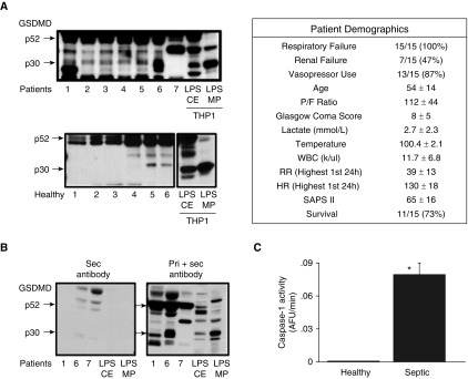
Lung endothelial cell apoptosis and injury occur throughout all stages of acute lung injury/acute respiratory distress syndrome and impact disease progression. Caspases 1, 4, and 5 are essential for completion of the apoptotic program known as pyroptosis that also involves proinflammatory cytokines. Because gasdermin D (GSDMD) mediates pyroptotic death and is essential for pore formation, we hypothesized that it might direct caspase 1-encapsulated microparticle (MP) release and mediate endothelial cell death. Our present work provides evidence that GSDMD is released by LPS-stimulated THP-1 monocytic cells, where it is packaged into microparticles together with active caspase 1. Furthermore, only MP released from stimulated monocytic cells that contain both cleaved GSDMD and active caspase 1 induce endothelial cell apoptosis. MPs pretreated with caspase 1 inhibitor Y-VAD or pan-caspase inhibitor Z-VAD do not contain cleaved GSDMD. MPs from caspase 1-knockout cells are also deficient in p30 active GSDMD, further confirming that caspase 1 regulates GSDMD function. Although control MPs contained cleaved GSDMD without caspase 1, these fractions were unable to induce cell death, suggesting that encapsulation of both caspase 1 and GSDMD is essential for cell death induction. Release of microparticulate active caspase 1 was abrogated in GSDMD knockout cells, although cytosolic caspase 1 activation was not impaired. Last, higher concentrations of microparticulate GSDMD were detected in the plasma of septic patients with acute respiratory distress syndrome than in that of healthy donors. Taken together, these findings suggest that GSDMD regulates the release of microparticulate active caspase 1 from monocytes essential for induction of cell death and thereby may play a critical role in sepsis-induced endothelial cell injury.
Figures
References
-
- Hugel B, Martínez MC, Kunzelmann C, Freyssinet JM. Membrane microparticles: two sides of the coin. Physiology (Bethesda) 2005;20:22–27. - PubMed
-
- Distler JH, Huber LC, Gay S, Distler O, Pisetsky DS. Microparticles as mediators of cellular cross-talk in inflammatory disease. Autoimmunity. 2006;39:683–690. - PubMed
-
- Martínez MC, Tesse A, Zobairi F, Andriantsitohaina R. Shed membrane microparticles from circulating and vascular cells in regulating vascular function. Am J Physiol Heart Circ Physiol. 2005;288:H1004–H1009. - PubMed
-
- Diamant M, Tushuizen ME, Sturk A, Nieuwland R. Cellular microparticles: new players in the field of vascular disease? Eur J Clin Invest. 2004;34:392–401. - PubMed
-
- Martin S, Tesse A, Hugel B, Martínez MC, Morel O, Freyssinet JM, et al. Shed membrane particles from T lymphocytes impair endothelial function and regulate endothelial protein expression. Circulation. 2004;109:1653–1659. - PubMed
Publication types
MeSH terms
Substances
Grants and funding
LinkOut - more resources
Full Text Sources
Other Literature Sources
