. 2018 Jan 23;2(2):76-84.
doi: 10.1182/bloodadvances.2017012971.

The human IL-15 superagonist ALT-803 directs SIV-specific CD8+ T cells into B-cell follicles

Affiliations

The human IL-15 superagonist ALT-803 directs SIV-specific CD8+ T cells into B-cell follicles

Gabriela M Webb et al. Blood Adv. .

Abstract

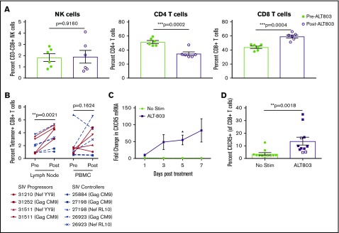
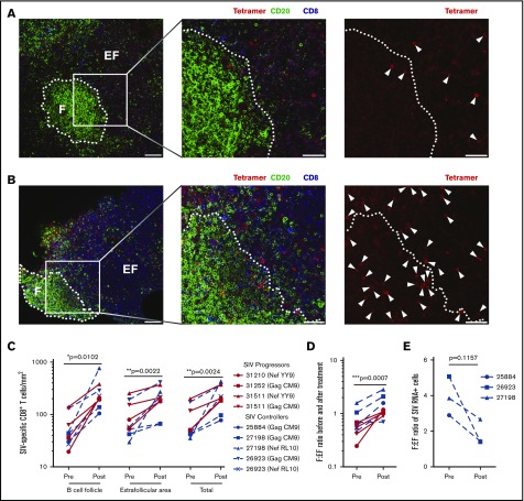
Sequestering of latent HIV in follicular helper T cells within B-cell follicles that largely exclude cytotoxic T cells is a major barrier to cellular immune-based approaches to eradicate HIV. Here, we show that the clinical-grade human interleukin-15 (IL-15) superagonist ALT-803 activates and redirects simian immunodeficiency virus (SIV)-specific CD8+ T cells from the peripheral blood into B-cell follicles. In agreement with the increased trafficking of SIV-specific cytotoxic T cells to sites of cryptic viral replication, lymph nodes of elite controlling macaques contained fewer cells expressing SIV RNA or harboring SIV DNA post-ALT-803 treatment. These data establish ALT-803 as an immunotherapeutic for HIV and other chronic viral pathogens that evade host immunity by persisting in B-cell follicles.


Conflict of interest statement

Conflict-of-interest disclosure: E.K.J. and H.C.W. are employees of Altor Bioscience Corporation. The remaining authors declare no competing financial interests.

Figures

None
Graphical abstract
Figure 1.
Figure 1.
In vivo administration of ALT-803 in SIV-naive RMs. SIV-naive RMs (n = 4) were injected IV at day 0 with 6 μg/kg of ALT-803. Blood was collected before injection and at days 1, 3, 5, 7, and 14 postinjection. (A) White blood cell (WBC) count, lymphocytes, and neutrophils as well as (B) CD4+ T cell, CD8+ T cells, and CD16+ NK cells were analyzed from the blood. Proliferation of (C) CD16+ NK cells, (D) CD4+ and CD8+ T cells as well as naive, effector memory (EM) and central memory (CM) populations of (E) CD4+ and (F) CD8+ T-cell subsets determined as a percentage of Ki67+ cells of that particular lymphocyte population. (G) Whole blood was further evaluated for classical monocytes (CD14+, CD16), intermediate monocytes (CD14+, CD16+), and nonclassical monocytes (CD14, CD16+) as a percentage of CD45+ cells. (H) Proliferation of monocyte subsets was determined as a percentage of Ki67+ cells of that particular population. (I) Absolute counts of myeloid dendritic cells (mDC, defined as Lin, HLA-DR+, CD11c+) and plasmacytoid dendritic cells (pDC, defined as Lin, HLA-DR+, CD123+) were also determined. Absolute counts were calculated based on the percentage of the particular cell subset and the WBC count. Data shown are means (± standard errors of the means) of combined data from all 4 animals. *P < .05; **P < .01; ***P < .001 comparing time points to time point zero.
Figure 2.
Figure 2.
In vivo administration of ALT-803 in SIV-infected RMs. (A-G) SIV-infected RMs (n = 4) were injected IV with 6 mg/kg ALT-803 at days 0, 7, and 14 (indicated by thin dashed lines) and subsequently with 100 mg/kg ALT-803 at day 49 (indicated by thick dashed line). (A) White blood cell (WBC), lymphocytes, and neutrophils as well as (B) CD4+ T cells, CD8+ T cells, and CD16+ NK cells were longitudinally evaluated in the peripheral blood. Proliferation of (C) CD16+ NK cells, (D) CD4+ and CD8+ T cells as well as of naive, effector memory (EM), and central memory (CM) populations of (E) CD4+ T cell and (F) CD8+ T-cell subsets were determined as a percentage of Ki-67+ cells of that particular lymphocyte population. (G) Whole blood was evaluated for classical monocytes (CD14+CD16), intermediate monocytes (CD14+CD16+), and nonclassical monocytes (CD14CD16+) as a percentage of CD45+ cells. (H) Proliferation of monocyte subsets was determined as a percentage of Ki67+ cells of that particular population. Absolute counts were calculated based on the percentage of the particular cell subset and the WBC count. Data shown are means (± standard errors of the means) of combined data from 4 animals. (I) SIV-infected RMs were injected IV with BrdU (60 mg/kg) and 100 mg/kg ALT-803 (n = 5) or PBS (n = 3). Animals were then taken to necropsy 5 days later. CD16+ NK cells, CD4+ T cells, and CD8+ T cells were assessed for BrdU incorporation in the peripheral blood (PBMC), bronchoalveolar lavage (BAL) fluid, bone marrow, colon, liver, spleen, axillary lymph nodes (AxLN), mesenteric lymph nodes (MesLN), and inguinal lymph nodes (IngLN). *P < .05; **P < .01; ***P < .001 comparing time points to time point zero.
Figure 3.
Figure 3.
ALT-803 drives SIV-specific CD8+T cells to lymph nodes in vivo and triggers upregulation of CXCR5 in vitro. SIV-infected RMs received 100 μg/kg of ALT-803. Lymph nodes were sampled before ALT-803 treatment and 5 days posttreatment. (A) The percentage of CD3-CD8+ NK cells, CD4+ T cells, and CD8+ T cells were determined in single-cell suspensions derived from AxLN before and after treatment with 100 μg/kg ALT-803. (B) Percent of SIV-specific CD8+ T cells as measured by MHC class I tetramer staining in lymph nodes and PBMCs. Animal identification numbers and MHC-I tetramer used are indicated. SIV progressors (n = 3) are indicated by solid red lines, and SIV controllers (n = 3) are indicated by dashed blue lines. (C) CD8β-sorted T cells from 6 RMs were cultured in vitro for 7 days with or without 15 nM ALT-803 and CXCR5 messenger RNA levels were determined via quantitative RT-PCR at days 1, 3, 5, and 7 posttreatment. (D) PBMCs from 9 RMs (filled) and 3 cynomolgus macaques (open) were cultured in vitro for 5 days with or without 15 nM ALT-803. CD8+ T cells were then assessed for surface expression of CXCR5. *P < .05; **P < .01; ***P < .001.
Figure 4.
Figure 4.
ALT-803 sends SIV-specific CD8+T cells to the B-cell follicles. SIV-infected RMs received 100 μg/kg of ALT-803. Lymph nodes were sampled before ALT-803 treatment and 5 days posttreatment. (A-B) Representative images of Mamu-A*01 Gag181-189CM9-specific tetramer–positive cells (red), CD20+ cells (green), and CD8+ cells (blue) in lymph node sections taken from SIV-infected animal 31252 (A) before and (B) 5 days after ALT-803 treatment. CD20 staining is used to define B-cell follicles (F) and extrafollicular regions (EF) outside B-cell follicles. The images on the far right show the same field as presented in the middle panel with only the red tetramer staining shown. Each tetramer-binding cell is indicated with a white arrow. In panels A and B, scale bars indicate 100 μm (left panels) and 50 μm (2 enlarged right panels). (C) Numbers of tetramer-positive CD8+ T cells per millimeter squared inside and outside of the B-cell follicle as well as total tissue before and after ALT-803 treatment. Animal identification numbers and the MHC-I tetramer used are indicated for both SIV progressors (red solid lines) and controllers (dashed blue lines). (D) F:EF ratio of tetramer-positive CD8+ T cells per millimeter squared before and after ALT-803 treatment. (E) F:EF ratio of SIV-producing cells evaluated via RNAscope analysis in lymph nodes of SIV-infected RMs before and after ALT-803 treatment. (C-E) *P < .05; **P < .01; ***P < .001.

References

    1. Connick E, Mattila T, Folkvord JM, et al. . CTL fail to accumulate at sites of HIV-1 replication in lymphoid tissue. J Immunol. 2007;178(11):6975-6983. - PubMed
    1. Connick E, Folkvord JM, Lind KT, et al. . Compartmentalization of simian immunodeficiency virus replication within secondary lymphoid tissues of rhesus macaques is linked to disease stage and inversely related to localization of virus-specific CTL. J Immunol. 2014;193(11):5613-5625. - PMC - PubMed
    1. Leong YA, Chen Y, Ong HS, et al. . CXCR5(+) follicular cytotoxic T cells control viral infection in B cell follicles. Nat Immunol. 2016;17(10):1187-1196. - PubMed
    1. He R, Hou S, Liu C, et al. . Follicular CXCR5-expressing CD8(+) T cells curtail chronic viral infection. Nature. 2016;537(7620):412-428. - PubMed
    1. Fukazawa Y, Lum R, Okoye AA, et al. . B cell follicle sanctuary permits persistent productive simian immunodeficiency virus infection in elite controllers. Nat Med. 2015;21(2):132-139. - PMC - PubMed

Publication types