Marek's disease vaccine activates chicken macrophages
- PMID: 29366301
- PMCID: PMC5974519
- DOI: 10.4142/jvs.2018.19.3.375
Marek's disease vaccine activates chicken macrophages
Abstract
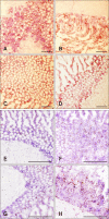
To provide insights into the role of innate immune responses in vaccine-mediated protection, we investigated the effect of Marek's disease (MD) vaccine, CVI988/Rispens, on the expression patterns of selected genes associated with activation of macrophages in MD-resistant and MD-susceptible chicken lines. Upregulation of interferon γ, interleukin (IL)-1β, IL-8, and IL-12 at different days post-inoculation (dpi) revealed activation of macrophages in both chicken lines. A strong immune response was induced in cecal tonsils of the susceptible line at 5 dpi. The highest transcriptional activities were observed in spleen tissues of the resistant line at 3 dpi. No increase in the population of CD3⁺ T cells was observed in duodenum of vaccinated birds at 5 dpi indicating a lack of involvement of the adaptive immune system in the transcriptional profiling of the tested genes. There was, however, an increase in the number of macrophages in the duodenum of vaccinated birds. The CVI988/Rispens antigen was detected in the duodenum and cecal tonsils of the susceptible line at 5 dpi but not in the resistant line. This study sheds light on the role of macrophages in vaccine-mediated protection against MD and on the possible development of new recombinant vaccines with enhanced innate immune system activation properties.
Keywords: CVI988/Rispens; Marek’s disease; cecal tonsils; duodenum; macrophages.
Conflict of interest statement
Figures


Similar articles
-
Early Immune Responses to Marek's Disease Vaccines.Viral Immunol. 2017 Apr;30(3):167-177. doi: 10.1089/vim.2016.0126. Epub 2017 Mar 27. Viral Immunol. 2017. PMID: 28346793
-
Suspension culture of Marek's disease virus and evaluation of its immunological effects.Avian Pathol. 2019 Jun;48(3):183-190. doi: 10.1080/03079457.2018.1556385. Epub 2019 Apr 3. Avian Pathol. 2019. PMID: 30518239
-
Protection provided by Rispens CVI988 vaccine against Marek's disease virus isolates of different pathotypes and early prediction of vaccine take and MD outcome.Avian Pathol. 2016;45(1):26-37. doi: 10.1080/03079457.2015.1110850. Avian Pathol. 2016. PMID: 26503904
-
History of the First-Generation Marek's Disease Vaccines: The Science and Little-Known Facts.Avian Dis. 2016 Dec;60(4):715-724. doi: 10.1637/11429-050216-Hist. Avian Dis. 2016. PMID: 27902902 Review.
-
Vaccinal control of Marek's disease: current challenges, and future strategies to maximize protection.Vet Immunol Immunopathol. 2006 Jul 15;112(1-2):78-86. doi: 10.1016/j.vetimm.2006.03.014. Epub 2006 May 8. Vet Immunol Immunopathol. 2006. PMID: 16682084 Review.
Cited by
-
An Anti-Tumor Vaccine Against Marek's Disease Virus Induces Differential Activation and Memory Response of γδ T Cells and CD8 T Cells in Chickens.Front Immunol. 2021 Feb 15;12:645426. doi: 10.3389/fimmu.2021.645426. eCollection 2021. Front Immunol. 2021. PMID: 33659011 Free PMC article.
-
Revisiting cellular immune response to oncogenic Marek's disease virus: the rising of avian T-cell immunity.Cell Mol Life Sci. 2020 Aug;77(16):3103-3116. doi: 10.1007/s00018-020-03477-z. Epub 2020 Feb 20. Cell Mol Life Sci. 2020. PMID: 32080753 Free PMC article. Review.
-
The immune cell landscape and response of Marek's disease resistant and susceptible chickens infected with Marek's disease virus.Sci Rep. 2023 Apr 1;13(1):5355. doi: 10.1038/s41598-023-32308-x. Sci Rep. 2023. PMID: 37005445 Free PMC article.
-
Fiction and Facts about BCG Imparting Trained Immunity against COVID-19.Vaccines (Basel). 2022 Jun 23;10(7):1006. doi: 10.3390/vaccines10071006. Vaccines (Basel). 2022. PMID: 35891168 Free PMC article. Review.
-
Cellular MicroRNA Expression Profile of Chicken Macrophages Infected with Newcastle Disease Virus Vaccine Strain LaSota.Pathogens. 2019 Aug 9;8(3):123. doi: 10.3390/pathogens8030123. Pathogens. 2019. PMID: 31405004 Free PMC article.
References
-
- Bacon LD, Hunt HD, Cheng HH. A review of the development of chicken lines to resolve genes determining resistance to diseases. Poult Sci. 2000;79:1082–1093. - PubMed
-
- Baigent S, Davison T. Marek's disease virus: biology and life cycle. In: Davison F, Nair V, editors. Marek's Disease: An Evolving Problem. Oxford: Elsevier Academic Press; 2004. pp. 62–77.
-
- Biron CA. Initial and innate responses to viral infections-pattern setting in immunity or disease. Curr Opin Microbiol. 1999;2:374–381. - PubMed
MeSH terms
Substances
LinkOut - more resources
Full Text Sources
Other Literature Sources

