. 2018 Feb:28:261-273.
doi: 10.1016/j.ebiom.2018.01.002. Epub 2018 Jan 9.

Enhanced Immunosuppressive Properties of Human Mesenchymal Stem Cells Primed by Interferon-γ

Affiliations

Enhanced Immunosuppressive Properties of Human Mesenchymal Stem Cells Primed by Interferon-γ

Dae Seong Kim et al. EBioMedicine. 2018 Feb.

Abstract

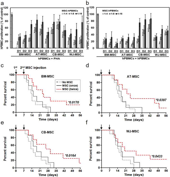
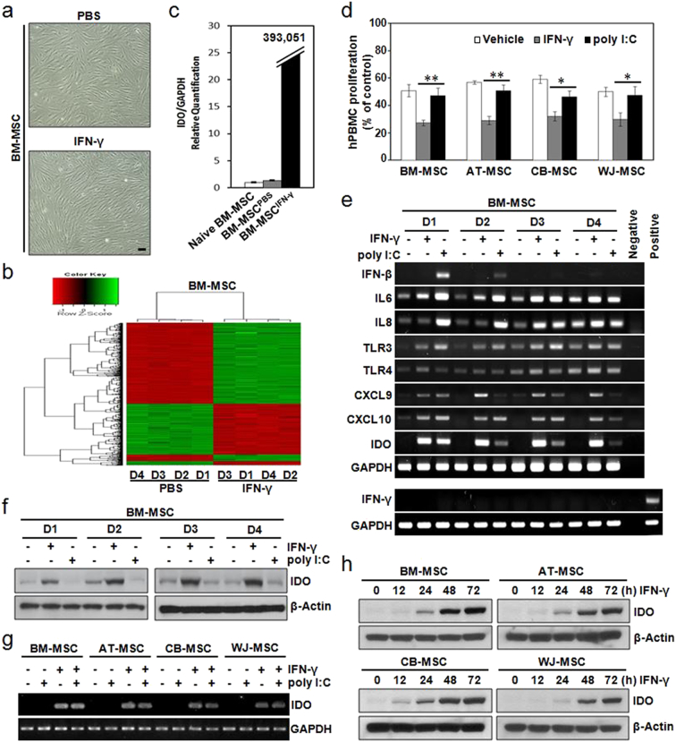
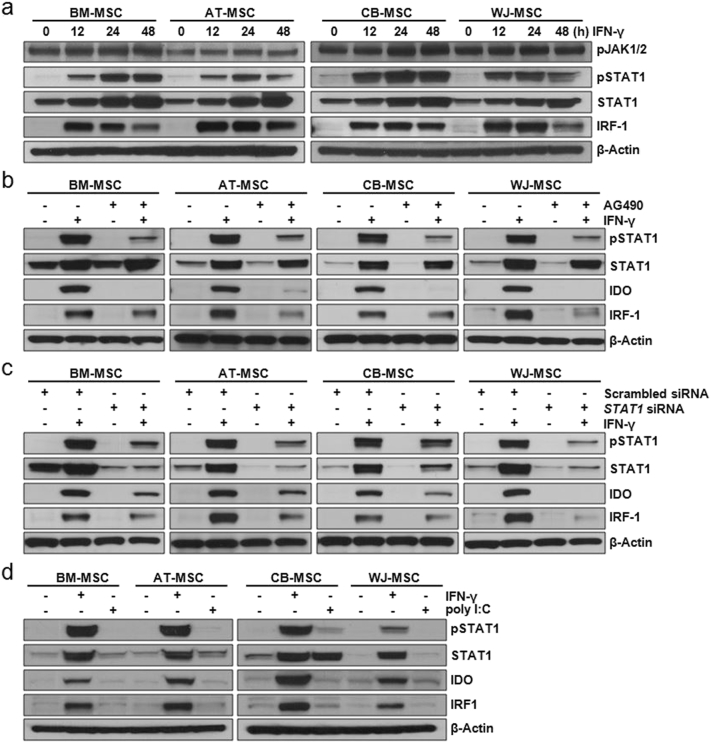
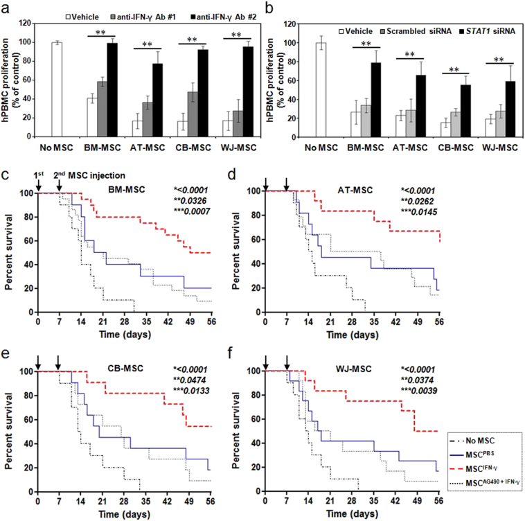
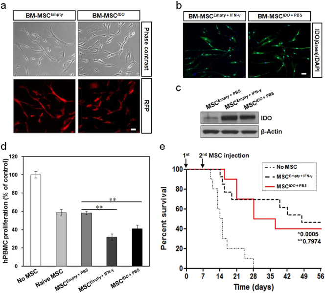
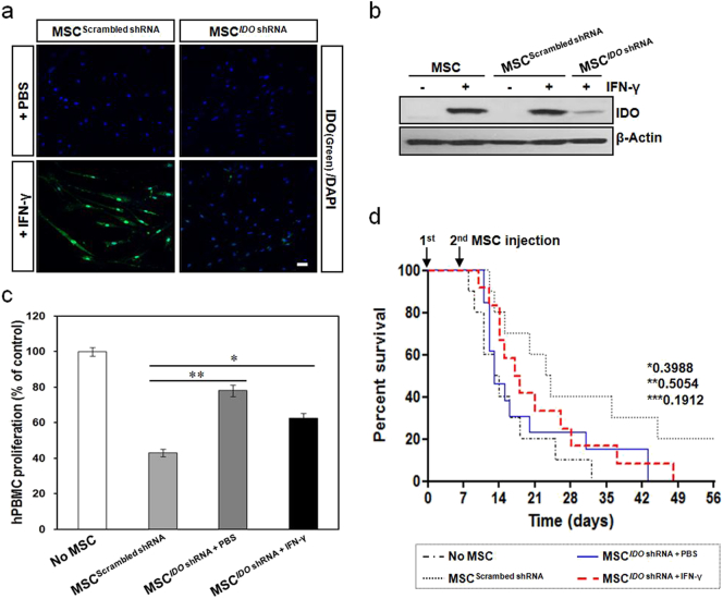
Mesenchymal stem cells (MSCs) are of particular interest for the treatment of immune-related diseases owing to their immunosuppressive properties. In this study, we aimed to identify the effect of interferon (IFN)-γ priming on immunomodulation by MSCs and elucidate the possible mechanism underlying their properties for the clinical treatment of allogeneic conflicts. Infusion of MSCs primed with IFN-γ significantly reduced the symptoms of graft-versus-host disease (GVHD) in NOD-SCID mice, thereby increasing survival rate when compared with naïve MSC-infused mice. However, infusion of IFN-γ-primed MSCs in which indoleamine 2,3-dioxygenase (IDO) was downregulated did not elicit this effect. The IDO gene was expressed in MSCs via the IFN-γ-Janus kinase (JAK)-signal transducer and activator of transcription 1 (STAT1) pathway, and the infusion of IDO-over-expressing MSCs increased survival rate in an in vivo GVHD model, similar to infusion of IFN-γ-primed MSCs. These data indicate that IFN-γ production by activated T-cells is correlated with the induction of IDO expression in MSCs via the IFN-γ-JAK-STAT1 pathway, which in turn results in the suppression of T-cell proliferation. Our findings also suggest that cell therapy based on MSCs primed with IFN-γ can be used for the clinical treatment of allogeneic conflicts, including GVHD.

Keywords: Cell therapy; Graft-versus-host disease; Indoleamine 2,3-dioxygenase; Interferon-γ; Mesenchymal stem cell.


Figures

Fig. 1
Fig. 1
Immunosuppressive properties of MSCs derived from different tissues. MSCs derived from four different tissues (BM-, AT-, CB-, and WJ-MSC) and three different donors (D1–D3) were used. (a) PHA-induced hPBMC proliferation in the absence or presence of different numbers of MSCs. hPBMC proliferation was evaluated on day 3 as the percentage of BrdU+ cells. (b) hPBMC proliferation using a mixed lymphocyte reaction in the absence or presence of different numbers of MSCs. Data are expressed as the percentage of hPBMC proliferation in the absence of MSCs and represent the mean ± SD of three separate experiments. Increased survival of GVHD mice in response to BM-MSCs (c), AT-MSCs (d), CB-MSCs (e), and WJ-MSCs (f). MSCs were administered once (black solid line), or twice (red dotted line) with a 7-day interval, via intravenous injection, and the survival rates of GVHD mice were determined. The survival rate increased when MSCs were administered twice. (c) No MSC (n = 10), MSC (once) (n = 7), and MSC (twice) (n = 10). (d) No MSC (n = 10), MSC (once) (n = 7), and MSC (twice) (n = 8). (e) No MSC (n = 10), MSC (once) (n = 7), and MSC (twice) (n = 8). (f) No MSC (n = 10), MSC (once) (n = 7), and MSC (twice) (n = 9). *, P-value of the MSC (twice) group versus the No MSC group.
Fig. 2
Fig. 2
IFN-γ priming commonly induces IDO expression in MSCs, but TLR3 activation rarely does. (a–c) BM-MSCs were treated with 200 IU/mL IFN-γ for 24 h prior to harvest. (a) Morphological appearance of BM-MSCs, with or without, IFN-γ priming. (b) Microarray data were filtered by applying two criteria for significant changes, i.e. P < 0.05 and a fold change > 2, between PBS-treated and IFN-γ-primed BM-MSCs. Hierarchical cluster analysis of genes differentially expressed between PBS-treated and IFN-γ-primed MSCs. Four samples (D1–D4) were analyzed per culture condition. (c) Quantitative real-time (qRT)-PCR analysis of IDO mRNA, which was up-regulated in IFN-γ-primed BM-MSCs. (d) PHA-induced hPBMC proliferation in the presence of MSCs pretreated with PBS (vehicle), 200 IU/mL IFN-γ, or 100 μg/mL poly I:C. hPBMC proliferation was evaluated on day 3 and is expressed as the percentage of BrdU+ cells. Data are expressed as the percentage of hPBMC proliferation in the absence of MSCs and represent the mean ± SD of three separate experiments. *P < 0.05, **P < 0.01. (e–f) MSCs derived from four different donors (D1–D4) were treated with 200 IU/mL IFN-γ or 100 μg/mL poly I:C for 24 h. (e) mRNA levels of the indicated genes were determined by semi-quantitative RT-PCR. PHA-treated hPBMCs were used as a positive control. (f) Protein levels of IDO were determined by immunoblotting. (g–h) MSCs derived from four different tissues (BM-, AT-, CB- and WJ-MSC) were used. (g) MSCs were treated with 200 IU/mL IFN-γ and/or 100 μg/mL poly I:C for 24 h. IDO mRNA levels were determined by semi-quantitative RT-PCR. (h) MSCs were treated with 200 IU/mL IFN-γ for the indicated amounts of time. IDO protein levels were determined by immunoblotting. IDO protein expression increased in a time-dependent manner in IFN-γ-primed MSCs. GAPDH and β-Actin were used as loading controls for PCR and western blotting, respectively.
Fig. 3
Fig. 3
IDO expression in IFN-γ-primed MSCs via a JAK-STAT1 signaling pathway. MSCs derived from four different tissues (BM-, AT-, CB-, and WJ-MSC) were used. (a) MSCs were incubated with 200 IU/mL IFN-γ for the indicated amounts of time. The expression levels of phospho-JAK1/2, phospho-STAT1, STAT1, and IRF-1 in these MSCs were detected by immunoblotting. (b) To inhibit the activity of JAK, an intracellular domain of the IFN-γ receptor, MSCs were incubated with 1 μM AG490 (a JAK inhibitor) for 24 h before IFN-γ priming. The expression levels of phospho-STAT1, STAT1, IDO, and IRF-1 in AG490-treated MSCs were detected by immunoblotting. AG490 treatment induced the down-regulation of STAT1 activity and IDO expression. (c) To down-regulate STAT1 activity, MSCs were transfected with a scrambled siRNA or with an siRNA targeting STAT1. The expression levels of phospho-STAT1, STAT1, IDO, and IRF-1 in these transfected MSCs were detected by immunoblotting. Down-regulation of STAT1 activity effectively induced a decrease in IDO expression in IFN-γ-primed MSCs. (d) MSCs were treated with 200 IU/mL IFN-γ or 100 μg/mL poly I:C for 24 h. The expression levels of phospho-STAT1, STAT1, IDO, and IRF-1 in these MSCs were detected by immunoblotting. β-Actin was used as a loading control for all western blots.
Fig. 4
Fig. 4
Enhanced immunosuppressive properties of IFN-γ-primed MSCs. (a–b) 200 IU/mL IFN-γ was added to MSCs, and the cells were incubated for 24 h. PHA-stimulated hPBMCs were incubated in the absence or presence of PBS-treated, or IFN-γ-primed MSCs. (a) Pretreatment with an anti-IFN-γ antibody (#1, once; #2, twice) before IFN-γ priming significantly decreased the suppressive effect of MSCs on PHA-induced T-cell proliferation. (b) Down-regulation of STAT1 activity using an siRNA before IFN-γ priming significantly decreased the suppressive effect of MSCs on PHA-induced T-cell proliferation. hPBMC proliferation was evaluated on day 3 and is expressed as the percentage of BrdU+ cells. Data are expressed as the percentage of hPBMC proliferation in the absence of MSCs and represent the mean ± SD of three separate experiments. **P < 0.01. (c–f) Increase in the survival rates of GVHD mice after infusion of IFN-γ-primed BM-MSCs (c), AT-MSCs (d), CB-MSCs (e), and WJ-MSCs (f). MSCs pretreated with or without IFN-γ were intravenously administered MSCs twice, with a 7-day interval. The survival rate was increased more when IFN-γ-primed MSCs were administered than when PBS-treated MSCs were administered. There was no difference in the survival rate of mice between the group co-transplanted with hPBMCs and PBS-treated MSCs, and the group co-transplanted with hPBMCs and the JAK inhibitor AG490- plus IFN-γ-primed MSCs. (c) No MSC (n = 10), MSCPBS (n = 10), MSCIFN-γ (n = 20), and MSCAG490 + IFN-γ (n = 22). (d) No MSC (n = 10), MSCPBS (n = 11), MSCIFN-γ (n = 12), and MSCAG490 + IFN-γ (n = 14). (e) No MSC (n = 10), MSCPBS (n = 11), MSCIFN-γ (n = 11), and MSCAG490 + IFN-γ (n = 11). (f) No MSC (n = 10), MSCPBS (n = 12), MSCIFN-γ (n = 12), and MSCAG490 + IFN-γ (n = 12). *, P-value of MSCIFN-γ group versus No MSC group; **, P-value of MSCIFN-γ group versus MSCPBS group; and ***, P-value of MSCIFN-γ group versus MSCAG490 + IFN-γ group.
Fig. 5
Fig. 5
Enhancement of immunosuppressive properties in IDO-overexpressing MSCs. A lentiviral vector carrying IDO and RFP was transduced into BM-MSCs. (a) MSCs with stable IDO expression were established and RFP expression was observed under a fluorescence microscope. Scale bar: 50 μm. (b) Immunocytochemistry showing the expression of IDO in IDO-overexpressing MSCs. IFN-γ-primed MSCs were used as a positive control. Scale bar: 50 μm. (c) Immunoblot analysis of IDO protein expression in IDO-overexpressing MSCs. (d) PHA-induced hPBMC proliferation in the presence of IDO-overexpressing MSCs. hPBMC proliferation was evaluated on day 3 and is expressed as the percentage of BrdU+ cells. Data are expressed as the percentage of hPBMC proliferation in the absence of MSCs and represent the mean ± SD of three separate experiments. **P < 0.01. (e) Survival rates of GVHD mice after infusion of IDO-overexpressing MSCs. MSCs were intravenously administered twice, with a 7-day interval, into GVHD mice. The survival rate in the IDO-overexpressing MSC group was as high as that in the IFN-γ-primed MSC group. *, P-value of MSCIDO + PBS group versus No MSC group; **, P-value of MSCIDO + PBS group versus MSCEmpty + IFN-γ group. No MSC (n = 10), MSCIDO + PBS (n = 10), and MSCEmpty + IFN-γ (n = 13).
Fig. 6
Fig. 6
Decrease in the immunosuppressive properties of MSCs after down-regulation of IDO expression. BM-MSCs were transduced with lentiviral particles containing an IDO-targeting shRNA or a scrambled shRNA. (a) Immunocytochemistry showing low expression of IDO in IDO-down-regulated MSCs. Scale bar: 50 μm. (b) Immunoblot analysis of IDO protein expression in IDO-down-regulated MSCs. (c) PHA-induced hPBMC proliferation in the presence of IDO-down-regulated MSCs that were primed by IFN-γ. hPBMC proliferation was evaluated on day 3 and is expressed as the percentage of BrdU+ cells. Data are expressed as the percentage of hPBMC proliferation in the absence of MSCs and represent the mean ± SD of three separate experiments. *P < 0.05, **P < 0.01. (d) Survival rates for GVHD mice after infusion of IDO-down-regulated MSCs. MSCs were prepared with or without IFN-γ pretreatment and were intravenously administered twice, with a 7-day interval, into GVHD mice. There was no statistically significant difference in the survival rate of mice between the group co-transplanted with hPBMCs plus IDO-down-regulated MSCs and the group transplanted with hPBMCs only. *, P-value of MSCIDO shRNA + PBS group versus No MSC group; **, P-value of MSCIDO shRNA + PBS group versus MSCIDO shRNA + IFN-γ group; ***, P-value of MSCIDO shRNA + IFN-γ group versus MSCScrambled shRNA group. No MSC (n = 10), MSCScrambled shRNA (n = 10), MSCIDO shRNA + PBS (n = 13), and MSCIDO shRNA + IFN-γ (n = 12).

References

    1. Ankrum J.A., Ong J.F., Karp J.M. Mesenchymal stem cells: immune evasive, not immune privileged. Nat. Biotechnol. 2014;32:252–260. - PMC - PubMed
    1. Asavaroengchai W., Wang H., Wang S., Wang L., Bronson R., Sykes M., Yang Y.G. An essential role for IFN-gamma in regulation of alloreactive CD8 T cells following allogeneic hematopoietic cell transplantation. Biol. Blood Marrow Transplant. 2007;13:46–55. - PMC - PubMed
    1. Augello A., Tasso R., Negrini S.M., Amateis A., Indiveri F., Cancedda R., Pennesi G. Bone marrow mesenchymal progenitor cells inhibit lymphocyte proliferation by activation of the programmed death 1 pathway. Eur. J. Immunol. 2005;35:1482–1490. - PubMed
    1. Baksh D., Song L., Tuan R.S. Adult mesenchymal stem cells: characterization, differentiation, and application in cell and gene therapy. J. Cell. Mol. Med. 2004;8:301–316. - PMC - PubMed
    1. Barry F.P., Murphy J.M. Mesenchymal stem cells: clinical applications and biological characterization. Int. J. Biochem. Cell Biol. 2004;36:568–584. - PubMed

MeSH terms