

Synergistic interplay between the two major bone minerals, hydroxyapatite and whitlockite nanoparticles, for osteogenic differentiation of mesenchymal stem cells
- PMID: 29366976
- PMCID: PMC5839653
- DOI: 10.1016/j.actbio.2018.01.016
Synergistic interplay between the two major bone minerals, hydroxyapatite and whitlockite nanoparticles, for osteogenic differentiation of mesenchymal stem cells
Erratum in
-
Corrigendum to "Synergistic interplay between the two major bone minerals, hydroxyapatite and whitlockite nanoparticles, for osteogenic differentiation of mesenchymal stem cells" [Acta Biomater. 69 (2018) 342-351].Acta Biomater. 2018 Nov;81:328-329. doi: 10.1016/j.actbio.2018.09.028. Epub 2018 Sep 26. Acta Biomater. 2018. PMID: 30268828 No abstract available.
Abstract
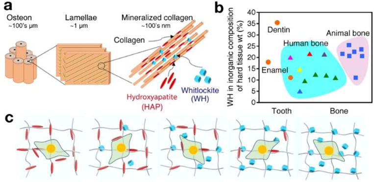
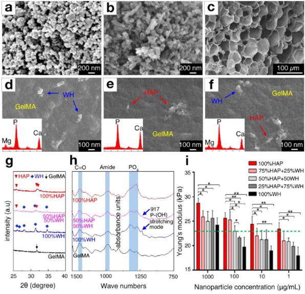
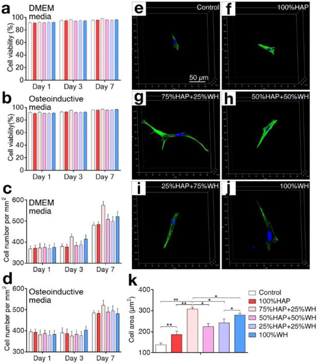
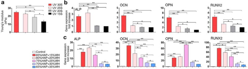
The inorganic part of human bone is mainly composed of hydroxyapatite (HAP: Ca10(PO4)6(OH)2) and whitlockite (WH: Ca18Mg2(HPO4)2(PO4)12) minerals, where the WH phase occupies up to 20-35% of total weight. These two bone minerals have different crystal structures and physicochemical properties, implying their distinguished role in bone physiology. However, until now, the biological significance of the presence of a certain ratio between HAP and WH in bone is unclear. To address this fundamental question, bone mimetic scaffolds are designed to encapsulate human mesenchymal stem cells (MSCs) for assessing their osteogenic activity depending on different ratios of HAP and WH. Interestingly, cellular growth and osteogenic differentiation are significantly promoted when MSCs are grown with a 3-1 ratio of HAP and WH nanoparticles, which is similar to bone. One of the reasons for this synergism between HAP and WH in hydrogel scaffolds is that, while WH nanoparticles can enhance osteogenic differentiation of MSCs compared to HAP, WH counterintuitively decreases the mechanical stiffness of nanocomposite hydrogels and hinders the osteogenic activity of cells. Taken together, these findings identify the optimal ratio between two major minerals in bone mimetic scaffolds to maximize the osteogenic differentiation of MSCs.
Statement of significance: Human bone minerals are composed of HAP and WH inorganic nanoparticles which have different material properties. However, the reason for the coexistence of HAP and WH in human bone is not fully identified, and HAP and WH composite biomaterial has not been utilized in the clinic. In this study, we have developed bone mimetic HAP and WH nanocomposite hydrogel scaffolds with various ratios. Importantly, we found out that HAP can promote the mechanical stiffness of the composite hydrogel scaffolds while WH can enhance the osteogenic activity of stem cells, which together induced synergism to maximize osteogenic differentiation of stem cells when mixed into 3-1 ratio that is similar to human bone.
Keywords: Bone materials; Hydroxyapatite; Mesenchymal stem cells; Osteogenic differentiation; Whitlockite.
Copyright © 2018 Acta Materialia Inc. Published by Elsevier Ltd. All rights reserved.
Conflict of interest statement
Authors declare no conflict of interest.
Figures

Similar articles
-
Comparative study of porous hydroxyapatite/chitosan and whitlockite/chitosan scaffolds for bone regeneration in calvarial defects.Int J Nanomedicine. 2017 Apr 4;12:2673-2687. doi: 10.2147/IJN.S131251. eCollection 2017. Int J Nanomedicine. 2017. PMID: 28435251 Free PMC article.
-
In Vitro and In Vivo Evaluation of Whitlockite Biocompatibility: Comparative Study with Hydroxyapatite and β-Tricalcium Phosphate.Adv Healthc Mater. 2016 Jan 7;5(1):128-36. doi: 10.1002/adhm.201400824. Epub 2015 May 12. Adv Healthc Mater. 2016. PMID: 25963732
-
Synthesis and formation mechanism of bone mineral, whitlockite nanocrystals in tri-solvent system.J Colloid Interface Sci. 2020 Jun 1;569:1-11. doi: 10.1016/j.jcis.2020.02.072. Epub 2020 Feb 17. J Colloid Interface Sci. 2020. PMID: 32092600
-
Hydroxyapatite Nanoparticles Promote the Development of Bone Microtissues for Accelerated Bone Regeneration by Activating the FAK/Akt Pathway.ACS Biomater Sci Eng. 2024 Jul 8;10(7):4463-4479. doi: 10.1021/acsbiomaterials.4c00574. Epub 2024 Jun 7. ACS Biomater Sci Eng. 2024. PMID: 38848471
-
The role of calcium phosphate surface structure in osteogenesis and the mechanisms involved.Acta Biomater. 2020 Apr 1;106:22-33. doi: 10.1016/j.actbio.2019.12.034. Epub 2020 Jan 9. Acta Biomater. 2020. PMID: 31926336 Review.
Cited by
-
Natural bone-mimicking nanopore-incorporated hydroxyapatite scaffolds for enhanced bone tissue regeneration.Biomater Res. 2022 Feb 25;26(1):7. doi: 10.1186/s40824-022-00253-x. Biomater Res. 2022. PMID: 35216625 Free PMC article.
-
Nanohydroxyapatite, Nanosilicate-Reinforced Injectable, and Biomimetic Gelatin-Methacryloyl Hydrogel for Bone Tissue Engineering.Int J Nanomedicine. 2021 Aug 16;16:5603-5619. doi: 10.2147/IJN.S321387. eCollection 2021. Int J Nanomedicine. 2021. PMID: 34429602 Free PMC article.
-
Resorbable Mg2+-Containing Phosphates for Bone Tissue Repair.Materials (Basel). 2021 Aug 26;14(17):4857. doi: 10.3390/ma14174857. Materials (Basel). 2021. PMID: 34500951 Free PMC article. Review.
-
Enhanced bone regeneration by osteoinductive and angiogenic zein/whitlockite composite scaffolds loaded with levofloxacin.RSC Adv. 2024 May 2;14(21):14470-14479. doi: 10.1039/d4ra00772g. eCollection 2024 May 2. RSC Adv. 2024. PMID: 38708116 Free PMC article.
-
The Effect of Whitlockite as an Osteoconductive Synthetic Bone Substitute Material in Animal Bony Defect Model.Materials (Basel). 2022 Mar 4;15(5):1921. doi: 10.3390/ma15051921. Materials (Basel). 2022. PMID: 35269154 Free PMC article.
References
-
- Einhorn TA. Enhancement of fracture-healing. J Bone Joint Surg. 1995;77:940–956. - PubMed
-
- Meisinger C, Wildner M, Stieber J, Heier M, Sangha O, Döring A. Epidemiology of limb fractures. Orthopade. 2002;31:92–99. - PubMed
-
- Calori GM, Colombo M, Mazza E, Ripamonti C, Mazzola S, Marelli N, Mineo GV. Monotherapy vs. polytherapy in the treatment of forearm non-unions and bone defects. Injury. 2013;44:S63–S69. - PubMed
Publication types
MeSH terms
Substances
Grants and funding
LinkOut - more resources
Full Text Sources
Other Literature Sources
Research Materials
