Review
. 2018 May;67(5):963-972.
doi: 10.1136/gutjnl-2017-315691. Epub 2018 Jan 24.

Non-alcoholic steatohepatitis pathogenesis: sublethal hepatocyte injury as a driver of liver inflammation

Affiliations
Review

Non-alcoholic steatohepatitis pathogenesis: sublethal hepatocyte injury as a driver of liver inflammation

Samar H Ibrahim et al. Gut. 2018 May.

Abstract

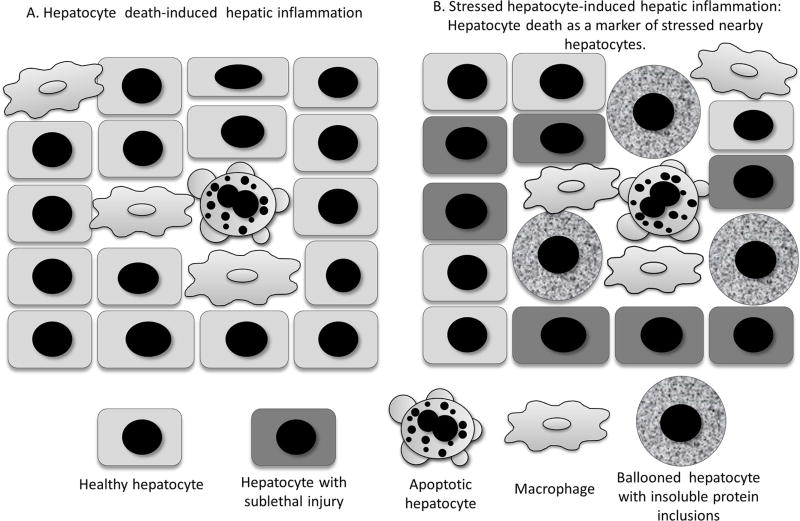
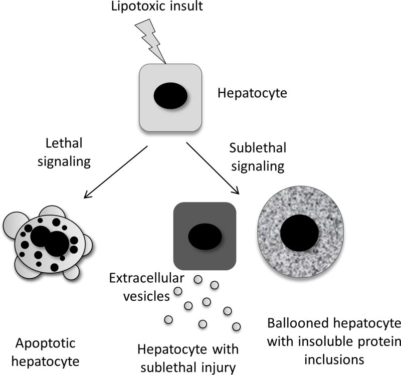
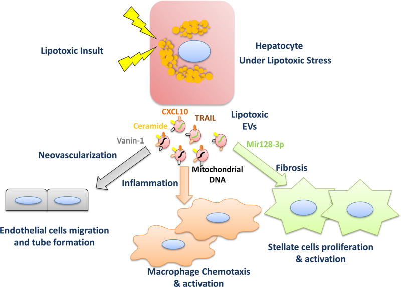
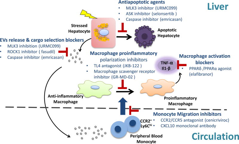
A subset of patients with non-alcoholic fatty liver disease develop an inflammatory condition, termed non-alcoholic steatohepatitis (NASH). NASH is characterised by hepatocellular injury, innate immune cell-mediated inflammation and progressive liver fibrosis. The mechanisms whereby hepatic inflammation occurs in NASH remain incompletely understood, but appear to be linked to the proinflammatory microenvironment created by toxic lipid-induced hepatocyte injury, termed lipotoxicity. In this review, we discuss the signalling pathways induced by sublethal hepatocyte lipid overload that contribute to the pathogenesis of NASH. Furthermore, we will review the role of proinflammatory, proangiogenic and profibrotic hepatocyte-derived extracellular vesicles as disease biomarkers and pathogenic mediators during lipotoxicity. We also review the potential therapeutic strategies to block the feed-forward loop between sublethal hepatocyte injury and liver inflammation.

Keywords: inflammation; nonalcoholic steatohepatitis.


Conflict of interest statement

Competing interests: None declared.

Figures

Figure 1
Figure 1
Two concepts of cell death role in NASH-associated inflammation. (A) Apoptotic hepatocytes can directly initiate inflammation via apoptotic bodies engulfed by macrophages. (B) Apoptotic hepatocytes serve as a marker for widespread proapoptotic/stress signalling occurring in the majority of nearby stressed hepatocytes. These stressed cells with sublethal injury may promote inflammation, for example, via release of proinflammatory extracellular vesicles. (Modified from Hirsova et al). NASH, non-alcoholic steatohepatitis.
Figure 2
Figure 2
Lipotoxic lethal and sublethal injury in hepatocytes. Toxic lipid-induced lethal signalling in hepatocytes causes apoptotic cell death (lipoapoptosis). Sublethal proapoptotic signalling induced by lipotoxicity results in release of extracellular vesicles. Sublethal stress also occurs in ‘undead’ hepatocytes which include ballooned hepatocytes with Mallory-Denk bodies’ inclusions.
Figure 3
Figure 3
Signalling events mediated by lipotoxic hepatocyte-derived EVs. Hepatocytes under lipotoxic conditions release increased amount of extracellular vesicles of distinct cargo. Recent in vitro and in vivo studies have described important roles of lipotoxic EVs in NASH pathogenesis through intercellular communication. CXCL10 and ceramide-enriched EVs mediate monocyte/macrophage hepatic trafficking and infiltration. Mitochondrial DNA and TRAIL-enriched EVs promote macrophage activation. VNN1-enriched EVs mediate endothelial cell migration and neovascularisation. miR-128–3 p-laden EVs induce HSC proliferation and activation. (Modified from Hirsova et al). CXCL10, C-X-C motif chemokine ligand 10; EV, extracellular vesicle; HSC, hepatic stellate cells; NASH, non-alcoholic steatohepatitis; TRAIL, tumour necrosis factor-like apoptosis-inducing ligand; VNN1, vanin-1.
Figure 4
Figure 4
Therapeutic agents that target hepatocyte injury and the sterile inflammatory response in NASH. Current therapeutic strategies are outlined and include antiapoptotic agents, inhibitors of vesicle release and pathogenic cargoes sorting, inhibitors of macrophage chemotaxis, proinflammatory polarisation and activation. ASK, apoptosis signal-regulating kinase; CCR, C-C chemokine receptor; EVs, extracellular vesicles; MLK3, mixed lineage kinase 3; NASH, non-alcoholic steatohepatitis; PPAR, peroxisome proliferator-activated receptor; ROCK1, Rho-associated, coiled-coil containing protein kinase 1.

References

    1. Younossi ZM, Koenig AB, Abdelatif D, et al. Global epidemiology of nonalcoholic fatty liver disease-meta-analytic assessment of prevalence, incidence, and outcomes. Hepatology. 2016;64:73–84. - PubMed
    1. Machado MV, Diehl AM. Pathogenesis of Nonalcoholic Steatohepatitis. Gastroenterology. 2016;150:1769–77. - PMC - PubMed
    1. Arab JP, Karpen SJ, Dawson PA, et al. Bile acids and nonalcoholic fatty liver disease: molecular insights and therapeutic perspectives. Hepatology. 2017;65:350–62. - PMC - PubMed
    1. Ahmed A, Wong RJ, Harrison SA. Nonalcoholic fatty liver disease review: diagnosis, treatment, and outcomes. Clin Gastroenterol Hepatol. 2015;13:2062–70. - PubMed
    1. Bonci E, Chiesa C, Versacci P, et al. Association of nonalcoholic fatty liver disease with subclinical cardiovascular changes: a systematic review and meta-analysis. Biomed Res Int. 2015;2015:1–11. - PMC - PubMed

Publication types