. 2018 Jan;15(138):20170593.
doi: 10.1098/rsif.2017.0593.

Stresses and strains on the human fetal skeleton during development

Affiliations

Stresses and strains on the human fetal skeleton during development

Stefaan W Verbruggen et al. J R Soc Interface. 2018 Jan.

Abstract

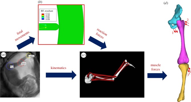
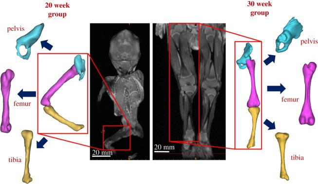
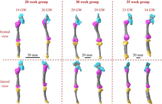
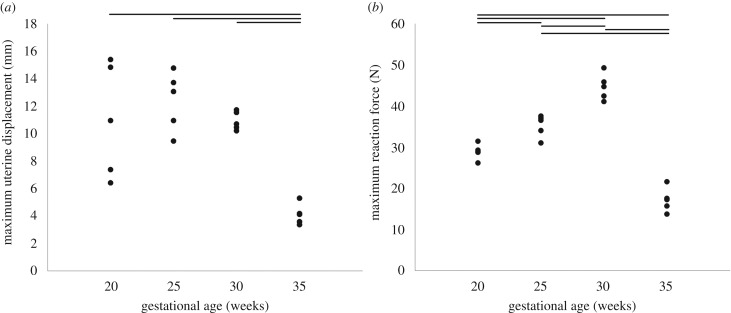
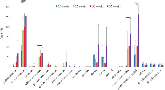
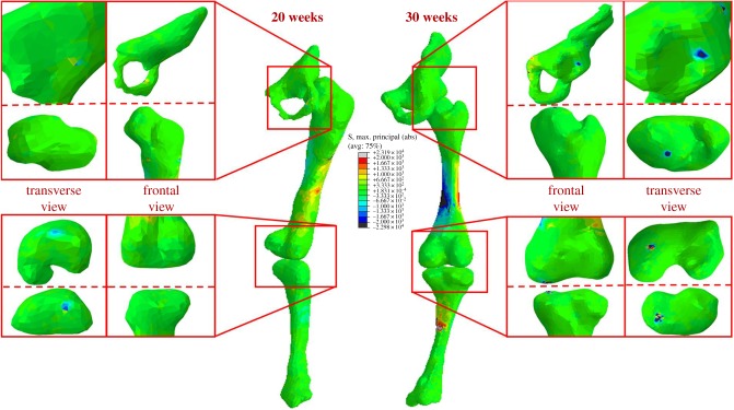
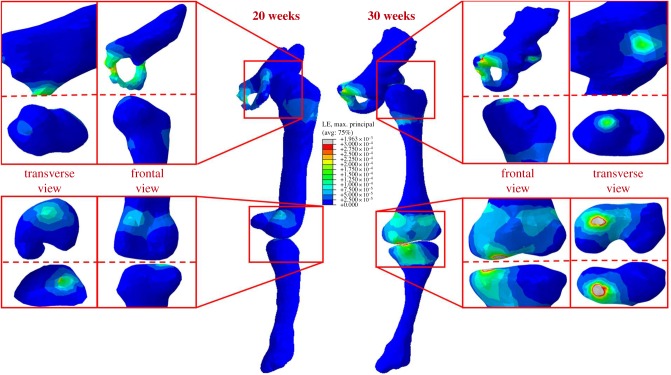
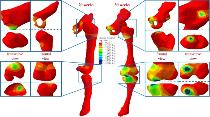
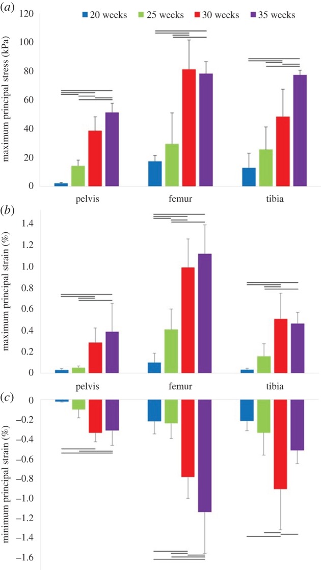
Mechanical forces generated by fetal kicks and movements result in stimulation of the fetal skeleton in the form of stress and strain. This stimulation is known to be critical for prenatal musculoskeletal development; indeed, abnormal or absent movements have been implicated in multiple congenital disorders. However, the mechanical stress and strain experienced by the developing human skeleton in utero have never before been characterized. Here, we quantify the biomechanics of fetal movements during the second half of gestation by modelling fetal movements captured using novel cine-magnetic resonance imaging technology. By tracking these movements, quantifying fetal kick and muscle forces, and applying them to three-dimensional geometries of the fetal skeleton, we test the hypothesis that stress and strain change over ontogeny. We find that fetal kick force increases significantly from 20 to 30 weeks' gestation, before decreasing towards term. However, stress and strain in the fetal skeleton rises significantly over the latter half of gestation. This increasing trend with gestational age is important because changes in fetal movement patterns in late pregnancy have been linked to poor fetal outcomes and musculoskeletal malformations. This research represents the first quantification of kick force and mechanical stress and strain due to fetal movements in the human skeleton in utero, thus advancing our understanding of the biomechanical environment of the uterus. Further, by revealing a potential link between fetal biomechanics and skeletal malformations, our work will stimulate future research in tissue engineering and mechanobiology.

Keywords: biomechanical stimuli; cine-MRI; finite element analysis; joint biomechanics; musculo-skeletal development.


Conflict of interest statement

We declare we have no competing interests.

Figures

Figure 1.
Figure 1.
Flowchart outlining the computational pipeline developed for this study. Computational methods applied comprise (a) tracking of fetal joint movements, (b) finite element modelling of reaction force resulting from fetal movements against the uterine wall, (c) musculoskeletal modelling to predict muscle forces, (d) application of muscle forces to finite element models of fetal geometries (forces for adductor magnus (1), gluteus maximus (2) and biceps femoris (3)).
Figure 2.
Figure 2.
Fetal geometries obtained from post-mortem MRI. Post-mortem MRI scans at 20 and 30 weeks' gestational age allow three-dimensional reconstruction of both mineralized and cartilaginous components of the pelvis, femur and tibia.
Figure 3.
Figure 3.
Fetal leg bone geometries grouped by gestational age. Three-dimensional geometries were reconstructed from post-mortem MRI scans, two each at approximately 20, 30 and 35 weeks. Fetal geometries increased in both size and complexity with advancing gestational age, with later gestational ages demonstrating larger, flatter iliac crests, more prominent greater trochanters and femoral condyles, and wider proximal tibia with more pronounced tibial condyles. Mineralized regions are shown in grey.
Figure 4.
Figure 4.
Maximum observed uterine displacements and resulting fetal kick forces. Average results for 20, 25, 30 and 35 weeks' gestational age, for (a) uterine wall displacement and (b) uterine reaction force. Horizontal lines indicate statistical significance between groups (p ≤ 0.05).
Figure 5.
Figure 5.
Average muscle forces at full-leg extension for 20, 25, 30 and 35 weeks' gestational age. The means and standard deviation of four groups of five kicks each are plotted; horizontal lines indicate statistical significance (p ≤ 0.05).
Figure 6.
Figure 6.
Maximum principal stress stimulation in fetal leg bones increases with gestational age. Average stress results for 20 and 30 week fetal geometries, demonstrating increased stress concentrations in mineralized regions and at joint surfaces over gestation.
Figure 7.
Figure 7.
Maximum principal strain stimulation in fetal leg bones increases with gestational age. Average maximum principal strain results for 20 and 30 week fetal geometries, demonstrating increased strain concentrations in cartilage and at joint surfaces over gestation.
Figure 8.
Figure 8.
Minimum principal strain stimulation in fetal leg bones increases with gestational age. Average minimum principal strain results for 20 and 30 week fetal geometries, demonstrating increased strain concentrations in cartilage and at joint surfaces over gestation.
Figure 9.
Figure 9.
Biomechanical stress and strain in fetal leg bones over second half of gestation. Average results for 20, 25, 30 and 35 weeks' gestational age, for (a) maximum principal stress, (b) maximum principal strain, (c) minimum principal strain. The means and standard deviation of four groups of five kicks each are plotted. Horizontal lines indicate statistical significance (p ≤ 0.05).

References

    1. de Vries JIP, Fong BF. 2006. Normal fetal motility: an overview. Ultrasound Obstet. Gynecol. 27, 701–711. ( 10.1002/uog.2740) - DOI - PubMed
    1. de Vries JIP, Visser GHA, Prechtl HFR. 1982. The emergence of fetal behaviour. I. Qualitative aspects. Early Hum. Dev. 7, 301–322. ( 10.1016/0378-3782(82)90033-0) - DOI - PubMed
    1. Hayat TTA, Nihat A, Martinez-Biarge M, McGuinness A, Allsop JM, Hajnal JV, Rutherford MA. 2011. Optimization and initial experience of a multisection balanced steady-state free precession cine sequence for the assessment of fetal behavior in utero. Am. J. Neuroradiol. 32, 331–338. ( 10.3174/ajnr.A2295) - DOI - PMC - PubMed
    1. Natale R, Nasello-Paterson C, Turliuk R. 1985. Longitudinal measurements of fetal breathing, body movements, heart rate, and heart rate accelerations and decelerations at 24 to 32 weeks of gestation. Am. J. Obstet. Gynecol. 151, 256–263. ( 10.1016/0002-9378(85)90022-5) - DOI - PubMed
    1. Ten Hof J, Nijhuis I, Nijhuis J, Narayan H, Taylor D, Visser G, Mulder E. 1999. Quantitative analysis of fetal general movements: methodological considerations. Early Hum. Dev. 56, 57–73. ( 10.1016/S0378-3782(99)00035-3) - DOI - PubMed

Publication types