. 2018 Mar 14;92(7):e02182-17.
doi: 10.1128/JVI.02182-17. Print 2018 Apr 1.

Genetic Analysis of Serum-Derived Defective Hepatitis C Virus Genomes Revealed Novel Viral cis Elements for Virus Replication and Assembly

Affiliations

Genetic Analysis of Serum-Derived Defective Hepatitis C Virus Genomes Revealed Novel Viral cis Elements for Virus Replication and Assembly

Qingchao Li et al. J Virol. .

Abstract

Defective viral genomes (DVGs) of hepatitis C virus (HCV) exist, but their biological significances have not been thoroughly investigated. Here, we analyzed HCV DVGs circulating in patient sera that possess deletions in the structural protein-encoding region. About 30% of 41 HCV clinical isolates possess DVGs that originated from the full-length genome in the same patients. No correlation between DVGs, viremia, and alanine aminotransferase (ALT) levels was found. Sequencing analysis of DVGs revealed the existence of deletion hot spots, with upstream sites in E1 and downstream sites in E2 and NS2. Interestingly, the coding sequences for the core protein and the C-terminal protease domain of NS2 were always intact in DVGs despite the fact that both proteins are dispensable for HCV genome replication. Mechanistic studies showed that transmembrane segment 3 (TMS3) of NS2, located immediately upstream of its protease domain, was required for the cleavage of NS2-NS3 and the replication of DVGs. Moreover, we identified a highly conserved secondary structure (SL750) within the core domain 2-coding region that is critical for HCV genome packaging. In summary, our analysis of serum-derived HCV DVGs revealed novel viral cis elements that play important roles in virus replication and assembly.IMPORTANCE HCV DVGs have been identified in vivo and in vitro, but their biogenesis and physiological significances remain elusive. In addition, a conventional packaging signal has not yet been identified on the HCV RNA genome, and mechanisms underlying the specificity in the encapsidation of the HCV genome into infectious particles remain to be uncovered. Here, we identified new viral cis elements critical for the HCV life cycle by determining genetic constraints that define the boundary of serum-derived HCV DVGs. We found that transmembrane segment 3 of NS2, located immediately upstream of its protease domain, was required for the cleavage of NS2-NS3 and the replication of DVGs. We identified a highly conserved secondary structure (SL750) within the core-coding region that is critical for HCV genome packaging. In summary, our analysis of serum-derived HCV DVGs revealed previously unexpected novel cis elements critical for HCV replication and morphogenesis.

Keywords: NS2; cis elements; core; defective genome; hepatitis C virus; virus packaging.


Figures

FIG 1
FIG 1
Identification of HCV DVGs from clinical isolates. (A) Schematic of the HCV genome and positions of primers used to detect DVGs in this study. (B) Agarose gel electrophoresis of PCR products to amplify DVGs. HCV prototype strains con1 (GT-1b) and JFH1 (GT-2a) were included as controls. The PCR products potentially derived from DVGs are labeled with asterisks. The gel graph was modified so that only the lanes with shorter PCR products are shown. The original gel graph is available upon request. M, molecular weight marker. (C) Frequencies of DVGs in 41 clinical isolates. (D) Correlation of DVGs with viral loads. (E) Correlation of DVGs with ALT levels. NS, nonsignificant.
FIG 2
FIG 2
Identification of hot deletion spots within DVGs. (A) Phylogenetic analysis of DVGs and FLGs. HCV prototype strains con1 (GT-1b) and JFH1 (GT-2a) were included for references. (B) Illustration of the deleted region within DVGs. Lines and black boxes represent deleted and remaining sequences, respectively. (C) Location of deletion sites on DVGs. The upstream and downstream deletion sites are labeled with black triangles pointing to the right and left, respectively. The hot deletion spots are grouped with boxes. The x axis indicates nucleotide positions starting from the beginning of core. (D) Lengths of the deleted sequences in DVGs. The average length of the deletions was 1,632 nt.
FIG 3
FIG 3
Packaging of DVGs by ectopically expressed structural proteins. (A) Schematic of four JFH1-based DVGs, JD286, JD533, JD745, and JD835, that possess representative deletion patterns of the clinical DVGs (patients 286, 533, 745, and 835). The residual p7 portion in DVG from patient 835 was deleted in JD835. The numbers indicate the HCV nucleotide sequence starting from the beginning of the 5′ UTR of the viral genome. (B and C) Intracellular HCV RNA levels (B) and extracellular infectivity titers (C) in naive Huh7.5.1 cells and Huh7.5.1 packaging cells transfected with RNA of JD286, JD533, JD745, and JD835. The HCV RNA levels were determined by RT-qPCR and expressed as values relative to the cellular actin mRNA levels. The dotted line is the detection limit of the titration assay. The error bars represent standard deviations from three independent experiments.
FIG 4
FIG 4
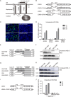
The transmembrane domain upstream of the NS2 cysteine protease domain is essential for DVG replication. (A) Schematic of three transmembrane segments of NS2. The numbers start with the first amino acid of NS2. Six DVGs that have a deletion site in NS2 are marked. (B) Schematics of the JFH1-based DVG JD533 and its NS2 protease-defective mutant (JD533-C184A) and of the JFH1-based DVG JD835 and its mutant with a deletion of the entire TMD (JD835-ΔTMD). (C) NS5A (green) immunostaining and nuclei (blue) in Huh7.5.1 cells transfected with in vitro-transcribed DVG RNA at day 2 posttransfection. (D) HCV RNA levels in Huh7.5.1 cells transfected with in vitro-transcribed DVG RNAs. The HCV RNA levels were determined by RT-qPCR and are expressed as values relative to the cellular actin mRNA levels. The error bars represent standard deviations from three independent experiments. *, P < 0.05. (E) Schematic of plasmids expressing core-NS3 of JD835 (835), JD835 with wild-type NS2 (835-TMD), or JD835 with TMD-deleted NS2 (835-ΔTMD). (F) Western blot analysis of NS2-NS3 cleavage in HEK293 cells. (G) Schematic of plasmids expressing core-NS3 of JD835 (835), JD835 with wild-type NS2 (835-TMD), JD835 with TMS1-deleted NS2 (835-ΔTMS1), or JD835 with TMD-deleted NS2 (835-ΔTMD). (H) Western blot analysis of NS2-NS3 cleavage in HEK293 cells. (I) Schematic of JFH1-based DVG JD835 and its mutant with TMS1-deleted NS2 (JD835-ΔTMS1). (J) HCV RNA levels in Huh7.5.1 cells transfected with in vitro-transcribed DVG RNAs. The HCV RNA levels were determined by RT-qPCR and are expressed as values relative to the cellular actin mRNA levels. The error bars represent standard deviations from three independent experiments. *, P < 0.05.
FIG 5
FIG 5
The core domain 2-coding region is critical for DVG packaging. (A) Schematic of JD533, CΔD2, and CΔD1-D2. Domain 2 of core was deleted in CΔD2, and both domains 1 and 2 were deleted in CΔD1-D2. The numbers indicate the HCV nucleotide sequence starting from core. (B and C) HCV RNA (B) and viral protein (C) levels in Huh7.5.1 cells transfected with JD533, CΔD2, and CΔD1-D2. The HCV RNA levels were determined by RT-qPCR and are expressed as values relative to the cellular actin mRNA levels. The error bars represent standard deviations from three independent experiments. The NS3 protein levels were quantified by using ImageJ and normalized against the internal actin control and are presented below the blot. (D and E) Intracellular HCV RNA levels (D) and extracellular infectivity titers (E) in Huh7.5.1 packaging cells transfected with JD533, CΔD2, and CΔD1-D2. The HCV RNA levels were determined by RT-qPCR and expressed as values relative to the cellular actin mRNA levels. The error bars represent standard deviations from three independent experiments. NS, nonsignificant (P > 0.05); *, P < 0.05.
FIG 6
FIG 6
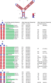
SL750 is a highly conserved structural element. A total of 3,326 HCV sequences from the HCV database of the U.S. Los Alamos National Laboratory (www.lanl.gov) were analyzed. (Top) Each nucleotide of SL750 was labeled with color according to its conservation in base pairing, and base-pairing nucleotides in more than 90% of HCV strains are highlighted in gray. (Bottom) Frequencies of base pairing at each position of the left arm (L), the right arm (R), and the central arm (C) of SL750 as well as detailed information on major and minor base pairings.
FIG 7
FIG 7
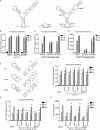
SL750 within domain 2 of the core-coding region contains a cis element critical for HCV genome packaging. (A) Secondary structure of SL750 in JFH1 (left), ΔSL750 (middle), and SL750FS (right). SL750FS had a 1-nucleotide deletion upstream of SL750 (nt 750) and a 1-nucleotide insertion downstream of SL750 (nt 825), which led to a frameshift of the SL750-coding sequence but maintained the Y-shaped RNA structure. To avoid stop codons introduced by the frameshift, A760U and A806C were also engineered into SL750FS. (B and C) HCV RNA levels in naive Huh7.5.1 cells (B) or Huh7.5.1 packaging cells (C) transfected with JD533, ΔSL750, and SL750FS. HCV RNA levels were determined by RT-qPCR and are expressed as values relative to the cellular actin mRNA levels. The error bars represent standard deviations from three independent experiments. (D) Infectivity titers in the supernatants of Huh7.5.1 packaging cells transfected with JD533, ΔSL750, and SL750FS. The error bars represent standard deviations from three independent experiments. NS, nonsignificant (P > 0.05); *, P < 0.05. (E) Schematic of unzipping and rezipping mutations on the left and right arms of SL750. Unzipping and rezipping mutations are marked with * and #, respectively. WT, wild type. (F and G) HCV RNA levels in Huh7.5.1 cells (F) or Huh7.5.1 packaging cells (G) transfected with JD533 and unzipping and rezipping mutants. The HCV RNA levels were determined by RT-qPCR and are expressed as values relative to the cellular actin mRNA levels. The error bars represent standard deviations from three independent experiments. (H) Infectivity titers in the supernatants of Huh7.5.1 packaging cells transfected with JD533 and unzipping and rezipping mutants. The error bars represent standard deviations from three independent experiments. NS, nonsignificant (P > 0.05); *, P < 0.05.
FIG 8
FIG 8
SL750 is critical for HCV FLG packaging. Huh7.5.1 cells were transfected with in vitro-transcribed wild-type JFH1, mutant JFH-1 containing unzipping and rezipping mutations in SL750R, wild-type Jc1, or mutant Jc1 containing unzipping and rezipping mutations in SL750. (A to C and E to G) Intracellular HCV RNA levels (A and E), viral protein levels (B and F), and extracellular infectivity titers (C and G) on days 1, 2, and 3 posttransfection were determined by RT-qPCR, Western blotting, and a titration assay, respectively. NS3 protein levels were quantified by using ImageJ and normalized against the internal actin control and are presented below the blot. The error bars represent standard deviations from three independent experiments. NS, nonsignificant (P > 0.05); *, P < 0.05. (D) Schematic of wild-type and unzipping and rezipping mutations on the left and right arms of SL750 in the Jc1 strain. Unzipping and rezipping mutations are marked by * and #, respectively.

Similar articles

Cited by

References

    1. Li D, Huang Z, Zhong J. 2015. Hepatitis C virus vaccine development: old challenges and new opportunities. Nat Sci Rev 2:285–295. doi:10.1093/nsr/nwv040. - DOI
    1. Klibanov OM, Gale SE, Santevecchi B. 2015. Ombitasvir/paritaprevir/ritonavir and dasabuvir tablets for hepatitis C virus genotype 1 infection. Ann Pharmacother 49:566–581. doi:10.1177/1060028015570729. - DOI - PubMed
    1. Hayes CN, Chayama K. 2015. Emerging treatments for chronic hepatitis C. J Formos Med Assoc 114:204–215. doi:10.1016/j.jfma.2014.09.001. - DOI - PubMed
    1. Lohmann V, Körner F, Koch J, Herian U, Theilmann L, Bartenschlager R. 1999. Replication of subgenomic hepatitis C virus RNAs in a hepatoma cell line. Science 285:110–113. doi:10.1126/science.285.5424.110. - DOI - PubMed
    1. Tsukiyama-Kohara K, Iizuka N, Kohara M, Nomoto A. 1992. Internal ribosome entry site within hepatitis C virus RNA. J Virol 66:1476–1483. - PMC - PubMed

Publication types

LinkOut - more resources