. 2018 Mar 14;92(7):e01318-17.
doi: 10.1128/JVI.01318-17. Print 2018 Apr 1.

Human Papillomavirus 16 E7 Stabilizes APOBEC3A Protein by Inhibiting Cullin 2-Dependent Protein Degradation

Affiliations

Human Papillomavirus 16 E7 Stabilizes APOBEC3A Protein by Inhibiting Cullin 2-Dependent Protein Degradation

Joseph A Westrich et al. J Virol. .

Abstract

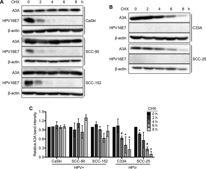
APOBEC3 (A3) mutation signatures have been observed in a variety of human cancer genomes, including those of cervical and head and neck cancers caused by human papillomavirus (HPV) infection. However, the driving forces that promote off-target A3 activity remain mostly unclear. Here, we report a mechanism for the dramatic increase of A3A protein levels in HPV-positive keratinocytes. We show that expression of the viral protein E7 from high-risk HPVs, but not E7 from low-risk HPVs, significantly prolongs the cellular half-life of A3A protein in human keratinocytes and HPV-positive cancer cell lines. We have mapped several residues within the cullin 2 (CUL2) binding motif of HPV16 E7 as being important for mediating A3A protein stabilization. Furthermore, we provide direct evidence that both A3A and HPV16 E7 interact with CUL2, suggesting that the E7-CUL2 complex formed during HPV infection may regulate A3A protein levels in the cell. Using an in vitro cytidine deaminase assay, we show that E7-stabilized A3A remains catalytically active. Taken together, our findings suggest that the HPV oncoprotein E7 dysregulates endogenous A3A protein levels and thus provides novel mechanistic insight into cellular triggers of A3 mutations in HPV-positive cancers.IMPORTANCE Human papillomavirus (HPV) is causally associated with over 5% of all human malignancies. Several recent studies have shown that a subset of cancers, including HPV-positive head and neck and cervical cancers, have distinct mutational signatures potentially caused by members of the APOBEC3 cytidine deaminase family. However, the mechanism that induces APOBEC3 activity in cancer cells is poorly understood. Here, we report that the HPV oncoprotein E7 stabilizes the APOBEC3A (A3A) protein in human keratinocytes by inhibiting ubiquitin-dependent protein degradation in a cullin-dependent manner. Interestingly, the HPV E7-stabilized A3A protein maintains its deaminase activity. These findings provide a new insight into cancer mutagenesis enhanced by virus-induced A3A protein stabilization.

Keywords: APOBEC3; cancer mutagenesis; cervical cancer; cullin; head and neck cancer; innate immunity; papillomavirus; somatic mutation.


Figures

FIG 1
FIG 1
HPV16 E7 increases A3A protein levels. Protein levels of endogenous A3A in NIKS, NIKS-16, and NIKS-16ΔE7 cells (A and B) and exogenous A3A in 293FT cells (C and D) were determined by Western blotting and densitometry. (C) 293FT cells were cotransfected with a fixed amount of pcDNA3.1-A3A-HA and pEGFP-N3 and increasing amounts of pCMV-16E7. (A and C) Cell lysates were prepared, and A3A (endogenous A3A or transfected A3A-HA), HPV16 E7, and β-actin were detected by Western blotting. (C) Transfection efficiency was determined by Western blotting of GFP. (B and D) The A3A band density was normalized to the β-actin band density. Data are shown as the fold changes of normalized A3A band density ± standard deviations relative to the results for NIKS cells (B) or 293FT cells without E7 expression (D). Shown are representative results of four independent experiments. P values were calculated by the Student t test. *, P < 0.05; n.s., not significant.
FIG 2
FIG 2
HPV16 prevents A3A protein degradation. (A and B) 293FT cells were cotransfected with pcDNA3.1-A3A-HA and pCMV-16E7 or a corresponding vector. Cotransfected 293FT cells (A, B, and E) and NIKS, NIKS-16 or NIKS-16ΔE7 cells (C, D, and F) were treated with 50 μg/ml cycloheximide (CHX) for the indicated times. (G and H) NIKS cells were treated with 20 μM MG132 for the indicated times. A3A protein expression was analyzed as described in the legend to Fig. 1. Transfected A3A-HA (A) or endogenous A3A (C and G) was detected by Western blotting using anti-HA or anti-A3A antibodies, respectively, and quantified by densitometry as described in the legend to Fig. 1 (B, D and H). The viability of CHX-treated 293FT cells (E) or NIKS, NIKS-16, and NIKS-16ΔE7 cells (F) was assessed using the CellTiter-Glo luminescent cell viability assay (Promega). Data are shown as percent cell viability ± standard deviation and normalized to the viability of untreated (0 h) cells (E and F). All experiments were repeated three times. Data are shown as the fold change relative to the results for cells at 0 h of treatment ± standard deviations. P values were determined by the Student t test (B, D, and H) or one-way ANOVA (E and F). *, P < 0.05; #, P < 0.005; ▽, P < 0.0005; n.s., not significant.
FIG 3
FIG 3
High-risk HPV E7s, but not low-risk HPV E7s, prevent A3A protein degradation. (A) NIKS cells stably expressing HPV6 (NIKS-6E7), HPV11 (NIKS-11E7), HPV16 (NIKS-16E7), or HPV18 (NIKS-18E7) or containing vector alone (NIKS-vector) were treated with 50 μg/ml CHX and analyzed by Western blotting using anti-A3A and anti-β-actin antibodies as described in the legend to Fig. 1. (B) The A3A band density was normalized to the β-actin band density. P values were determined by the Student t test. *, P < 0.05; #, P < 0.005; ▽, P < 0.0005.
FIG 4
FIG 4
A3A protein is stabilized in HPV-positive CxCa and HNC cell lines. (A and B) HPV-positive CxCa (CaSki) and HNC (SCC-90 and SCC-152) cell lines (A) and HPV-negative CxCa (C33A) and HNC (SCC-25) cell lines (B) were treated with 50 μg/ml CHX for the indicated times and analyzed by Western blotting using anti-A3A, anti-HPV16 E7, and anti-β-actin antibodies as described in the legend to Fig. 1. (C) The A3A band density was normalized to the β-actin band density. Data are shown as the fold change from the results for cells at 0 h of treatment ± standard deviations. Shown are representative results of at least two independent experiments. P values were calculated by the Student t test. *, P < 0.05; #, P < 0.005.
FIG 5
FIG 5
The CUL2 binding site in HPV16 E7 is important for A3A protein stabilization. (A) Schematic diagram of HPV16 E7 protein indicating mutations (^), conserved regions (CR1-3), and CUL2 binding domain (solid black bar). (B) NIKS cells stably expressing wild-type or mutant HPV16 E7 (H2P, ΔDLYC, CKII, CVQ, and ΔLEDLL) or containing the vector alone were generated by lentiviral transduction and 3 μg/ml puromycin selection. Total RNA was extracted from established NIKS cells, and the mRNA expression levels of wild-type and mutant HPV16 E7s were measured by RT-qPCR using specific primers and normalized to the β-actin mRNA levels. Data are shown as the HPV16 E7 mRNA copy number relative to the β-actin mRNA copy number ± standard deviations. N.D., not detected. (C, D) Established NIKS cells were treated with 50 μg/ml CHX, and A3A protein levels were analyzed as described in the legend to Fig. 1. (E) HPV16 E7 protein levels from CHX-treated NIKS-16, NIKS-16E7, NIKS-16E7(CVQ), and NIKS-16E7(ΔLEDLL) cells were analyzed as described in the legend to Fig. 1. Data are shown as the fold changes from the results for cells at 0 h of treatment ± standard deviations. Shown are representative results of two independent experiments. P values were determined by the Student t test. *, P < 0.05; #, P < 0.005; ▽, P < 0.0005. Wt, wild type.
FIG 6
FIG 6
A3A degradation requires CUL2 expression and neddylation. (A) NIKS cells were treated with MLN4924 at the indicated concentrations or vehicle for 8 h. Neddylated CUL2 (NEDD8-CUL2) and CUL2 proteins were analyzed by Western blotting using anti-CUL2 antibody. (B) NIKS cells were treated with 50 μg/ml CHX and either vehicle (dimethyl sulfoxide) or 330 nM MLN4924 for 0, 2, 4, 6, and 8 h. Endogenous A3A and β-actin proteins were analyzed by Western blotting using anti-A3A and anti-β-actin antibodies, respectively. (C) NIKS cells stably expressing scrambled shRNA (shRNA-Scr) or three clones of CUL2-specific shRNA (shRNA-CUL2 #1 to shRNA-CUL2 #3) were generated by lentiviral transduction and 3-μg/ml puromycin selection. CUL2 and β-actin protein expression was detected using anti-CUL2 and anti-β-actin antibodies, respectively. (D) Established NIKS cells were treated with 50 μg/ml CHX, and A3A and β-actin proteins were analyzed by Western blotting as described in the legend to Fig. 1. (E) NIKS-16 cells stably expressing CUL2 were generated by lentiviral transduction of the CUL2-V5 open reading frame (ORF), followed by 10 μg/ml blasticidin selection. Established NIKS-16 cells were treated with 50 μg/ml CHX for 8 h, and CUL2, A3A, and β-actin proteins were analyzed by Western blotting using anti-V5, anti-A3A, and anti-β-actin antibodies, respectively. Shown are representative results of two independent experiments.
FIG 7
FIG 7
CUL2 interacts with A3A and HPV16 E7 proteins. 293FT cells were cotransfected with expression plasmids of A3A-HA (A to C), CUL2-V5 (A to E), CUL3-V5 (F), HPV16 E7 (A to D), HPV16 E7(CVQ) (D), HPV16 E7(ΔLEDLL) (D), and/or HPV18 E7 (E and F). At 72 h posttransfection, cells were lysed and CUL-V5 and A3A-HA proteins were pulled down using anti-V5 (A, D to F) and anti-HA (B) antibodies, respectively. Each protein in the input and pulldown samples was detected by Western blotting using anti-V5, anti-HA, anti-HPV16 E7, and anti-HPV18 E7, as indicated. Shown are representative results of at least two independent experiments. IP, immunoprecipitation.
FIG 8
FIG 8
HPV16 E7-stabilized A3A maintains deaminase activity. (A and B) 293FT cells were transfected with vector only, wild-type A3A-HA, or A3A/E72Q-HA for 72 h. Phosphate-buffered saline was used for the no-lysate negative control. (B) Wild-type and mutant A3A expression was validated by Western blotting. (C) 293FT cells were transfected with A3A-HA alone or both A3A-HA and HPV16 E7 for 72 h and treated with vehicle or 50 μg/ml CHX as described in the legend to Fig. 1. (A and C) Cell lysates were prepared, and an in vitro cytidine deaminase assay was performed using TC-containing oligonucleotides as described in Materials and Methods. The reaction products were analyzed on 15% polyacrylamide-urea gel, and percent cleavage was determined by densitometry using the following formula: percent cleavage = [amount of product/(amount of substrate + amount of product)] × 100. (D) A3A expression was validated by Western blotting. Shown are representative results of two independent experiments.
FIG 9
FIG 9
A3A protein stabilization by high-risk HPV E7 is independent of TRIB3. (A) Endogenous TRIB3 protein in NIKS, NIKS-16, and NIKS-16ΔE7 cells was detected by Western blotting using anti-TRIB3 antibody. NIKS cells stably expressing scrambled shRNA (shRNA-Scr) or two TRIB3-specific shRNA clones (shRNA-TRIB3 #1 and shRNA-TRIB3 #2) were generated by lentiviral transduction and 3 μg/ml puromycin selection. (B) TRIB3, A3A, and β-actin proteins were detected using anti-TRIB3, anti-A3A, and anti-β-actin antibodies, respectively. (C) NIKS cells with TRIB3 knockdown were treated with 50 μg/ml CHX, and A3A and β-actin proteins were analyzed by Western blotting.

Similar articles

Cited by

References

    1. Smith JS, Lindsay L, Hoots B, Keys J, Franceschi S, Winer R, Clifford GM. 2007. Human papillomavirus type distribution in invasive cervical cancer and high-grade cervical lesions: a meta-analysis update. Int J Cancer 121:621–632. doi: 10.1002/ijc.22527. - DOI - PubMed
    1. Parkin DM. 2006. The global health burden of infection-associated cancers in the year 2002. Int J Cancer 118:3030–3044. doi: 10.1002/ijc.21731. - DOI - PubMed
    1. Chen H-C, Schiffman M, Lin C-Y, Pan M-H, You S-L, Chuang L-C, Hsieh C-Y, Liaw K-L, Hsing AW, Chen C-J. 2011. Persistence of type-specific human papillomavirus infection and increased long-term risk of cervical cancer. J Natl Cancer Inst 103:1387–1396. doi: 10.1093/jnci/djr283. - DOI - PMC - PubMed
    1. Münger K, Howley PM. 2002. Human papillomavirus immortalization and transformation functions. Virus Res 89:213–228. doi: 10.1016/S0168-1702(02)00190-9. - DOI - PubMed
    1. Jabbar SF, Park S, Schweizer J, Berard-Bergery M, Pitot HC, Lee D, Lambert PF. 2012. Cervical cancers require the continuous expression of the human papillomavirus type 16 E7 oncoprotein even in the presence of the viral E6 oncoprotein. Cancer Res 72:4008–4016. doi: 10.1158/0008-5472.CAN-11-3085. - DOI - PMC - PubMed

Publication types

MeSH terms

LinkOut - more resources