. 2018 Mar 28;92(8):e01852-17.
doi: 10.1128/JVI.01852-17. Print 2018 Apr 15.

Purification of Highly Active Alphavirus Replication Complexes Demonstrates Altered Fractionation of Multiple Cellular Membranes

Affiliations

Purification of Highly Active Alphavirus Replication Complexes Demonstrates Altered Fractionation of Multiple Cellular Membranes

Maija K Pietilä et al. J Virol. .

Abstract

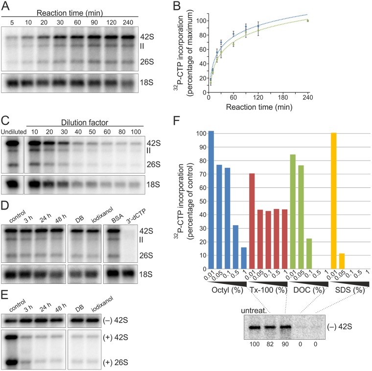
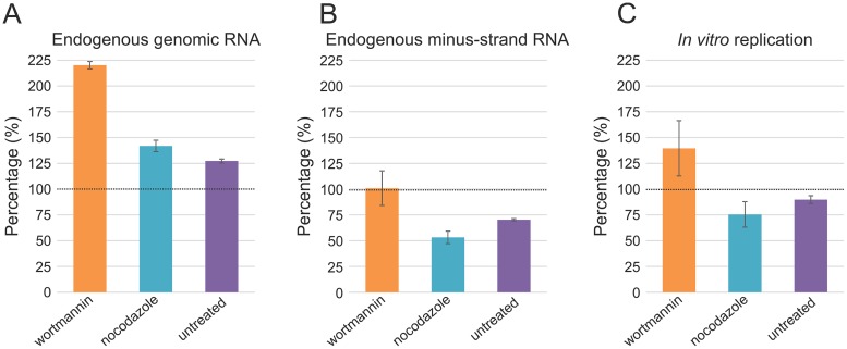
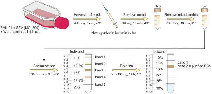
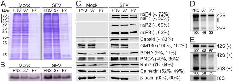
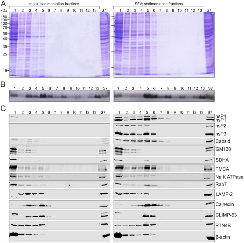
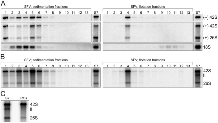
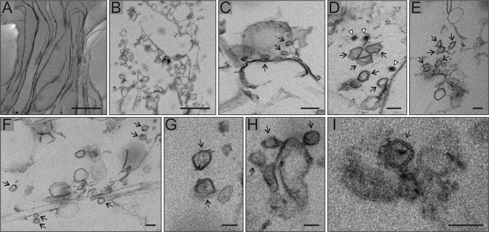
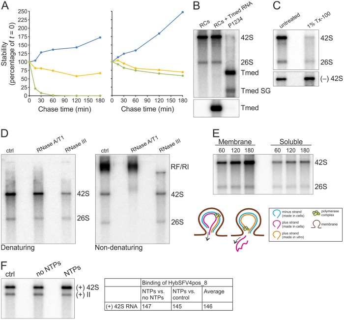
Positive-strand RNA viruses replicate their genomes in membrane-associated structures; alphaviruses and many other groups induce membrane invaginations called spherules. Here, we established a protocol to purify these membranous replication complexes (RCs) from cells infected with Semliki Forest virus (SFV). We isolated SFV spherules located on the plasma membrane and further purified them using two consecutive density gradients. This revealed that SFV infection strongly modifies cellular membranes. We removed soluble proteins, the Golgi membranes, and most of the mitochondria, but plasma membrane, endoplasmic reticulum (ER), and late endosome markers were retained in the membrane fraction that contained viral RNA synthesizing activity, replicase proteins, and minus- and plus-strand RNA. Electron microscopy revealed that the purified membranes displayed spherule-like structures with a narrow neck. This membrane enrichment was specific to viral replication, as such a distribution of membrane markers was only observed after infection. Besides the plasma membrane, SFV infection remodeled the ER, and the cofractionation of the RC-carrying plasma membrane and ER suggests that SFV recruits ER proteins or membrane to the site of replication. The purified RCs were highly active in synthesizing both genomic and subgenomic RNA. Detergent solubilization destroyed the replication activity, demonstrating that the membrane association of the complex is essential. Most of the newly made RNA was in double-stranded replicative molecules, but the purified complexes also produced single-stranded RNA as well as released newly made RNA. This indicates that the purification established here maintained the functionality of RCs and thus enables further structural and functional studies of active RCs.IMPORTANCE Similar to all positive-strand RNA viruses, the arthropod-borne alphaviruses induce membranous genome factories, but little is known about the arrangement of viral replicase proteins and the presence of host proteins in these replication complexes. To improve our knowledge of alphavirus RNA-synthesizing complexes, we isolated and purified them from infected mammalian cells. Detection of viral RNA and in vitro replication assays revealed that these complexes are abundant and highly active when located on the plasma membrane. After multiple purification steps, they remain functional in synthesizing and releasing viral RNA. Besides the plasma membrane, markers for the endoplasmic reticulum and late endosomes were enriched with the replication complexes, demonstrating that alphavirus infection modified cellular membranes beyond inducing replication spherules on the plasma membrane. We have developed here a gentle purification method to obtain large quantities of highly active replication complexes, and similar methods can be applied to other positive-strand RNA viruses.

Keywords: RNA replication; Semliki Forest virus; alphavirus; membrane fractionation; purification; replication complex.


Figures

FIG 1
FIG 1
SFV RCs are stable and robust in RNA synthesis in vitro. (A) BHK cells were infected with SFV at an MOI of 50, and PNS was prepared at 4 h p.i. In vitro replication was studied by the incorporation of [32P]CTP into SFV RNA, indicated by 26S, II, and 42S. After the incubation times indicated, RNA was isolated and analyzed in a denaturing agarose gel. Ribosomal 18S RNA (18S rRNA) was detected by in-gel hybridization from the same gel. (B) Kinetics of the incorporation of [32P]CTP into 26S (blue) and 42S (green) RNA. Percentages indicate the incorporation compared to the last time point (100%). Data are presented as means ± standard deviations from two independent experiments. (C) Replication assay performed with either undiluted PNS or PNS diluted as indicated. 18S rRNA was detected as described for panel A. For the undiluted sample, 1/10 of the amount of RNA used for other samples was used. (D) Stability of replication activity. PNS was preincubated at 4°C for 3, 24, and 48 h, or PNS was diluted in dilution buffer (DB) or iodixanol and preincubated at 4°C for 24 h before a replication assay. The last two lanes show samples containing 1 mg/ml BSA or 100 μM 3′-dCTP. 18S rRNA was detected by in-gel hybridization from the same gel. (E) Stability of endogenous RNA. To detect endogenous SFV minus and plus strands, total RNA was isolated after the same preincubations as those described for panel D and analyzed by in-gel hybridization with specific probes. (F) Detergent sensitivity. PNS samples containing the indicated detergent concentrations were incubated at 4°C for 1 h and used for either a replication assay or total RNA isolation, followed by in-gel hybridization. After the replication assay, the incorporation of [32P]CTP into 42S RNA was quantified, and percentages indicate the incorporation compared to the untreated PNS. The lower panel shows the presence of endogenous minus-strand RNA in the untreated and 1% detergent-treated samples detected by in-gel hybridization. Numbers below the lanes indicate the percentage of [32P]signal compared to that of the untreated sample. Octyl, n-octylglucoside.
FIG 2
FIG 2
Spherule location and replication activity. At 4 h p.i., PNS samples were prepared from SFV-infected (MOI of 500) BHK cells, treated with 100 nM wortmannin at 1.5 h p.i. or 5 μM nocodazole at 0 h p.i., or left untreated. These samples were compared to the PNS samples prepared from SFV-infected (MOI of 50), untreated cells to give the percentages indicated in panels A to C. Error bars represent standard deviations from two independent experiments. (A and B) Total RNA was isolated from the PNS samples and plus- and minus-strand RNA were detected by in-gel hybridization with specific probes, followed by quantification of 42S RNA. (C) In vitro incorporation of [32P]CTP into viral RNA. 42S RNA was quantified.
FIG 3
FIG 3
Schematic of RC isolation and purification. Overview of the purification from cell harvesting to iodixanol density gradient centrifugations. From the sedimentation gradient, band 3 was collected for further purification by flotation. Band 2 in the flotation gradient represents the purified RCs.
FIG 4
FIG 4
Distribution of proteins, lipids, and RNA in the S7 and P7 fractions. PNS was prepared from mock- or SFV-infected (MOI of 500) BHK cells treated with wortmannin and separated into an S7 supernatant and P7 pellet. (A and B) Protein and lipid profiles of PNS, S7, and P7 in an SDS-polyacrylamide gel stained with Coomassie blue (A) or Sudan black B (B). Numbers on the left indicate the molecular masses (kDa) of marker proteins. (C) Distribution of viral and cellular markers as studied by Western blotting. PMCA was used as a marker for the plasma membrane, GM130 for the Golgi membrane, calnexin for the endoplasmic reticulum, SDHA for the mitochondria, Rab7 for the late endosomes, and β-actin for the cytosol. Numbers in parentheses indicate the percentage of the protein present in the S7 fraction compared to that in PNS. The first number is from mock-infected samples and the second one from virus-infected samples. (D) Incorporation of [32P]CTP into SFV RNA in PNS, S7, and P7 prepared from SFV-infected cells. Numbers below the lanes indicate the amount of incorporation into 42S RNA as a percentage of the label incorporated in PNS. (E) Distribution of endogenous minus- and plus-strand RNA analyzed by in-gel hybridization from PNS, S7, and P7 prepared from SFV-infected cells. As a control, a probe against 18S rRNA was used. Numbers below the lanes indicate the amount of 42S RNA as a percentage of that in PNS.
FIG 5
FIG 5
Infection modulates cellular membranes. S7 was prepared from mock- and SFV-infected (MOI of 500) BHK cells treated with wortmannin, and RCs were purified by a 3-h sedimentation in a 10 to 20% iodixanol step gradient. After centrifugation, gradients were fractionated (fr. 1 to 12, from top to bottom; fr. 13, the pellet) and compared to S7. On the left, fractions are from mock-infected sample, and on the right, fractions are from SFV-infected sample. Protein (A) and lipid (B) patterns were studied in an SDS-polyacrylamide gel stained with Coomassie blue or Sudan black B. Numbers on the left indicate the molecular masses (kDa) of marker proteins. (C) Western blotting shows the distribution of viral and cellular markers as described for Fig. 4. Furthermore, Na,K ATPase was used as an additional marker for the plasma membrane, LAMP-2 for the late endosomes and lysosomes, and CLIMP-63 and RTN4B as additional markers for the endoplasmic reticulum (sheets and tubules, respectively). The sample volumes loaded from the fractions in the Coomassie- or Sudan-stained gels (A and B) and Western blot gels (C) were about five and seven times larger, respectively, than the sample volume loaded from the original S7, shown on the right.
FIG 6
FIG 6
SFV RCs concentrate with the plasma membrane, ER, and late endosome markers. Light-scattering band 3 from the sedimentation gradient was further purified by an 18-h flotation in a 10 to 30% iodixanol step gradient. After centrifugation, gradients were fractionated (fr. 1 to 12, from top to bottom; fr. 13, the pellet) and compared to S7. Fractions from mock-infected samples are on the left, and fractions from SFV-infected sample are on the right. Protein (A) and lipid (B) patterns were analyzed in an SDS-polyacrylamide gel stained with Coomassie blue or Sudan black B. Numbers on the left indicate the molecular masses (kDa) of marker proteins. (C) Distribution of viral and cellular markers studied by Western blotting as described for Fig. 4. (D) Distribution of Na,K ATPase (plasma membrane), LAMP-2 (late endosomes and lysosomes), and CLIMP-63 and RTN4B (endoplasmic reticulum sheets and tubules, respectively) between flotation fractions 3 to 5 was analyzed and compared to S7 by Western blotting. The sample volumes are the same as those for Fig. 5.
FIG 7
FIG 7
Purified RCs remain active in RNA synthesis. (A) Distribution of endogenous minus- and plus-strand RNAs between SFV sedimentation (left) and flotation (right) fractions studied by in-gel hybridization. 18S rRNA was detected from the same fractions (fr. 1 to 12, from top to bottom; fr. 13 is the pellet), and S7 is shown for comparison. (B) In vitro replication activity of the same fractions as those used for panel A, studied by [32P]CTP incorporation. (C) Comparison of in vitro replication activity of S7 and purified RCs after replication reaction mixtures were incubated 4 h. S7 and RCs indicate [32P]CTP incorporation by the S7 fraction and the purified RCs, respectively. In panel A the volume from the fractions used for RNA isolation was about ∼7.5 times larger than the S7 sample volume, and in panels B and C 20-fold-diluted S7 was used.
FIG 8
FIG 8
Purified RC membranes contain spherule-like structures. SFV RCs were purified in subsequent sedimentation and flotation gradients, fixed, and pelleted by differential centrifugation for thin-section electron microscopy. Mock-infected samples served as a control. All micrographs shown are from SFV samples. (A) Membrane sheets observed in both SFV and mock sample. (B) Membranes with spherical invaginations of about 50 nm in diameter typical of SFV only. This most likely represents plasma membrane sheets sectioned in a different orientation than that of panel A, resulting in vesicle-like appearance. (C) Membrane sheet showing smaller round vesicles that may represent neck parts of spherule-like structures, indicated by arrows. (D to I) Close-ups of spherule-like structures, indicated by arrows. In panel D, white arrowheads indicate nucleocapsids. Scale bars, 1,000 nm (A and B), 100 nm (C to F), and 50 nm (G to I).
FIG 9
FIG 9
Characterization of the in vitro replication activity of the purified RCs. (A) Stability of newly synthesized SFV genomic RNA. After a 60-min replication assay, [32P]CTP incorporation was blocked by the addition of 100 μM 3′-dCTP, and incubation at 30°C was continued for an additional 180 min. The graph on the left represents an assay with 10-fold-diluted S7 and on the right with the purified RCs. At the indicated chase times radioactivity in 42S was quantified (yellow). As a control, a reaction without 3′-dCTP was quantified (blue). In addition, in vitro transcript of Tmed added in the reaction at a 0-min chase was quantified by in-gel hybridization (green). All values are presented as percentages of the values at the 0-min chase. (B) Purified RCs and additional exogenous template. Replication assay reaction mixtures were incubated for 2 h. RCs indicates [32P]CTP incorporation by the purified RCs. RCs + Tmed RNA shows incorporation by the purified RCs after an exogenous Tmed RNA transcript was added. As a control, [32P]CTP incorporation by the P15 membrane fraction from cells transfected with the P1234 polyprotein and Tmed template plasmids is shown, and genomic and subgenomic (SG) Tmed are indicated. The lower panel shows the presence of Tmed, detected by in-gel hybridization. (C) Detergent stability and sensitivity. Purified RCs were treated with 1% Tx-100, followed by an assay to detect replication (upper) and in-gel hybridization to detect the minus-strand template RNA (lower). (D) ssRNA and RF/RI forms of in vitro-synthesized RNAs. After a 2-h replication assay with the purified RCs, RNA was isolated and treated with RNase A/T1 or III under high-salt conditions to specifically digest ssRNA or dsRNA, respectively, and analyzed under denaturing (left) or nondenaturing (right) conditions. (E) Release of newly made RNA. A replication assay was performed with the concentrated purified RCs, and after 60-, 120-, and 180-min replication reactions, aliquots were removed to prepare pellet and supernatant fractions, followed by RNA isolation and analysis in a denaturing agarose gel. The schematic shows how [32P]CTP is incorporated into viral RNA during in vitro RNA synthesis, resulting in dsRNA containing a plus strand synthesized both in cells (indicated by magenta) and in vitro (indicated by orange). After a round of replication and release of the previous plus strand, an RC contains only the in vitro-synthesized plus strand if replication is semiconservative. (F) Increase in the amount of RNA during in vitro replication. After a 4-h replication assay with the purified RCs and unlabeled NTPs, RNA was isolated and genomic RNA was detected by in-gel hybridization. No NTPs indicates a reaction without added NTPs, and ctrl indicates a sample without any incubations before RNA isolation. 42S RNA was quantified, and average percentages from two independent experiments are shown in the table.

Similar articles

Cited by

References

    1. Yang X, Yang H, Zhou G, Zhao GP. 2008. Infectious disease in the genomic era. Annu Rev Genomics Hum Genet 9:21–48. doi:10.1146/annurev.genom.9.081307.164428. - DOI - PubMed
    1. Koonin EV, Dolja VV, Krupovic M. 2015. Origins and evolution of viruses of eukaryotes: the ultimate modularity. Virology 479-480:2–25. - PMC - PubMed
    1. Strauss JH, Strauss EG. 1994. The alphaviruses: gene expression, replication, and evolution. Microbiol Rev 58:491–562. - PMC - PubMed
    1. den Boon JA, Ahlquist P. 2010. Organelle-like membrane compartmentalization of positive-strand RNA virus replication factories. Annu Rev Microbiol 64:241–256. doi:10.1146/annurev.micro.112408.134012. - DOI - PubMed
    1. Paul D, Bartenschlager R. 2013. Architecture and biogenesis of plus-strand RNA virus replication factories. World J Virol 2:32–48. doi:10.5501/wjv.v2.i2.32. - DOI - PMC - PubMed

Publication types

LinkOut - more resources