. 2018 Mar 19;221(Pt 6):jeb168419.
doi: 10.1242/jeb.168419.

Biogenesis of zinc storage granules in Drosophila melanogaster

Affiliations

Biogenesis of zinc storage granules in Drosophila melanogaster

Carlos Tejeda-Guzmán et al. J Exp Biol. .

Abstract

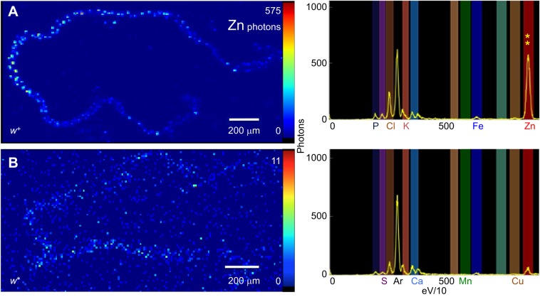
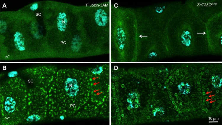
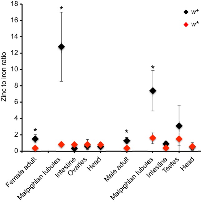
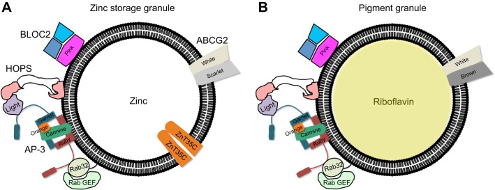
Membrane transporters and sequestration mechanisms concentrate metal ions differentially into discrete subcellular microenvironments for use in protein cofactors, signalling, storage or excretion. Here we identify zinc storage granules as the insect's major zinc reservoir in principal Malpighian tubule epithelial cells of Drosophila melanogaster The concerted action of Adaptor Protein-3, Rab32, HOPS and BLOC complexes as well as of the white-scarlet (ABCG2-like) and ZnT35C (ZnT2/ZnT3/ZnT8-like) transporters is required for zinc storage granule biogenesis. Due to lysosome-related organelle defects caused by mutations in the homologous human genes, patients with Hermansky-Pudlak syndrome may lack zinc granules in beta pancreatic cells, intestinal paneth cells and presynaptic vesicles of hippocampal mossy fibers.

Keywords: AP-3 complex; Eye color mutants; ICP-OES; Malpighian tubules; Synchrotron; Zincosomes.


Conflict of interest statement

Competing interestsThe authors declare no competing or financial interests.

Figures

Fig. 1.
Fig. 1.
Meiotic recombination mapping strategy for poco-zinc in Drosophila melanogaster. The wild-type Tan3 chromosome is represented in green, whereas the mapping stock chromosome, carrying recessive alleles with visible phenotypes, is orange. (A) The first set of recombinant analysis situated the poco-zinc allele on the left part of the X-chromosome as it segregated 100% together with the cm gene. (B) Efforts to dissociate poco-zinc from cm were not successful, suggesting that poco-zinc is tightly linked (or identical) to the cm gene. (C) Efforts to map poco-zinc using a different mapping stock resulted in joint segregation of poco-zinc together with the w gene and far away from the cm gene. N is the total number of recombinants analysed.
Fig. 2.
Fig. 2.
Mutants in the LRO-biogenesis pathway have low body zinc content. (A) Linear regression between zinc determinations by AAS versus ICP-OES. The mean value determined for each genotype (Table 1), measured by both methods in biological replicates, is plotted. Stocks with normal, intermediate and low levels of zinc are readily identifiable. (B) Values for iron and copper are shown; here genotypes do not segregate. (C) Zinc (mg) to iron (mg) ratio was calculated for every independent measurement made (by either AAS or ICP-OES). Mean values and standard deviations are plotted; the different colors indicate statistically significant differences as revealed by Tukey’s post hoc test following one-way ANOVA.
Fig. 3.
Fig. 3.
Zinc storage is affected in w and cm mutants. Two control strains (wild-type Tan3 and y1) and two low-zinc mutants (w* and cm1) were grown on media with the zinc chelator TPEN (red bars) or supplemented with zinc sulfate (green bars) prior to measuring zinc and iron by AAS in whole flies from these populations. The control strains respond to the zinc treatments by changing body zinc stores, whereas this response is impaired in low-zinc mutants. Iron was unaffected. Two-way ANOVA showed differences by diet and by genotype; groups not different from each other in a Tukey's post hoc analysis are marked by the same lower case letter.
Fig. 4.
Fig. 4.
Mutants in the LRO-biogenesis pathway fail to accumulate riboflavin in the Malpighian tubules. The Malpighian tubules of adult females are shown for the indicated genotypes along with the concentration of total body zinc in mg Zn g−1 dry mass. (A) Riboflavin in wild-type Malpighian tubules gives them their characteristic yellow-orange color (Nickla, 1972). (B–D) Three representative LRO-biogenesis mutants are all colorless and low in zinc. A comparison between isogenic (E) w+ and (F) w* suggests that the w gene is required for the accumulation of both riboflavin and zinc. (G) The bw mutants are severely reduced in their coloration and less so in their zinc content. (H) The st mutants show riboflavin coloration, but severely reduced zinc concentration.
Fig. 5.
Fig. 5.
Synchrotron X-ray fluorescence microscopy demonstrates the presence of zinc in Malpighian tubules from adult female flies. Pixel resolution was 10 µm2. (A) Representative heat-map image for the zinc signal is shown for w+ Malpighian tubules. The spectral maximum for zinc emission is 575 photons per 100 ms (also indicated by the two yellow asterisks along with the full spectrum in the right-hand panel). (B) Almost no zinc is detectable in Malpighian tubules from the w mutant.
Fig. 6.
Fig. 6.
Zinc storage granules are present in the Malpighian tubules. Confocal images of Fluozin-3AM fluorescence, indicative of labile zinc in the Malpighian tubule of (A) a w* female adult or (B) an isogenic w+ female adult. Red arrows point to a subset of zinc storage granules. (C) The protein trap line y,w;ZnT35CGFP was used to monitor the subcellular localization of ZnT35C, confirming previous observations that it associates with the plasma membrane (white arrows). (D) The same reporter in the w+ background clearly marks the zinc storage granule membrane (red arrows). All flies were grown on a diet supplemented with 5 mmol l−1 zinc sulfate. SC, stellate cell; PC, principal cell; N, nucleus.
Fig. 7.
Fig. 7.
Zinc accumulates in the Malpighian tubules of both sexes and in testes. Zinc (mg) to iron (mg) ratio was calculated for every independent measurement made by ICP-OES. Mean values and standard deviations are plotted (w+ in black and w* in red). Two-way ANOVA indicated statistically significant differences both by tissue and by genotype; asterisks denote difference between genotypes for any given tissue from a Tukey’s post hoc analysis.
Fig. 8.
Fig. 8.
Schematic representation of a zinc storage granule and a riboflavin pigment granule, in principal cells of the Malpighian tubules. (A) Zinc storage granule. (B) Riboflavin pigment granule. On the left of each vesicle known players in protein trafficking are shown, common for the biogenesis of both types of LROs. Transporters are on the right-hand side. It is unclear how cells differentially give rise to the two LROs.

Similar articles

Cited by

References

    1. Afshar N., Argunhan B., Bettedi L., Szular J. and Missirlis F. (2013). A recessive X-linked mutation causes a threefold reduction of total body zinc accumulation in Drosophila melanogaster laboratory strains. FEBS Open Bio 3, 302-304. 10.1016/j.fob.2013.07.003 - DOI - PMC - PubMed
    1. Ammann S., Schulz A., Krageloh-Mann I., Dieckmann N. M., Niethammer K., Fuchs S., Eckl K. M., Plank R., Werner R., Altmuller J. et al. (2016). Mutations in AP3D1 associated with immunodeficiency and seizures define a new type of Hermansky-Pudlak syndrome. Blood 127, 997-1006. 10.1182/blood-2015-09-671636 - DOI - PMC - PubMed
    1. Anderson P. R., Kirby K., Hilliker A. J. and Phillips J. P. (2005). RNAi-mediated suppression of the mitochondrial iron chaperone, frataxin, in Drosophila. Hum. Mol. Genet. 14, 3397-3405. 10.1093/hmg/ddi367 - DOI - PubMed
    1. Assoum M., Philippe C., Isidor B., Perrin L., Makrythanasis P., Sondheimer N., Paris C., Douglas J., Lesca G., Antonarakis S. et al. (2016). Autosomal-recessive mutations in AP3B2, adaptor-related protein complex 3 β2 subunit, cause an early-onset epileptic encephalopathy with optic atrophy. Am. J. Hum. Genet. 99, 1368-1376. 10.1016/j.ajhg.2016.10.009 - DOI - PMC - PubMed
    1. Atanesyan L., Günther V., Celniker S. E., Georgiev O. and Schaffner W. (2011). Characterization of MtnE, the fifth metallothionein member in Drosophila. J. Biol. Inorg Chem. 16, 1047-1056. 10.1007/s00775-011-0825-4 - DOI - PubMed

Publication types