Review
. 2018 Jan 24;38(4):767-775.
doi: 10.1523/JNEUROSCI.1105-17.2017.

How Forces Fold the Cerebral Cortex

Affiliations
Review

How Forces Fold the Cerebral Cortex

Christopher D Kroenke et al. J Neurosci. .

Abstract

Improved understanding of the factors that govern folding of the cerebral cortex is desirable for many reasons. The existence of consistent patterns in folding within and between species suggests a fundamental role in brain function. Abnormal folding patterns found in individuals affected by a diverse array of neurodevelopmental disorders underline the clinical relevance of understanding the folding process. Recent experimental and computational efforts to elucidate the biomechanical forces involved in cerebral cortical folding have converged on a consistent approach. Brain growth is modeled with two components: an expanding outer zone, destined to become the cerebral cortex, is mechanically coupled to an inner zone, destined to become white matter, that grows at a slower rate, perhaps in response to stress induced by expansion from the outer layer. This framework is consistent with experimentally observed internal forces in developing brains, and with observations of the folding process in physical models. In addition, computational simulations based on this foundation can produce folding patterns that recapitulate the characteristics of folding patterns found in gyroencephalic brains. This perspective establishes the importance of mechanical forces in our current understanding of how brains fold, and identifies realistic ranges for specific parameters in biophysical models of developing brain tissue. However, further refinement of this approach is needed. An understanding of mechanical forces that arise during brain development and their cellular-level origins is necessary to interpret the consequences of abnormal brain folding and its role in functional deficits as well as neurodevelopmental disease.Dual Perspectives Companion Paper: How Cells Fold the Cerebral Cortex, by Víctor Borrell.

Keywords: biomechanics; development; fetal brain; gyrus; morphology.


Figures

Figure 1.
Figure 1.
Cerebral cortical folding, as approximated in physical models, and in relation to developmental changes in tissue zones identified by light microscopy. a, Lateral views of models of the rhesus macaque cerebral cortex obtained from fetal MRI measurements at gestation ages G85, G110, and G135. Gestation term is 168 d for rhesus macaques. The mean normalized curvature, a measure of folding, increases from 23% to 87% of the adult value from G85 to G135 (Wang et al., 2017). b, Average coronal T2-weighted fetal MRI from 17 G85 brains, 8 G110 brains, and 16 G135 brains (manuscript in preparation). Scale bar, 1 cm. Illustrations of the outer (blue) and inner (yellow) zones for one hemisphere are overlaid on the hemisphere portrayed on the right at each age. c, Nissl-stained images spanning from the ventricular zone to the pial surface in the inferior temporal lobe of a G90, G110, and G135 brain. For the G135 brain, cerebral tissue is shown for a sulcal and gyral region. Lamina are labeled according to Smart et al. (2002). Notable changes in thickness for several layers, including the cortical plate, occur over this period. MZ, Marginal zone; CP, cortical plate; SP, subplate; OFL, outer fibrous layer; OSVZ, outer subventricular zone; IFL, inner fibrous layer; ISVZ, inner subventricular zone; VZ, ventricular zone. Scale bar, 1 mm.
Figure 2.
Figure 2.
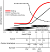
In gyroencephalic species, gyrogenesis is temporally coincident with rapid linear surface area expansion and morphological differentiation, subsequent to cortical pyramidal cell neurogenesis and migration. Workman et al. (2013) have defined a species-independent CNS event time, shown on the abscissa. Lower axes represent corresponding postnatal ages (days) for ferret, gestational ages (days) for rhesus macaques, and gestation weeks for humans. Common trajectories of folding (red curve), surface area expansion (solid line), and reduction in cerebral cortical diffusion anisotropy (hyphenated line) (Wang et al., 2017), relative to event time, have been observed for these three species.

Similar articles

Cited by

References

    1. Altman J, Bayer SA (2002) Regional differences in the stratified transitional field and the honeycomb matrix of the developing human cerebral cortex. J Neurocytol 31:613–632. 10.1023/A:1025787427576 - DOI - PubMed
    1. Barkovich AJ, Raybaud C (2012) Pediatric neuroimaging. Philadelphia, PA: Lippincott Williams and Wilkins.
    1. Barkovich AJ, Kuzniecky RI, Jackson GD, Guerrini R, Dobyns WB (2005) A developmental and genetic classification for malformations of cortical development. Neurology 65:1873–1887. 10.1212/01.wnl.0000183747.05269.2d - DOI - PubMed
    1. Barkovich AJ, Guerrini R, Kuzniecky RI, Jackson GD, Dobyns WB (2012) A developmental and genetic classification for malformations of cortical development: update 2012. Brain 135:1348–1369. 10.1093/brain/aws019 - DOI - PMC - PubMed
    1. Bartley AJ, Jones DW, Weinberger DR (1997) Genetic variability of human brain size and cortical gyral patterns. Brain 120:257–269. 10.1093/brain/120.2.257 - DOI - PubMed

Publication types