

How Cells Fold the Cerebral Cortex
- PMID: 29367288
- PMCID: PMC6596235
- DOI: 10.1523/JNEUROSCI.1106-17.2017
How Cells Fold the Cerebral Cortex
Abstract
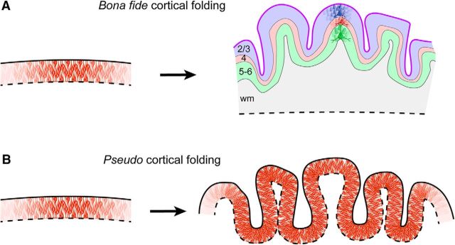
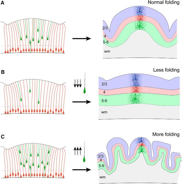
Folding of the cerebral cortex is as highly intriguing as poorly understood. At first sight, this may appear as simple tissue crumpling inside an excessively small cranium, but the process is clearly much more complex and developmentally predetermined. Whereas theoretical modeling supports a critical role for biomechanics, experimental evidence demonstrates the fundamental role of specific progenitor cell types, cellular processes, and genetic programs on cortical folding.Dual Perspectives Companion Paper: How Forces Fold the Cerebral Cortex, by Christopher D. Kroenke and Philip V. Bayly.
Keywords: OSVZ; Pax6; basal Radial Glia; ferret; neurogenesis; primate.
Copyright © 2018 the authors 0270-6474/18/380776-08$15.00/0.
Figures
References
-
- Arcila ML, Betizeau M, Cambronne XA, Guzman E, Doerflinger N, Bouhallier F, Zhou H, Wu B, Rani N, Bassett DS, Borello U, Huissoud C, Goodman RH, Dehay C, Kosik KS (2014) Novel primate miRNAs coevolved with ancient target genes in germinal zone-specific expression patterns. Neuron 81:1255–1262. 10.1016/j.neuron.2014.01.017 - DOI - PMC - PubMed
-
- Baala L, Briault S, Etchevers HC, Laumonnier F, Natiq A, Amiel J, Boddaert N, Picard C, Sbiti A, Asermouh A, Attié-Bitach T, Encha-Razavi F, Munnich A, Sefiani A, Lyonnet S (2007) Homozygous silencing of T-box transcription factor EOMES leads to microcephaly with polymicrogyria and corpus callosum agenesis. Nat Genet 39:454–456. 10.1038/ng1993 - DOI - PubMed
-
- Bahi-Buisson N, Souville I, Fourniol FJ, Toussaint A, Moores CA, Houdusse A, Lemaitre JY, Poirier K, Khalaf-Nazzal R, Hully M, Leger PL, Elie C, Boddaert N, Beldjord C, Chelly J, Francis F (2013) New insights into genotype-phenotype correlations for the doublecortin-related lissencephaly spectrum. Brain 136:223–244. 10.1093/brain/aws323 - DOI - PMC - PubMed
-
- Bayer SA, Altman J (2005) The human brain during the second trimester, Ed 1 Boca Raton, FL: CRC.
Publication types
MeSH terms
LinkOut - more resources
Full Text Sources
Other Literature Sources