. 2018 Feb;30(2):397-414.
doi: 10.1105/tpc.17.00420. Epub 2018 Jan 24.

Role of the Nod Factor Hydrolase MtNFH1 in Regulating Nod Factor Levels during Rhizobial Infection and in Mature Nodules of Medicago truncatula

Affiliations

Role of the Nod Factor Hydrolase MtNFH1 in Regulating Nod Factor Levels during Rhizobial Infection and in Mature Nodules of Medicago truncatula

Jie Cai et al. Plant Cell. 2018 Feb.

Abstract

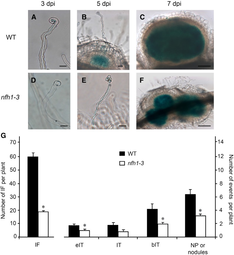
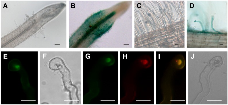
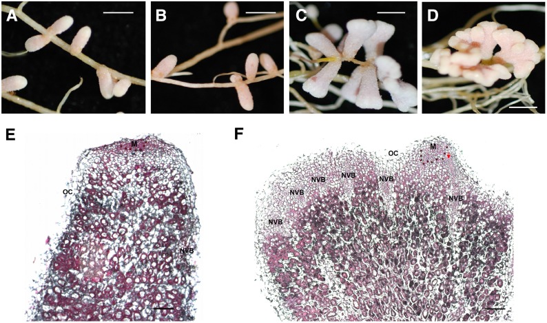
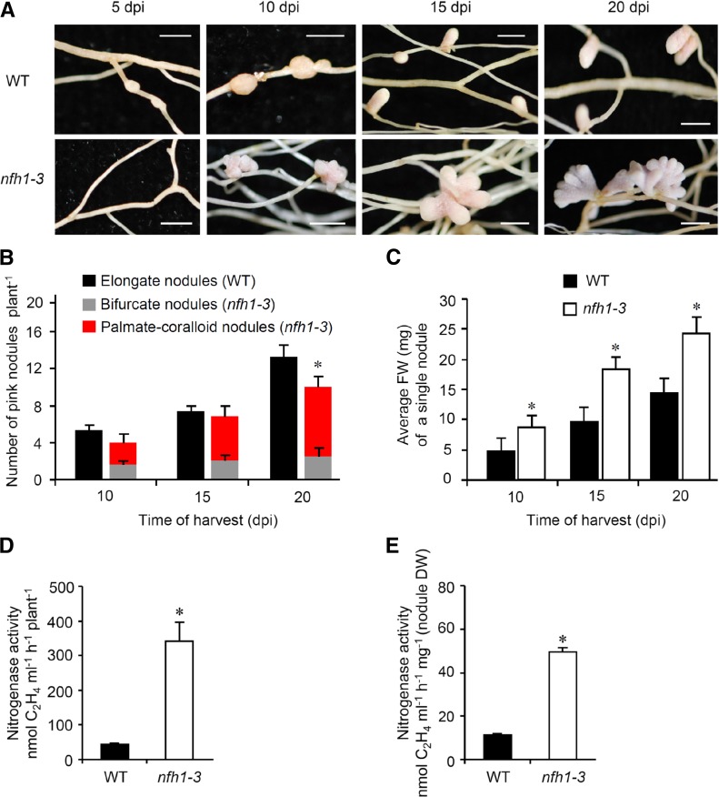
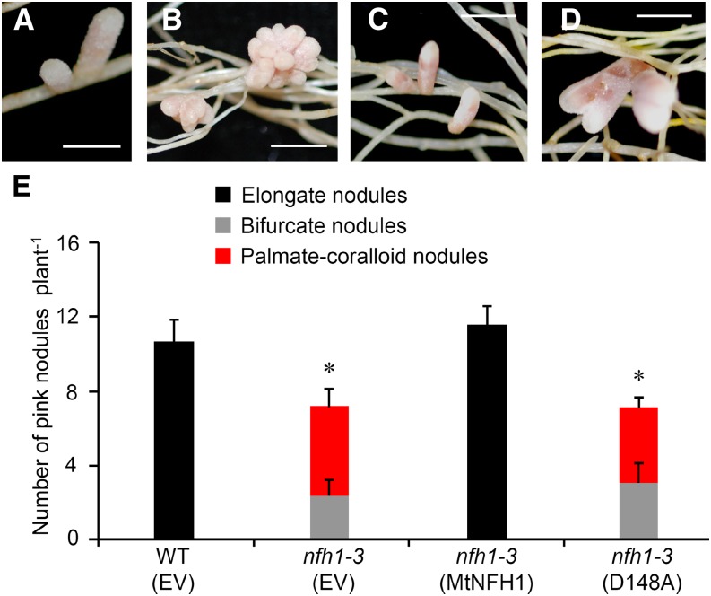
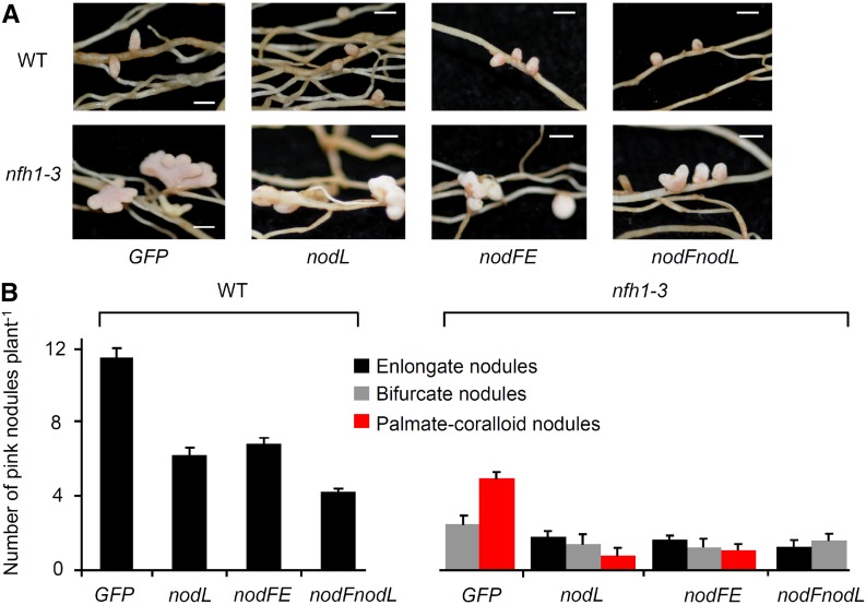
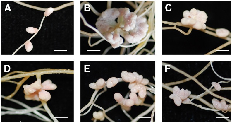
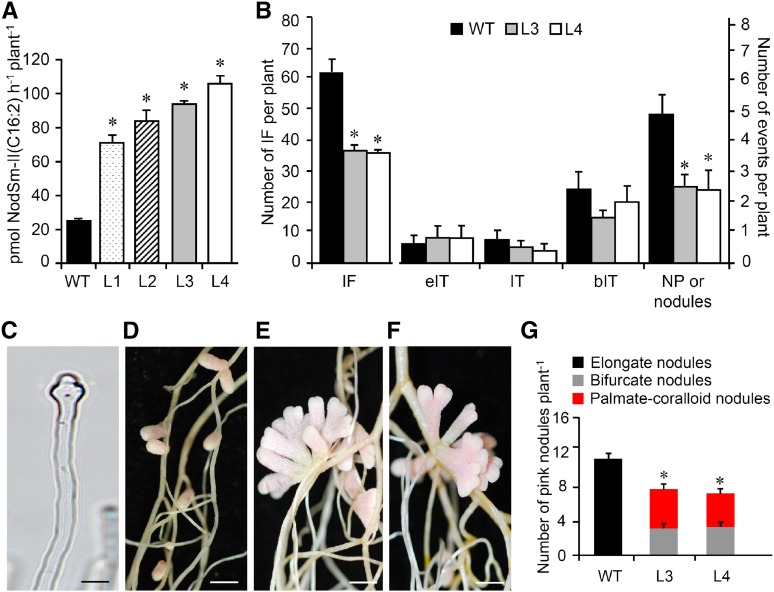
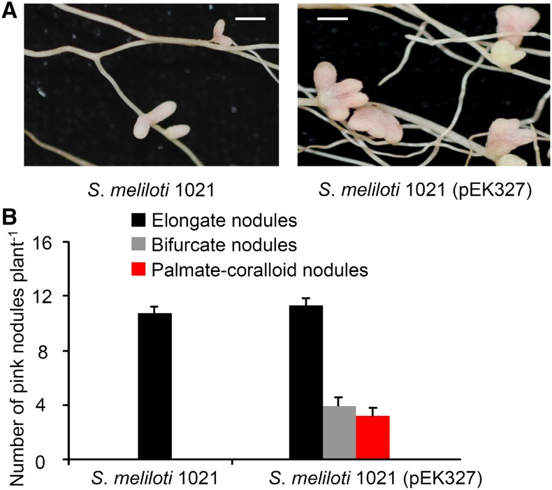
Establishment of symbiosis between legumes and nitrogen-fixing rhizobia depends on bacterial Nod factors (NFs) that trigger symbiosis-related NF signaling in host plants. NFs are modified oligosaccharides of chitin with a fatty acid moiety. NFs can be cleaved and inactivated by host enzymes, such as MtNFH1 (MEDICAGO TRUNCATULA NOD FACTOR HYDROLASE1). In contrast to related chitinases, MtNFH1 hydrolyzes neither chitin nor chitin fragments, indicating a high cleavage preference for NFs. Here, we provide evidence for a role of MtNFH1 in the symbiosis with Sinorhizobium meliloti Upon rhizobial inoculation, MtNFH1 accumulated at the curled tip of root hairs, in the so-called infection chamber. Mutant analysis revealed that lack of MtNFH1 delayed rhizobial root hair infection, suggesting that excess amounts of NFs negatively affect the initiation of infection threads. MtNFH1 deficiency resulted in nodule hypertrophy and abnormal nodule branching of young nodules. Nodule branching was also stimulated in plants expressing MtNFH1 driven by a tandem CaMV 35S promoter and plants inoculated by a NF-overproducing S. meliloti strain. We suggest that fine-tuning of NF levels by MtNFH1 is necessary for optimal root hair infection as well as for NF-regulated growth of mature nodules.


Figures

Figure 1.
Figure 1.
Characterization of nfh1 Mutants. (A) Tnt1 insertion sites of the M. truncatula R108 mutants nfh1-1 (NF16587), nfh1-2 (NF12841), and nfh1-3 (NF11260). The box indicates the coding region of MtNFH1. The location of Tnt1 insertions in the promoter region are marked by indicated arrows. (B) Analysis of MtNFH1 expression by RT-qPCR in wild-type and nfh1 mutant seedlings (nfh1-1, nfh1-2, and nfh1-3). Roots of seedlings were immersed in 1-mL syringes filled with Jensen medium containing 0.1 μM NodSm-IV(C16:2, Ac, S) for 18 h. Roots from 20 seedlings were used for each RNA extraction (3 RNA extractions per genotype; n = 3). Data indicate means ± se of normalized expression values (mean value of wild-type plants set to one). Statistically different transcript levels of mutants compared with wild-type plants are marked by asterisks (Student’s t test, P ≤ 0.05; Supplemental File 1). (C) Formation of the lipodisaccharide NodSm-II(C16:2, Ac) released from NodSm-IV(C16:2, Ac, S) in the rhizosphere of wild-type and nfh1 mutant seedlings. Data indicate means ± se (1 plant per sample; n ≥ 9). Asterisks indicate significantly reduced hydrolytic activity in nfh1 mutants compared with wild-type plants (Kruskal-Wallis test, P ≤ 0.05; Supplemental File 1).
Figure 2.
Figure 2.
MtNFH1 Transcript Levels and NF Hydrolysis Activity in the Rhizosphere of Nodulation Signaling Mutants. (A) Analysis of MtNFH1 expression by RT-qPCR in M. truncatula Jemalong A17 wild-type plants and nodulation signaling mutants (dmi1, dmi2, dmi3, and hcl). Roots of seedlings were immersed in Jensen medium containing 0.1 μM NodSm-IV(C16:2, Ac, S) for 18 h. Roots from 20 plants were combined for each RNA extraction (3 RNA extractions per genotype; n = 3). Data indicate means ± se of normalized expression values (mean value of wild-type plants set to one). Statistically different transcript levels of mutants compared with wild-type plants are marked by asterisks (Student’s t test, P ≤ 0.05; Supplemental File 1). (B) Corresponding NF hydrolysis tests: Roots of seedlings were pretreated with 0.1 μM NodSm-IV(C16:2, Ac, S) for 18 h and then incubated with 15 μM NodSm-IV(C16:2, Ac, S) for 3 h. The amounts of NodSm-II(C16:2, Ac) formed were deduced from HPLC chromatograms. Data indicate means ± se (1 plant per sample, n ≥ 9). Asterisks indicate significantly reduced hydrolytic activity in nodulation signaling mutants compared with wild-type plants (Kruskal-Wallis test, P ≤ 0.05; Supplemental File 1). WT, M. truncatula wild-type (ecotype Jemalong A17); dmi1, dmi1-3 mutant (Y6); dmi2, dmi2-1 mutant (TR25); dmi3, dmi3-1 mutant (TRV25); hcl, hcl-1 mutant (lyk3, B56).
Figure 3.
Figure 3.
Stimulation of MtNFH1 Expression and MtNFH1 Activity in Response to NF Treatments. (A) Time-dependent stimulation of MtNFH1 expression by NFs: Roots of M. truncatula R108 seedlings were immersed in Jensen medium containing 1 μM NodSm-IV(C16:2, S) for the indicated time periods. Control plants without NF treatment were incubated under the same conditions. RNA from harvested roots (30 roots per RNA extraction; 3 RNA extractions; n = 3) was used for RT-qPCR analysis. Data indicate means ± se of normalized expression values (mean value of control plants at the 2-h time point set to one). (B) Time-dependent stimulation of MtNFH1 activity by NFs: Roots of seedlings (Jemalong A17) were pretreated with 1 µM NodSm-IV(C16:2, S) for the indicated time periods. After pretreatment, plants were transferred to Jensen medium containing 5 µM NodSm-IV(C16:2, S) and incubated for 3 h. Plants without NF pretreatment were grown under the same conditions. The NodSm-II(C16:2) formed in samples (three to four plants per sample) was analyzed by reverse-phase HPLC. Data indicate means ± se from three independent experiments (n = 3). (C) Concentration-dependent stimulation of MtNFH1 expression by NFs: Roots of R108 seedlings were immersed in Jensen medium containing NodSm-IV(C16:2, S) at indicated concentrations for 18 h. Plants without NF treatment served as a control. RNA was isolated from harvested roots (20 roots per RNA extraction; 3 RNA extractions; n = 3). Data indicate means ± se of normalized expression values (mean value of control plants set to one). MtNFH1 transcript levels of NF-treated plants significantly different from control plants are marked by asterisks (Student’s t test, P ≤ 0.05; Supplemental File 1). (D) Concentration-dependent stimulation of MtNFH1 activity by NFs: Roots of R108 seedlings were pretreated with Jensen medium containing NodSm-IV(C16:2, S) at indicated concentrations for 18 h. MtNFH1 activity was then assayed with 5 μM NodSm-IV(C16:2, S) for 3 h. Formed NodSm-II(C16:2) (3 plants per sample) was analyzed by reverse-phase HPLC. Data indicate means ± se (3 samples; n = 3). Asterisks indicate a significant difference compared with the control (Kruskal-Wallis test, P ≤ 0.05; Supplemental File 1).
Figure 4.
Figure 4.
Analysis of Early Symbiotic Stages in Wild-Type and nfh1-3 Mutant Plants. Roots of seedlings inoculated with S. meliloti 2011 carrying pXLGD4 (lacZ) were harvested at indicated time points and bacteria visualized with X-Gal. (A) to (C) Root hairs and formed nodule primordia of wild-type seedlings at 3 (A), 5 (B), and 7 (C) dpi. Bars = 20 µm in (A) and (B) and 100 µm in (C). (D) to (F) Abnormal root hair deformation and formed nodule primordia of nfh1-3 mutant seedlings at 3 (D), 5 (E), and 7 (F) dpi. Bars = 20 µm in (D) and (E) and 100 µm in (F). (G) Quantification of different symbiotic stages at 7 dpi. Data indicate means (±se) for 12 plants. Significant differences between wild-type and nfh1-3 mutant plants are marked with asterisks (Kruskal-Wallis test, P ≤ 0.05; Supplemental File 1). IF, infection foci; eIT, elongating infection thread in root hair; IT, fully elongated infection thread in root hair; bIT, branched infection thread in cortex; NP, nodule primordium.
Figure 5.
Figure 5.
Expression of MtNFH1 in Response to Rhizobial Inoculation and Accumulation of MtNFH1:GFP in the Infection Chamber. (A) to (D) Analysis of M. truncatula R108 roots transformed with a MtNFH1pro-GUS construct. Plants were inoculated with S. meliloti Rm41 (3 dpi; [B] and [D]) or left noninoculated ([A] and [C]). Roots were stained with X-Gluc and then cleared with diluted NaClO solution. Bars = 50 µm in (A) and (B) and 20 µm in (C) and (D). (E) to (J) Analysis of curled root hairs of R108 roots expressing MtNFH1:GFP driven by the MtNFH1 promoter (3 dpi) under green fluorescence ([E] and [G]), red fluorescence (H), and bright-field conditions ([F] and [J]). Roots were inoculated with S. meliloti Rm41 ([E] and [F]) or S. meliloti 1021 (pQDN03) constitutively expressing mCherry ([G] to [J]). GFP fluorescence signals reflecting the presence of MtNFH1:GFP protein are increased in the infection chamber. Colocalization with bacteria is indicated in yellow (merged image; [I]). Bars = 20 µm.
Figure 6.
Figure 6.
Nodulation Phenotype of the nfh1-3 Mutant. Plants were inoculated with S. meliloti Rm41. (A) Unbranched nodules formed on M. truncatula R108 wild-type roots (20 dpi). (B) Wild-type nodules formed on roots of a wild-type sibling line of nfh1-3 (20 dpi). (C) and (D) Bifurcate (C) or palmate-coralloid (D) nodules formed on roots of the nfh1-3 mutant (20 dpi). (E) and (F) Microscopy analysis of a wild-type (E) and nfh1-3 (F) nodule (20 dpi). Sections were stained with ruthenium red. M, meristem (indicated with asterisks); NVB, nodule vascular bundle; OC, outer cortex; PVM, provascular meristem (red arrow). Bars = 2 mm in (A) to (D) and 200 µm in (E) and (F).
Figure 7.
Figure 7.
Time-Course Analysis of Nodule Formation and Nitrogenase Activity in Nodules. M. truncatula R108 wild-type and nfh1-3 mutant plants were inoculated with S. meliloti Rm41 and harvested at indicated time points. (A) Photographs of roots and nodules at the time of harvest. Bars = 2 mm. (B) Quantification of different types of nodules. Wild-type plants formed elongate nodules while the nfh1-3 mutant formed either bifurcate or palmate-coralloid nodules. Data indicate means (±se) based on analysis of 10 plants of each genotype and for each time point. Compared with the wild type, a significantly reduced nodule number (marked by an asterisk) was observed for the nfh1-3 mutant at 20 dpi (Kruskal-Wallis test, P ≤ 0.05; Supplemental File 1). (C) Quantification of the fresh weight of an individual nodule formed by wild-type and nfh1-3 roots. Data indicate means (±se) based from 10 plants of each genotype and for each time point. Asterisks indicate significantly increased values for the nfh1-3 mutant as compared with the wild-type (Kruskal-Wallis test, P ≤ 0.05; Supplemental File 1). (D) and (E) Nitrogenase activity per plant (D) and per nodule biomass (dry weight) (E) at 20 dpi. Asterisks indicate significantly increased activities in nfh1-3 nodules as compared with wild-type nodules at this time point (Kruskal-Wallis test, P ≤ 0.05; Supplemental File 1).
Figure 8.
Figure 8.
Expression of MtNFH1 and MtNFH1(D148A) in the nfh1-3 Mutant. Roots of the nfh1-3 mutant plants were transformed with the MtNFH1pro-MtNFH1 or MtNFH1pro-MtNFH1(D148A) construct. For comparison, wild-type (R108) plants and the nfh1-3 mutant were transformed with the empty vector pCAMBIA1302. Plants with transgenic roots were then inoculated with S. meliloti Rm41. Nodule formation was analyzed at 20 dpi. (A) Elongate nodules formed on wild-type roots transformed with the empty vector. (B) Palmate-coralloid nodules formed on nfh1-3 roots transformed with the empty vector. (C) Elongate nodules formed on nfh1-3 roots transformed with MtNFH1pro-MtNFH1. (D) Palmate-coralloid nodules formed on nfh1-3 roots transformed with MtNFH1pro-MtNFH1(D148A). (E) Quantification of different types of nodules. Data from individually analyzed plants (n ≥ 12) indicate means (±se). The nfh1-3 mutant transformed with the empty vector or the MtNFH1pro-MtNFH1(D148A) construct formed fewer nodules than wild-type plants transformed with the empty vector (asterisks; Kruskal-Wallis test, P ≤ 0.05; Supplemental File 1). EV, empty vector; MtNFH1, transformed with MtNFH1pro-MtNFH1; D148A, transformed with MtNFH1pro-MtNFH1(D148A). Bars = 2 mm.
Figure 9.
Figure 9.
Nodules Induced by S. meliloti 2011 Mutants Producing Modified NFs (20 dpi). M. truncatula R108 wild-type and nfh1-3 mutant plants were inoculated with S. meliloti strains that produce different NFs. (A) Photographs of nodules at the time of harvest (20 dpi). Bars = 2 mm. (B) Quantification of different types of nodules. Only elongate nodules were observed for wild-type plants. The nfh1-3 mutant formed elongate, bifurcate and palmate-coralloid nodules. Data indicate means (±se) based on analysis of 10 plants of each genotype. GFP, strain 2011 carrying pHC60 constitutively expressing GFP; nodL, strain 2011nodL::Tn5-GFP producing NFs without O-acetyl group at the nonreducing end; nodFE, strain 2011ΔnodFE-GFP producing NFs with vaccenic acid; nodFnodL, strain ΔnodFnodL::Tn5-GFP producing NFs with vaccenic acid and lacking an O-acetyl group.
Figure 10.
Figure 10.
Nodulation Phenotype of Various Genotypes with Reduced MtNFH1 Activity. Plants (≥9 per genotype) were inoculated with S. meliloti Rm41 and harvested at 20 dpi. Bars = 2 mm. (A) Nodules formed on wild-type R108 roots (normal MtNFH1 activity). (B) Nodules formed on roots of the nfh1-3 mutant (no MtNFH1 activity). (C) Nodules formed on roots of the nfh1-1 mutant (reduced MtNFH1 activity). (D) Nodules formed on roots of the nfh1-2 mutant (reduced MtNFH1 activity). (E) and (F) Nodules formed on roots of the RNAi lines L3 and L5 (reduced MtNFH1 activity; Tian et al., 2013).
Figure 11.
Figure 11.
Symbiotic Phenotype of M. truncatula Plants Expressing MtNFH1 Driven by a Tandem CaMV 35S Promoter. M. truncatula R108 plants were stably transformed with A. tumefaciens carrying pISV-MtNFH1. The symbiotic phenotype of two transgenic lines with increased NF-cleaving activity was compared with wild-type plants. (A) Hydrolysis of NFs by intact roots of M. truncatula lines constitutively expressing MtNFH1. Roots of seedlings from wild-type R108 plants and four independent lines constitutively expressing MtNFH1 driven by a tandem CaMV 35S promoter (named L1 to L4; T4 generation) were first individually pretreated with 0.1 μM NodSm-IV(C16:2, S) for 24 h and then incubated with 15 μM NodSm-IV(C16:2, S) for 18 h. Formation of NodSm-II(C16:2) was analyzed by reverse-phase HPLC (1 plant per sample). Data indicate means ± se. In total, 36 transgenic and nine wild-type plants were analyzed. Hydrolysis of NFs by the four lines was significantly elevated compared with wild-type plants (Kruskal-Wallis test; P < 0.05; Supplemental File 1). (B) Analysis of early symbiotic stages in wild-type, L3, and L4 lines inoculated with S. meliloti 2011 carrying pXLGD4 (lacZ). Roots were harvested at 7 dpi and stained with X-Gal to visualize bacteria. Data indicate means (±se) for 14 plants per genotype. Significant differences between the L3 or L4 lines and wild-type plants are marked with asterisks (Kruskal-Wallis test, P ≤ 0.05; Supplemental File 1). IF, infection foci; eIT, elongating infection thread in root hair; IT, fully elongated infection thread in root hair; bIT, branched infection thread in cortex; NP, nodule primordium. (C) Photograph of an abnormal root hair of L4 showing tip swelling induced by S. meliloti 2011 carrying pXLGD4 (3 dpi). Bar = 20 μm. (D) to (F) Photographs of nodules induced by S. meliloti Rm41 harvested at 20 dpi. Wild-type plants (D) formed elongate nodules while the L3 (E) and L4 (F) lines formed bifurcate or palmate-coralloid nodules. Bars = 2 mm. (G) Quantification of different types of nodules formed by wild-type, L3, and L4 plants. Data indicate means values (±se) from 10 plants per genotype. The number of nodules formed on L3 or L4 roots was significantly lower than on wild-type roots (differences marked by asterisks; Kruskal-Wallis test, P ≤ 0.05; Supplemental File 1).
Figure 12.
Figure 12.
Nodule Shape of M. truncatula Plants Inoculated with a NF-Overproducing S. meliloti Strain. (A) Photographs of nodules on M. truncatula R108 roots inoculated with S. meliloti 1021 or S. meliloti 1021 (pEK327). Plants were harvested at 20 dpi. Bars = 2 mm. (B) Quantification of different types of nodules. Data indicate means (±se) from 12 plants per strain. S. meliloti 1021 (pEK327) induced significantly more nodules than the parent strain 1021 (Kruskal-Wallis test, P ≤ 0.05).

Comment in

Similar articles

Cited by

References

    1. Ané J.M., et al. (2004). Medicago truncatula DMI1 required for bacterial and fungal symbioses in legumes. Science 303: 1364–1367. - PubMed
    1. Ardourel M., Demont N., Debellé F., Maillet F., de Billy F., Promé J.C., Dénarié J., Truchet G. (1994). Rhizobium meliloti lipooligosaccharide nodulation factors: different structural requirements for bacterial entry into target root hair cells and induction of plant symbiotic developmental responses. Plant Cell 6: 1357–1374. - PMC - PubMed
    1. Arrighi J.F., et al. (2006). The Medicago truncatula lysin [corrected] motif-receptor-like kinase gene family includes NFP and new nodule-expressed genes. Plant Physiol. 142: 265–279. - PMC - PubMed
    1. Auriac M.C., Timmers A.C.J. (2007). Nodulation studies in the model legume Medicago truncatula: advantages of using the constitutive EF1α promoter and limitations in detecting fluorescent reporter proteins in nodule tissues. Mol. Plant Microbe Interact. 20: 1040–1047. - PubMed
    1. Baev N., Endre G., Petrovics G., Banfalvi Z., Kondorosi A. (1991). Six nodulation genes of nod box locus 4 in Rhizobium meliloti are involved in nodulation signal production: nodM codes for D-glucosamine synthetase. Mol. Gen. Genet. 228: 113–124. - PubMed

Publication types

MeSH terms

LinkOut - more resources