. 2018 Mar 16;293(11):4085-4096.
doi: 10.1074/jbc.RA117.001530. Epub 2018 Jan 24.

Defining the binding determinants of Shewanella oneidensis OxyR: Implications for the link between the contracted OxyR regulon and adaptation

Affiliations

Defining the binding determinants of Shewanella oneidensis OxyR: Implications for the link between the contracted OxyR regulon and adaptation

Fen Wan et al. J Biol Chem. .

Abstract

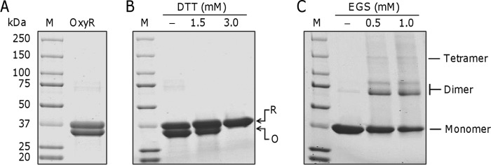
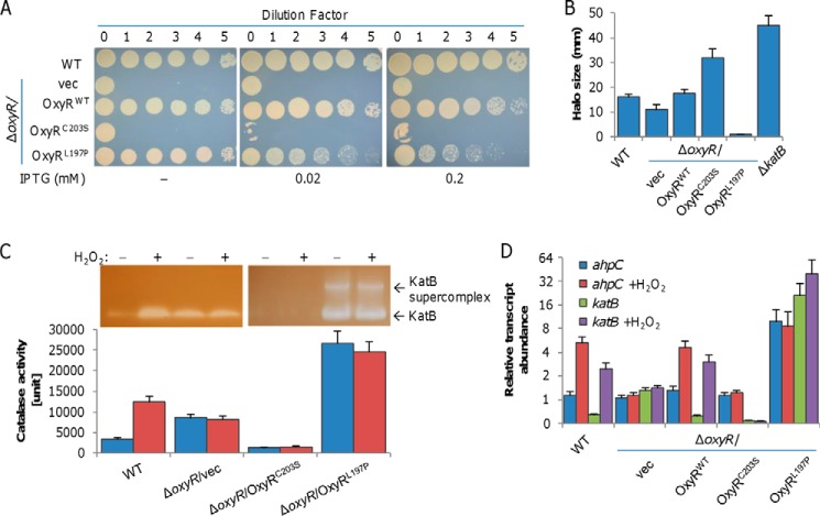
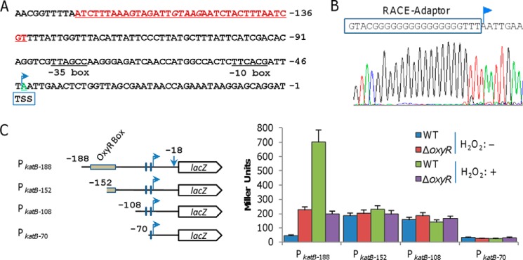
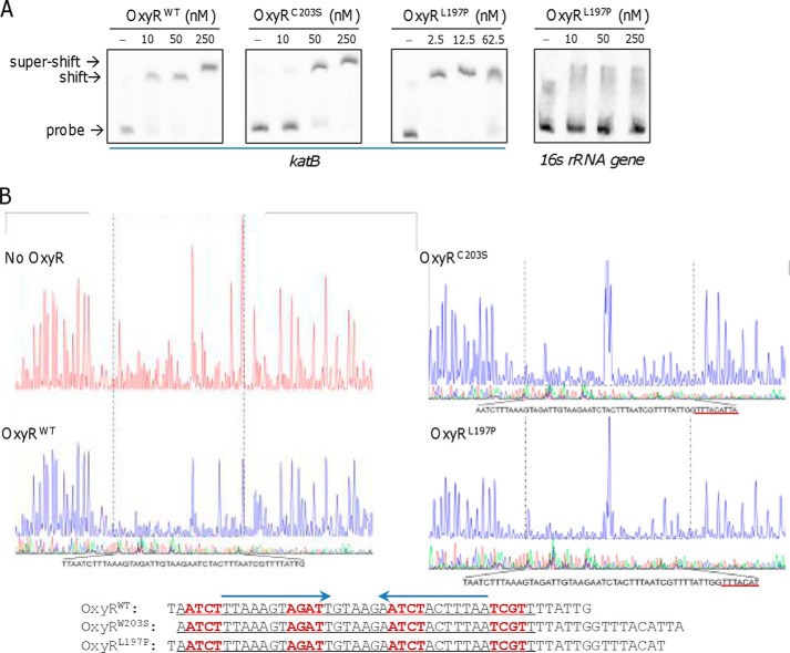
It is well-established that OxyR functions as a transcriptional activator of the peroxide stress response in bacteria, primarily based on studies on Escherichia coli Recent investigations have revealed that OxyRs of some other bacteria can regulate gene expression through both repression and activation or repression only; however, the underlying mechanisms remain largely unknown. Here, we demonstrated in γ-proteobacteriumShewanella oneidensis regulation of OxyR on expression of major catalase gene katB in a dual-control manner through interaction with a single site in the promoter region. Under non-stress conditions, katB expression was repressed by reduced OxyR (OxyRred), whereas when oxidized, OxyR (OxyRoxi) outcompeted OxyRred for the site because of substantially enhanced affinity, resulting in a graded response to oxidative stress, from repression to derepression to activation. The OxyR-binding motif is characterized as a combination of the E. coli motif (tetranucleotides spaced by heptanucleotide) and palindromic structure. We provided evidence to suggest that the S. oneidensis OxyR regulon is significantly contracted compared with those reported, probably containing only five members that are exclusively involved in oxygen reactive species scavenging and iron sequestering. These characteristics probably reflect the adapting strategy of the bacteria that S. oneidensis represents to thrive in redox-stratified microaerobic and anaerobic environments.

Keywords: DNA binding protein; bacterial genetics; gene regulation; hydrogen peroxide; oxidative stress.


Conflict of interest statement

The authors declare that they have no conflicts of interest with the contents of this article

Figures

Figure 1.
Figure 1.
Characteristics of purified S. oneidensis OxyR protein. A, SDS-PAGE analysis of Purified recombinant OxyR protein after gel-exclusion chromatography. B, SDS-PAGE analysis of OxyR treated with DTT. The reaction mixtures consisted of 5 μm OxyR protein and DTT at the indicated concentrations. R and O, reduced and oxidized forms of the protein, respectively. C, chemical cross-linking analyses of the purified OxyR protein. The EGS cross-linking reagent was added at the concentrations shown at the top. In all panels, M represents protein standard marker.
Figure 2.
Figure 2.
Physiological impacts of reduced and oxidized S. oneidensis OxyR proteins. A, droplet assays for viability and growth assessment. Cultures of the indicated strains prepared to contain ∼109 colony-forming units/ml were regarded as undiluted (dilution factor, 0) and were subjected to 10-fold series dilution. Five microliters of each dilution was dropped on LB plates containing IPTG at the indicated concentrations. Results were recorded after a 24-h incubation. Expression of OxyR variants was driven by IPTG-inducible Ptac. B, H2O2 susceptibility by disc diffusion assay. One hundred microliters of cultures was spread onto an agar plate containing 0.02 mm IPTG. Paper discs (diameter, 8 mm) containing 10 μl of 10 m H2O2 were placed on top of the agar. The plates were incubated at 30 °C for 16 h prior to analysis. The diameters of the zones of clearing (halo, in millimeters) generated by the peroxides were measured. C, catalase detected by staining and activity assay. Cells of the mid-log phase either directly used (non-treated) or incubated with 0.2 mm H2O2 for 30 min (treated) were used for the assay. Cell lysates containing the same amount of protein were subjected to 10% nondenaturing PAGE (top) and an activity assay (bottom). Staining was performed as described under “Experimental procedures.” For the catalase activity assay, decomposition of H2O2 was measured at 240 nm with absorbance readings taken at 15-s time intervals for a total time of 3.5 min. The unit of activity of each sample is expressed as catalase unit (μmol of H2O2 decomposed per min and per mg of protein). D, analysis of ahpC and katB transcripts by qRT-PCR. Cells of the mid-log phase incubated with 0.2 mm H2O2 for 2 min were used for total RNA extraction. The cycle threshold (CT) values were averaged and normalized against the CT value of the 16S rRNA gene. RTA, relative transcript abundance. In all panels, experiments were performed at least three times, with the average ± S.E. (error bars) of representative results presented.
Figure 3.
Figure 3.
Characterization of the S. oneidensis katB promoter region. A, promoter region of the katB gene. The binding site for OxyR is in red and underlined (discussed below in the legend to Fig. 6B), and the transcription start site (TSS) is in green and underlined. The number of nucleotides is relative to the translational starting code. B, determination of the katB transcriptional start site using 5′-RACE. The result of direct DNA sequencing of the 5′-RACE product of the katB gene is shown. C, deletion mapping of the katB regulatory region. Transcriptional fusion constructs are diagramed; coordinates indicate the extent of the regulatory katB region cloned in front of the lacZ reporter gene. The plasmids carrying the constructs were introduced into the relevant strains and integrated on the chromosome. After the antibiotic marker removal, promoter activity was measured by a β-galactosidase assay and is presented as Miller units. Experiments were performed at least three times, with the average ± S.E. (error bars) presented.
Figure 4.
Figure 4.
Both reduced and oxidized S. oneidensis OxyR proteins interact with the katB promoter region. A, in vitro interaction of His-tagged OxyR variants and the katB promoter sequence revealed by using EMSA. The digoxigenin-labeled DNA probes were prepared by PCR. The EMSA was performed with 1 μm probes and various amounts of proteins as indicated. Nonspecific competitor DNA (2 μm poly(dI·dC)) was included. B, DNase I footprinting analysis of OxyR variants. OxyR variants at 2 μm were used for binding to the 6-FAM–labeled katB promoter fragment. The regions protected by OxyR variants are indicated by a black dotted box and given below for clarity.
Figure 5.
Figure 5.
S. oneidensis OxyRoxi (OxyRL197P) outcompetes OxyRred (OxyRC203S) for binding to the katB promoter. A, fluorescence anisotropy change upon titration of a limiting concentration of 6-FAM–labeled 60-bp katB oligonucleotides (10 nm) with the indicated OxyR variants. Data are plotted as fluorescence anisotropy change values in millianisotrophy units (mA) as a function of protein concentration by using GraphPad Prism version 7 and fit to a model describing a 1:1 protein tetramer, and lines represent simulated curves produced from the average. B, impacts of OxyR variants at varying levels on growth of the wildtype with droplet assays. Production of OxyR variants was driven by Ptac from a single copy on the chromosome. Production level in the presence of 0.02 mm IPTG was equivalent to that of the native oxyR promoter. Catalase was added to differentiate defects in viability and in growth. Experiments were performed at least three times, with the average ± S.E. (error bars) of representative results presented.
Figure 6.
Figure 6.
Binding motifs of S. oneidensis OxyR. A, in vitro interaction of His-tagged OxyRL197P and the various promoter sequences revealed by using EMSA. Experiments were performed in the same manner as Fig. 4A. B, predicted OxyR-binding motifs in S. oneidensis based on the verified promoter sequences. The motifs of the 13- and 29-bp palindromic sequences predicted by AlignACE and MEME are marked with black and blue dashed lines, respectively. Tetranucleotide sequences are underlined based on the E. coli OxyR consensus. C, mutational analysis of the katB regulatory region. Transcriptional fusion constructs are diagramed as in Fig. 4C. In PkatB, the motif of 13 bp is in blue and extended to form the 29-bp motif, the additional nucleotides are in red, and tetranucleotide sequences are underlined. In PkatB mutants, a dot represents the corresponding nucleotide deleted, and nucleotides underlined and in boldface type were mutated. Promoter activity was measured by β-galactosidase assay and is presented as Miller units. Experiments were performed at least three times, with the average ± S.E. (error bars) presented.

Similar articles

Cited by

References

    1. Imlay J. A. (2013) The molecular mechanisms and physiological consequences of oxidative stress: lessons from a model bacterium. Nat. Rev. Microbiol. 11, 443–454 10.1038/nrmicro3032 - DOI - PMC - PubMed
    1. Imlay J. A. (2015) Transcription factors that defend bacteria against reactive oxygen species. Annu. Rev. Microbiol. 69, 93–108 10.1146/annurev-micro-091014-104322 - DOI - PMC - PubMed
    1. Mishra S., and Imlay J. (2012) Why do bacteria use so many enzymes to scavenge hydrogen peroxide? Arch. Biochem. Biophys. 525, 145–160 10.1016/j.abb.2012.04.014 - DOI - PMC - PubMed
    1. Maddocks S. E., and Oyston P. C. F. (2008) Structure and function of the LysR-type transcriptional regulator (LTTR) family proteins. Microbiology 154, 3609–3623 10.1099/mic.0.2008/022772-0 - DOI - PubMed
    1. Storz G., Tartaglia L. A., and Ames B. N. (1990) Transcriptional regulator of oxidative stress-inducible genes: direct activation by oxidation. Science 248, 189–194 10.1126/science.2183352 - DOI - PubMed

Publication types

MeSH terms

LinkOut - more resources