. 2018 Feb 21;38(8):2094-2105.
doi: 10.1523/JNEUROSCI.2457-17.2017. Epub 2018 Jan 24.

Cortical Neural Activity Predicts Sensory Acuity Under Optogenetic Manipulation

Affiliations

Cortical Neural Activity Predicts Sensory Acuity Under Optogenetic Manipulation

John J Briguglio et al. J Neurosci. .

Abstract

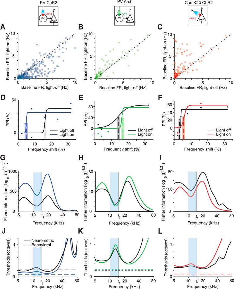
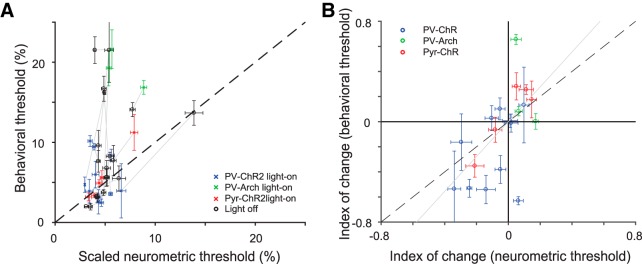
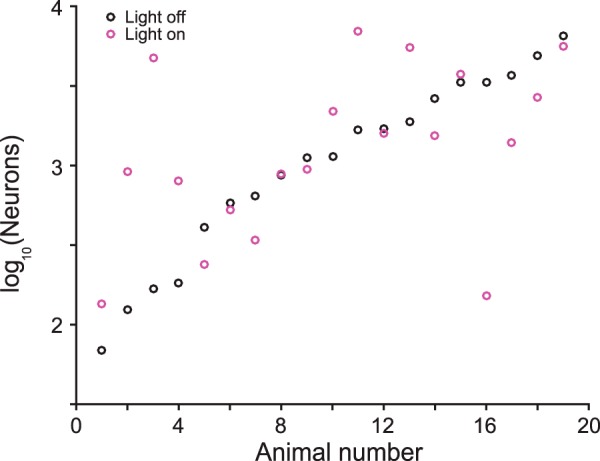
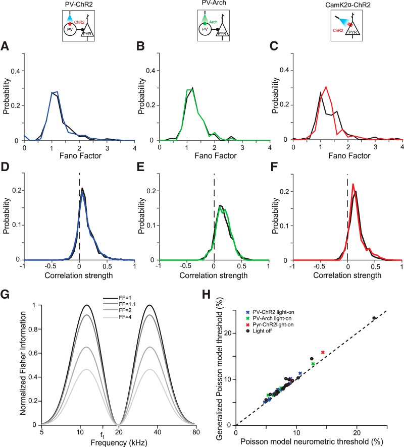
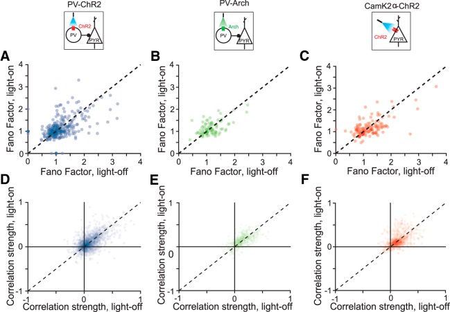
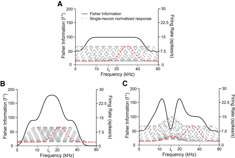
Excitatory and inhibitory neurons in the mammalian sensory cortex form interconnected circuits that control cortical stimulus selectivity and sensory acuity. Theoretical studies have predicted that suppression of inhibition in such excitatory-inhibitory networks can lead to either an increase or, paradoxically, a decrease in excitatory neuronal firing, with consequent effects on stimulus selectivity. We tested whether modulation of inhibition or excitation in the auditory cortex of male mice could evoke such a variety of effects in tone-evoked responses and in behavioral frequency discrimination acuity. We found that, indeed, the effects of optogenetic manipulation on stimulus selectivity and behavior varied in both magnitude and sign across subjects, possibly reflecting differences in circuitry or expression of optogenetic factors. Changes in neural population responses consistently predicted behavioral changes for individuals separately, including improvement and impairment in acuity. This correlation between cortical and behavioral change demonstrates that, despite the complex and varied effects that these manipulations can have on neuronal dynamics, the resulting changes in cortical activity account for accompanying changes in behavioral acuity.SIGNIFICANCE STATEMENT Excitatory and inhibitory interactions determine stimulus specificity and tuning in sensory cortex, thereby controlling perceptual discrimination acuity. Modeling has predicted that suppressing the activity of inhibitory neurons can lead to increased or, paradoxically, decreased excitatory activity depending on the architecture of the network. Here, we capitalized on differences between subjects to test whether suppressing/activating inhibition and excitation can in fact exhibit such paradoxical effects for both stimulus sensitivity and behavioral discriminability. Indeed, the same optogenetic manipulation in the auditory cortex of different mice could improve or impair frequency discrimination acuity, predictable from the effects on cortical responses to tones. The same manipulations sometimes produced opposite changes in the behavior of different individuals, supporting theoretical predictions for inhibition-stabilized networks.

Keywords: auditory cortex; behavior; computational modeling; excitatory–inhibitory circuits; frequency discrimination; optogenetics.


Figures

Figure 1.
Figure 1.
Measurement of behavioral frequency discrimination acuity. A, Schematic of measurement of frequency discrimination acuity in mouse. Left, Startle response measured as pressure the subject exerts on a platform. Right, Sound stimulus time course: an ongoing background tone (light gray, f1) is followed by a brief prepulse tone of different frequency (dark gray band, f2) and then by a startle noise (thin black band, SN). B, Normalized time course of platform pressure during the startle response to noise for different prepulse tones for an exemplar mouse. Time relative to SN onset. C, PPI measured as reduction in the acoustic startle response as a function of the frequency shift (Δf) between the background and prepulse tones (see Materials and Methods, Eq. 1) of an exemplar mouse. PPI does not reach 100% because, even with an easily identifiable prepulse tones, the animal still startles. Dots, Data; solid line, fit.
Figure 2.
Figure 2.
Measurement of neurometric frequency discrimination acuity. A, Left, Schematic of electrophysiological recording of neuronal responses in the primary auditory cortex (A1) in awake mouse. Right, Stimulus consisting of a pseudorandom sequence of pure tones at varying frequency and intensity levels. B, Representative frequency response function for a single neuron (f1 = background tone in Fig. 1). Black dots, Data; black line, fit. C, Fisher information computed as in Equation 5 for tone discrimination (black) computed on the basis of frequency response functions (gray dashed) of all frequency-tuned neurons (n = 14) recorded in the same mouse as in B. D, Neurometric threshold for decoding frequency (solid) computed on the basis of the inverse square root of Fisher information computed in C. Neurometric threshold based on the recorded population lies above behavioral threshold for discrimination around f1 (dashed line). Light blue band indicates the region in frequency space from which behavioral measurements were taken.
Figure 3.
Figure 3.
Optogenetic manipulation of PV activity shifts behavioral and neurometric frequency discrimination thresholds in individual subjects. AC, Baseline firing rate of light-on versus light-off trials for all frequency-tuned neurons pooled across subjects in PV-ChR2 (blue), PV-Arch (green), and CamK2a-ChR2 (red) mice, respectively. DF, PPI as a function of tone frequency shift for exemplar mice. Best estimated thresholds (dashed lines) and uncertainties (overlaid gray rectangle) are plotted for reference. Black, Light-off trials; blue, green, red, light-on trials. Dots, Data; solid lines, best fit curve. GI, Fisher information computed using tuning curves using neurons recorded from mice in DF. Frequencies used are indicated by the blue region. JL, Neurometric threshold estimate as inverse square root of Fisher information (solid) and behavioral threshold at f1 (horizontal dotted) for the same mice as DF. Light blue bands indicate the region in frequency space from which behavioral measurements were taken.
Figure 4.
Figure 4.
Changes in A1 tone responses due to optogenetic manipulations predict changes in behavioral frequency discrimination acuity across individuals. A, Behavioral versus scaled neurometric frequency discrimination thresholds (Table 4-1). Neurometric threshold (computed as inverse of Fisher information squared for tone-evoked responses from all neurons recorded in each mouse) is scaled to an effective population size of 1000 neurons to control for differences in numbers of measured neurons. Changing this scale factor is equivalent to changing y-axis labels. The scaled neurometric threshold based on the small recorded population was significantly (but weakly so, C = 0.37, p = 0.02) correlated with the behavioral threshold (computed as the shift in frequency between the background and prepulse tone that evoked 50% of the maximum PPI). Each of 19 mice contributes two data points, representing the threshold computed on the basis of light-on and light-off trials. Gray lines connect light-on and light-off estimates for each mouse. B, Index of change in neurometric threshold (difference between thresholds computed from data on light-on vs light-off trials divided by the sum) was strongly correlated with the behavioral frequency discrimination (C = 0.59, p = 0.007). There is one data point for each mouse. Gray line is the best fit line through the origin. Behavioral errors were computed as described in the Materials and Methods.
Figure 5.
Figure 5.
Number of frequency-tuned neurons required to account for behavioral sensitivity for each mouse (Eq. 11). Average of both light-on and light-off conditions is 1000 neurons.
Figure 6.
Figure 6.
Optogenetic manipulations do not change neuronal variability or correlations. A–C, Fano factor pooled across mice distributions are similar under light-on and light-off conditions. A, PV-ChR2; B, PV-Arch; C, CamK2a-ChR2. Black, Light-off trials; blue, green, red, light-on trials. D–F, Pairwise correlation distributions pooled across mice are similar under light-on and light-off conditions. D, PV-ChR2; E, PV-Arch; F, CamK2a-ChR2. Colors same as in A. G, Increasing Fano factor reduces Fisher information, shown here for a single neuron with Gaussian tuning curve (amplitude 8 spikes/s, center frequency 20 kHZ, tuning width 0.2 decades) with a constant baseline (2 spikes/s). H, Incorporating the measured Fano factors into our model of neuronal firing via a generalized Poisson model has a weak effect on the predicted threshold.
Figure 7.
Figure 7.
Fano factor and correlation scatter plots comparing light-on and light-off conditions. A–C, Fano factor with and without light on for PV-ChR2, PV-Arch, and Pyr-ChR2 mice, respectively. D–F, Pairwise correlations with and without light on for PV-ChR2, PV-Arch, and Pyr-ChR2 mice, respectively.
Figure 8.
Figure 8.
Overrepresenting a specific frequency can increase or reduce sensitivity to that frequency. A, Fisher information (black) computed from a homogeneous population of neurons (responses in gray) has an even sensitivity across a broad range of frequencies. A sample tuning curve (red) is used to illustrate neural transformations in B and C. Neurons have baseline activity of 2 spikes/s, peak response of 10 spikes/s, peak frequency spaced 1/20th of a decade apart, with an HWHM of 0.1 decades. B, Fisher information is plotted for a neural population overrepresenting frequency f1 by shifting peak frequencies halfway between their original location in A and f1. Fisher information approximately doubles near f1, but is reduced near the edges. C, Fisher information is plotted for a neural population overrepresenting frequency f1 by adding a Gaussian bump near f1 with an amplitude that diminishes with distance between the preferred frequency of the neuron and f1. Fisher information is diminished at f1, leading to reduced sensitivity at this frequency despite its overrepresentation within the population firing activity.

Similar articles

Cited by

References

    1. Ahrens MB, Linden JF, Sahani M (2008) Nonlinearities and contextual influences in auditory cortical responses modeled with multilinear spectrotemporal methods. J Neurosci 28:1929–1942. 10.1523/JNEUROSCI.3377-07.2008 - DOI - PMC - PubMed
    1. Aizenberg M, Geffen MN (2013) Bidirectional effects of auditory aversive learning on sensory acuity are mediated by the auditory cortex. Nat Neurosci 16:994–996. 10.1038/nn.3443 - DOI - PubMed
    1. Aizenberg M, Mwilambwe-Tshilobo L, Briguglio JJ, Natan RG, Geffen MN (2015) Bidirectional regulation of innate and learned behaviors that rely on frequency discrimination by cortical inhibitory neurons. PLoS Biol 13:e1002308. 10.1371/journal.pbio.1002308 - DOI - PMC - PubMed
    1. Atallah BV, Bruns W, Carandini M, Scanziani M (2012) Parvalbumin-expressing interneurons linearly transform cortical responses to visual stimuli. Neuron 73:159–170. 10.1016/j.neuron.2011.12.013 - DOI - PMC - PubMed
    1. Atasoy D, Aponte Y, Su HH, Sternson SM (2008) A FLEX switch targets channelrhodopsin-2 to multiple cell types for imaging and long-range circuit mapping. J Neurosci 28:7025–7030. 10.1523/JNEUROSCI.1954-08.2008 - DOI - PMC - PubMed

Publication types

LinkOut - more resources