. 2018 Feb 6;115(6):E1127-E1136.
doi: 10.1073/pnas.1714085115. Epub 2018 Jan 24.

Extracellular vesicle budding is inhibited by redundant regulators of TAT-5 flippase localization and phospholipid asymmetry

Affiliations

Extracellular vesicle budding is inhibited by redundant regulators of TAT-5 flippase localization and phospholipid asymmetry

Katharina B Beer et al. Proc Natl Acad Sci U S A. .

Abstract

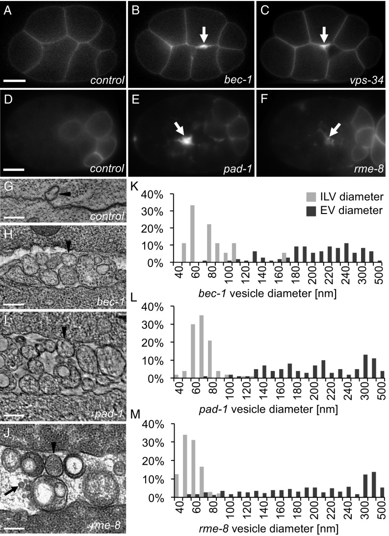
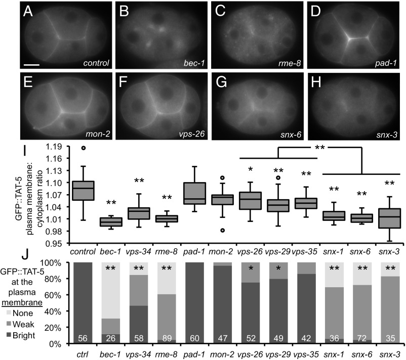
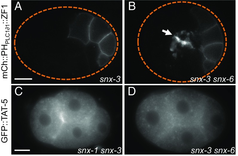
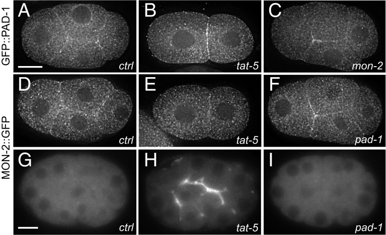
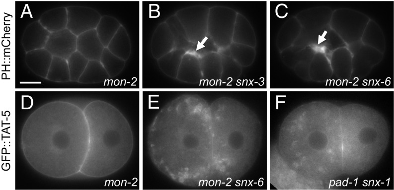
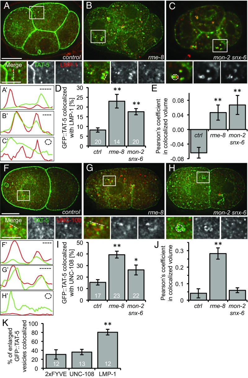
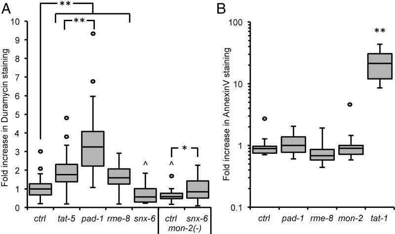
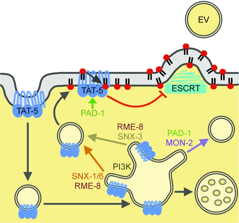
Cells release extracellular vesicles (EVs) that mediate intercellular communication and repair damaged membranes. Despite the pleiotropic functions of EVs in vitro, their in vivo function is debated, largely because it is unclear how to induce or inhibit their formation. In particular, the mechanisms of EV release by plasma membrane budding or ectocytosis are poorly understood. We previously showed that TAT-5 phospholipid flippase activity maintains the asymmetric localization of the lipid phosphatidylethanolamine (PE) in the plasma membrane and inhibits EV budding by ectocytosis in Caenorhabditis elegans However, no proteins that inhibit ectocytosis upstream of TAT-5 were known. Here, we identify TAT-5 regulators associated with retrograde endosomal recycling: PI3Kinase VPS-34, Beclin1 homolog BEC-1, DnaJ protein RME-8, and the uncharacterized Dopey homolog PAD-1. PI3Kinase, RME-8, and semiredundant sorting nexins are required for the plasma membrane localization of TAT-5, which is important to maintain PE asymmetry and inhibit EV release. PAD-1 does not directly regulate TAT-5 localization, but is required for the lipid flipping activity of TAT-5. PAD-1 also has roles in endosomal trafficking with the GEF-like protein MON-2, which regulates PE asymmetry and EV release redundantly with sorting nexins independent of the core retromer. Thus, in addition to uncovering redundant intracellular trafficking pathways, our study identifies additional proteins that regulate EV release. This work pinpoints TAT-5 and PE as key regulators of plasma membrane budding, further supporting the model that PE externalization drives ectocytosis.

Keywords: extracellular vesicle; flippase; lipid asymmetry; microvesicle; retromer.


Conflict of interest statement

The authors declare no conflict of interest.

Figures

Fig. 1.
Fig. 1.
Retrograde trafficking proteins inhibit microvesicle release. (A) PHPLC1∂1::mCh localizes primarily to the plasma membrane in control embryos at the eight-cell stage. (B and C) PH reporters localize to thickened membranes (arrow) in bec-1 (PHPLC1∂1::mCh) and vps-34 (PHPLC1∂1::GFP) maternal zygotic mutants. (D) In a 26-cell control embryo, GFP::ZF1::PHPLC1∂1 is degraded in most somatic cells, only persisting on the plasma membrane in a few posterior cells. (E and F) EVs labeled with GFP::ZF1::PHPLC1∂1 (arrow) accumulate between cells in pad-1 and rme-8 RNAi-treated embryos. (G) EVs are infrequently observed between wild-type N2 cells in a two-cell embryo tomogram. (HJ) Released EVs accumulate between cells in a tomogram from a three-cell bec-1 maternal-zygotic mutant, a three-cell embryo treated with pad-1 RNAi, and a 24-cell rme-8 RNAi embryo. Arrowheads point to microvesicle-sized EVs; arrows point to exosome-sized EVs. (KM) Histograms of EV and intraluminal vesicle (ILV) diameters measured from TEM images of bec-1 maternal-zygotic mutant embryos and pad-1 RNAi embryos demonstrate that the majority of EVs are microvesicles, because they are larger than ILVs. In rme-8 RNAi, EVs could be both exosomes and microvesicles, because they are the same size as or larger than ILVs. [Scale bars: A, 10 µm (also applies to B and C); D, 10 µm (also applies to E and F); GJ, 200 nm.]
Fig. 2.
Fig. 2.
TAT-5 is recycled by retromer-associated proteins. (A) GFP::TAT-5 localizes to the plasma membrane in a control four-cell embryo. (B) GFP::TAT-5 is mislocalized to large cytoplasmic structures in bec-1 maternal zygotic mutants. (C) GFP::TAT-5 shows a more dispersed localization to cytoplasmic structures in rme-8 RNAi. See Fig. S2 for reduced total levels of GFP::TAT-5. (D and E) GFP::TAT-5 localization is not altered after pad-1 or mon-2 RNAi. See Fig. S3 for GFP::TAT-5 localization to EVs. (F) GFP::TAT-5 still localizes to the plasma membrane in core retromer vps-26 mutants. (G and H) In contrast, GFP::TAT-5 plasma membrane localization is weak after snx-6 RNAi or in snx-3 deletion mutants. (I) Ratio of GFP::TAT-5 fluorescence intensity in the plasma membrane to the intensity of the cytoplasm. Disrupting PI3K subunits and RME-8 had highly significant effects on GFP::TAT-5 plasma membrane localization, as did SNX. More variability is seen in vps-34 mutants due to the presence of a mosaic rescuing transgene (Materials and Methods). There was no significant change in plasma membrane localization after pad-1 or mon-2 RNAi, but knocking down or deleting core retromer proteins resulted in mild, but significant, decreases. Student’s t test with Bonferroni correction was used for statistical analysis. *P < 0.05; **P < 0.001 (compared with control empty vector RNAi). (J) Embryos were categorized for the brightness of GFP::TAT-5 localization in the plasma membrane. PI3K subunits, RME-8, and sorting nexins prevented or decreased TAT-5 plasma membrane localization, while core retromer proteins resulted in weaker plasma membrane localization. *P < 0.01; **P < 0.0001 (Fisher’s exact test with Bonferroni correction). Number of embryos scored is indicated for each genotype. [Scale bar: A, 10 µm (also applies to BH).]
Fig. 3.
Fig. 3.
SNX-1/SNX-6 and SNX-3 redundantly inhibit EV release. (A) EV release was not increased in a 26-cell snx-3 mutant embryo expressing the mCherry::PHPLC1∂1::ZF1 plasma membrane reporter. (B) EVs labeled with mCherry::PHPLC1∂1::ZF1 (arrow) accumulate between cell contacts in a 26-cell snx-3 mutant treated with snx-6 RNAi. (C and D) GFP::TAT-5 is mislocalized to cytoplasmic compartments in snx-1 mutants treated with snx-3 RNAi, as well as in snx-3 mutants treated with snx-6 RNAi. [Scale bars: A, 10 μm (also applies to B); C, 10 μm (also applies to D).]
Fig. 4.
Fig. 4.
PAD-1 and MON-2 localize to the cell cortex and cytoplasm. (A) GFP::PAD-1 staining is found on cytoplasmic puncta as well as at the plasma membrane in a four-cell embryo. GFP was knocked into the endogenous pad-1 locus. (B) GFP::PAD-1 localization is not significantly altered after tat-5 RNAi treatment in a two-cell embryo. (C) GFP::PAD-1 localizes to the plasma membrane after mon-2 RNAi treatment in a four-cell embryo. (D) MON-2::GFP::3xFlag staining also localizes to cytoplasmic puncta as well as at the plasma membrane in a four-cell embryo. (E) MON-2::GFP::3xFlag localization is not significantly altered after tat-5 RNAi treatment in a two-cell embryo. (F) MON-2::GFP::3xFlag still localizes to the plasma membrane after pad-1 RNAi treatment in a four-cell embryo. (G) In live 26-cell embryos, MON-2::GFP::3xFlag puncta are barely visible. (H) MON-2::GFP::3xFlag is increased at the cell surface after tat-5 RNAi treatment, suggesting that cortical MON-2 is released outside cells in EVs. (I) MON-2::GFP::3xFlag is not released in EVs after pad-1 RNAi treatment, suggesting that PAD-1 is required for MON-2 localization in EVs. [Scale bars: A, 10 μm (also applies to BF); G, 10 μm (also applies to H and I).]
Fig. 5.
Fig. 5.
MON-2 inhibits EV release in snx-mutants. (A) The plasma membrane marker PHPLC1∂1::mCh appears normal in a 15-cell mon-2(xh22) mutant embryo. (B and C) Thickened patches of PHPLC1∂1::mCh (arrow) are observed in mon-2(xh22) mutants treated with snx-3 or -6 RNAi, indicating increased EV release. Similar phenotypes were also seen after snx-1 RNAi treatment (Table 1), demonstrating that sorting nexins and MON-2 redundantly inhibit EV release. (D) GFP::TAT-5 localizes to the plasma membrane in a two-cell mon-2(xh22) mutant embryo. (E) GFP::TAT-5 localizes to prominent cytoplasmic vesicles after snx-6 RNAi treatment in mon-2(xh22) mutants. Similar phenotypes were also seen after snx-1 or -3 RNAi treatment, suggesting that MON-2 acts redundantly with sorting nexins to control TAT-5 localization. (F) GFP::TAT-5 localizes to large cytoplasmic vesicles after pad-1 RNAi in snx-1(tm847) mutants, demonstrating that PAD-1 also acts redundantly with sorting nexins during TAT-5 trafficking. [Scale bar: A, 10 µm (also applies to B–F).]
Fig. 6.
Fig. 6.
TAT-5 localization to late endosomes increases in EV-releasing mutants. (A) GFP::TAT-5 (green) staining localizes primarily to the plasma membrane in control four-cell embryos. TAT-5 is also found in cytoplasmic vesicles, but they do not often colocalize with LMP-1 (red) staining in late endosomes or lysosomes, as shown in Insets. (B) Mislocalization of GFP::TAT-5 in rme-8 RNAi causes increased colocalization with LMP-1. (C) Large GFP::TAT-5-positive cytoplasmic vesicles colocalize with LMP-1 in a mon-2(xh22) mutant embryo treated with snx-6 RNAi. (A′C′) Line scans of GFP::TAT-5 and LMP-1 intensity next to the dotted lines or circle in Insets. (D) The percentage of TAT-5 colocalized with LMP-1 is significantly increased in 2- to 12-cell embryos after rme-8 RNAi and in mon-2(xh22) mutants treated with snx-6 RNAi compared with control embryos. Number of embryos scored is indicated for each genotype. (E) The Pearson’s coefficient of GFP::TAT-5 colocalization with LMP-1 increases significantly from a negative correlation in control embryos to a positive correlation in rme-8 RNAi as well as in mon-2(xh22) mutants treated with snx-6 RNAi. (F) Cytoplasmic GFP::TAT-5 vesicles (green) rarely colocalize with mCherry::UNC-108 staining in Golgi and endosomes (red) in two-cell embryos, as shown in Insets. (G) TAT-5 colocalization with the Rab2 homolog UNC-108 increases after rme-8 RNAi. (H) TAT-5 colocalization with UNC-108 increased slightly in mon-2 mutants treated with snx-6 RNAi. (F′H′) Line scans of GFP::TAT-5 and UNC-108 intensity next to the dotted lines or circle in Insets. (I) The percentage of TAT-5 colocalized with UNC-108 is significantly increased in 2- to 12-cell embryos after rme-8 RNAi and in mon-2(xh22) mutants treated with snx-6 RNAi compared with control embryos. Number of embryos scored is indicated for each genotype. (J) The Pearson’s coefficient of GFP::TAT-5 colocalization with LMP-1 increases significantly in rme-8 RNAi, but not in mon-2(xh22) mutants treated with snx-6 RNAi. (K) Enlarged GFP::TAT-5 vesicles colocalize significantly more often with LMP-1 (n = 54 vesicles) than the PI3P reporter 2xFYVE (n = 85) or UNC-108 (n = 89). Percentages are expressed in relation to the total number of large vesicles. Number of embryos scored is indicated for each genotype. Student’s t test with Bonferroni correction was used for statistical analysis. *P < 0.05; **P < 0.001. [Scale bars: A and F, 10 µm (also apply to B and C and G and H, respectively); Insets in A and F, 5 µm (also apply to Insets in B and C and G and H, respectively).]
Fig. 7.
Fig. 7.
PAD-1 and RME-8 are required to maintain PE asymmetry. (A) Duramycin staining is significantly increased on dissected gonads after pad-1, rme-8, or tat-5 RNAi treatment compared to control embryos (ctrl), indicating that PAD-1 and RME-8 are required for TAT-5 to maintain PE asymmetry. The increase after pad-1 RNAi is also significantly more than tat-5 knockdown, suggesting that PAD-1 may influence PE asymmetry through more than TAT-5 activity. Duramycin staining is significantly decreased after snx-6 RNAi treatment and in untreated mon-2(xh22) mutants. Duramycin staining is increased on dissected gonads after snx-6 RNAi treatment in mon-2(xh22) nonsense mutants, in comparison with untreated mon-2(xh22) mutants, demonstrating that MON-2 and SNX-6 are required redundantly for PE asymmetry. (B) Annexin V staining is not increased on dissected gonads after mon-2, pad-1, or rme-8 RNAi treatment (P > 0.05), indicating that PS is not externalized. Annexin V staining is strongly increased in tat-1(kr15) mutants, together indicating that MON-2, PAD-1, and RME-8 are not required for TAT-1 PS flippase activity. Student’s t test with Bonferroni correction was used for statistical analysis. Asterisks indicate significantly increased values, and carets indicate significantly decreased values. *P < 0.05; **P < 0.001; ^P < 0.05.
Fig. 8.
Fig. 8.
Model of TAT-5 trafficking to inhibit EV release. TAT-5 maintains PE asymmetry in the plasma membrane to inhibit recruitment of the ESCRT machinery to release EVs by plasma membrane budding. PAD-1 is required for TAT-5 flippase activity, which inhibits the externalization of PE and EV release. TAT-5 is endocytosed and needs to be recycled from sorting endosomes to the plasma membrane through both SNX-1–SNX-6–mediated and SNX-3–mediated tubulation and vesicle formation. RME-8 and PI3K mediate TAT-5 recycling through multiple pathways. MON-2 and PAD-1 regulate an unknown step of endosomal trafficking, here drawn as a third recycling pathway preventing membrane cargos from being delivered to multivesicular endosomes for degradation. When both SNX-mediated recycling and MON-2/PAD-1–mediated trafficking are lost, TAT-5 is mislocalized to late endosomes where it can no longer maintain plasma membrane asymmetry.

References

    1. Colombo M, Raposo G, Théry C. Biogenesis, secretion, and intercellular interactions of exosomes and other extracellular vesicles. Annu Rev Cell Dev Biol. 2014;30:255–289. - PubMed
    1. Beer KB, Wehman AM. Mechanisms and functions of extracellular vesicle release in vivo–What we can learn from flies and worms. Cell Adhes Migr. 2017;11:135–150. - PMC - PubMed
    1. Jimenez AJ, et al. ESCRT machinery is required for plasma membrane repair. Science. 2014;343:1247136. - PubMed
    1. Gong Y-N, et al. ESCRT-III acts downstream of MLKL to regulate necroptotic cell death and its consequences. Cell. 2017;169:286–300.e16. - PMC - PubMed
    1. Henne WM, Stenmark H, Emr SD. Molecular mechanisms of the membrane sculpting ESCRT pathway. Cold Spring Harb Perspect Biol. 2013;5:a016766. - PMC - PubMed

Publication types

MeSH terms

Substances

LinkOut - more resources