. 2018 Feb 6;115(6):E1239-E1248.
doi: 10.1073/pnas.1718197115. Epub 2018 Jan 24.

Chemotherapy induces enrichment of CD47+/CD73+/PDL1+ immune evasive triple-negative breast cancer cells

Affiliations

Chemotherapy induces enrichment of CD47+/CD73+/PDL1+ immune evasive triple-negative breast cancer cells

Debangshu Samanta et al. Proc Natl Acad Sci U S A. .

Abstract

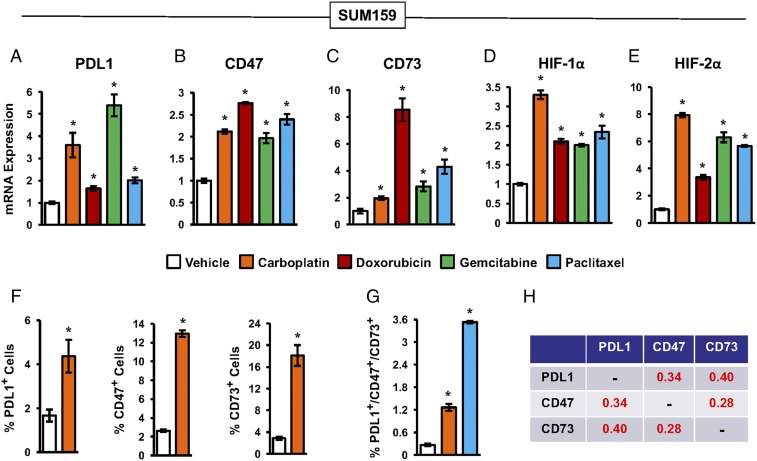
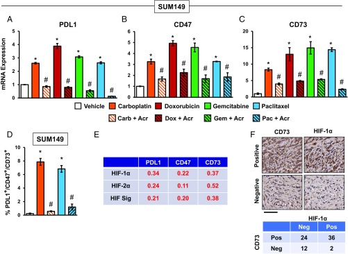
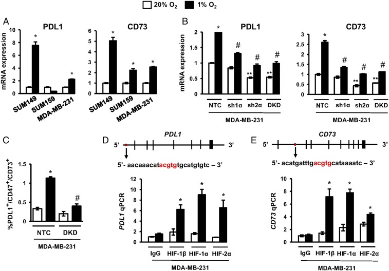
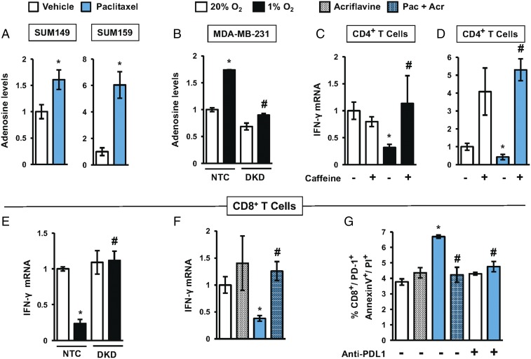
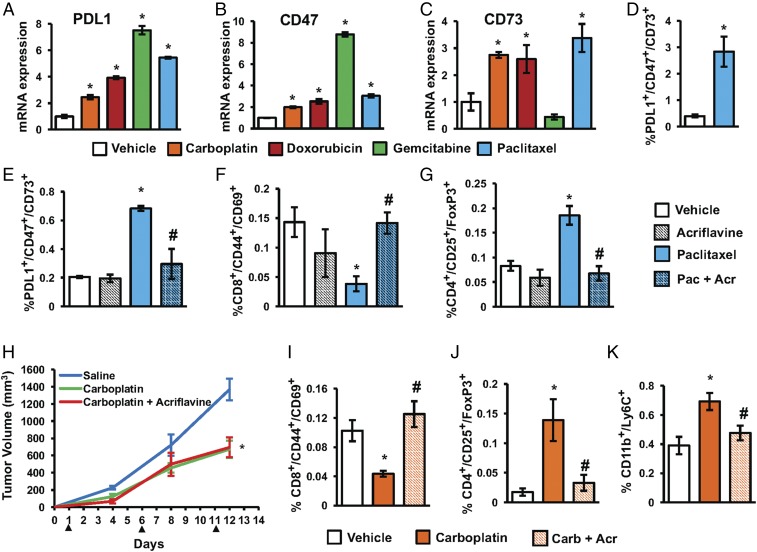
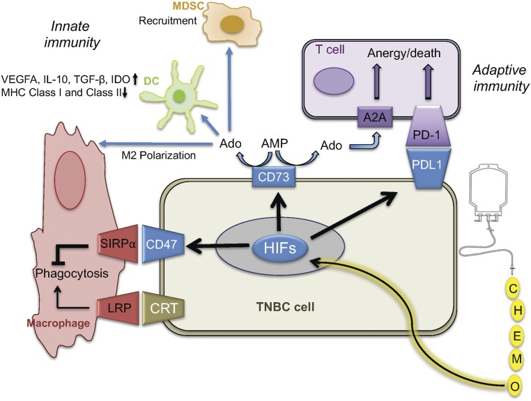
Triple-negative breast cancer (TNBC) is treated with cytotoxic chemotherapy and is often characterized by early relapse and metastasis. To form a secondary (recurrent and/or metastatic) tumor, a breast cancer cell must evade the innate and adaptive immune systems. CD47 enables cancer cells to evade killing by macrophages, whereas CD73 and PDL1 mediate independent mechanisms of evasion of cytotoxic T lymphocytes. Here, we report that treatment of human or murine TNBC cells with carboplatin, doxorubicin, gemcitabine, or paclitaxel induces the coordinate transcriptional induction of CD47, CD73, and PDL1 mRNA and protein expression, leading to a marked increase in the percentage of CD47+CD73+PDL1+ breast cancer cells. Genetic or pharmacological inhibition of hypoxia-inducible factors (HIFs) blocked chemotherapy-induced enrichment of CD47+CD73+PDL1+ TNBC cells, which were also enriched in the absence of chemotherapy by incubation under hypoxic conditions, leading to T cell anergy or death. Treatment of mice with cytotoxic chemotherapy markedly increased the intratumoral ratio of regulatory/effector T cells, an effect that was abrogated by HIF inhibition. Our results delineate an HIF-dependent transcriptional mechanism contributing to TNBC progression and suggest that combining chemotherapy with an HIF inhibitor may prevent countertherapeutic induction of proteins that mediate evasion of innate and adaptive antitumor immunity.

Keywords: HIF-1; PD-1; adenosine; effector T cells; regulatory T cells.


Conflict of interest statement

The authors declare no conflict of interest.

Figures

Fig. 1.
Fig. 1.
Chemotherapy induces expression of PDL1, CD73, and CD47. (AE) SUM159 cells were treated with 50 μM carboplatin, 50 nM doxorubicin, 10 nM gemcitabine, 10 nM paclitaxel, or vehicle for 4 d. RT-qPCR was performed to quantify PDL1 (A), CD47 (B), CD73 (C), HIF-1α (D), and HIF-2α (E) mRNA levels relative to 18S rRNA and normalized to vehicle-treated cells (mean ± SEM; n = 3). *P < 0.001 compared with vehicle (by one-way ANOVA with a Bonferroni posttest). (F) Cells were treated with vehicle or carboplatin. After 4 d, the percentage of cells with surface expression of PDL1 (Left), CD47 (Center), or CD73 (Right) was determined by flow cytometry (mean ± SEM; n = 3). *P < 0.001 compared with vehicle (by Student’s t test). (G) Cells were treated with vehicle, carboplatin, or paclitaxel as described above. After 4 d, the percentage of cells that were triple-positive for PDL1, CD73, and CD47 was determined (mean ± SEM; n = 3). *P < 0.001 compared with vehicle (by one-way ANOVA with a Bonferroni posttest). All experiments in this figure were performed using cells exposed to 20% O2 in a standard 95% air/5% CO2 incubator. (H) Analysis of gene expression data from primary human breast cancers. The Pearson correlation test was performed to compare coexpression of PDL1, CD73, and CD47 mRNA using data from 1,215 breast cancer samples from The Cancer Genome Atlas (TCGA) database. Pearson’s correlation (r) is shown; P < 0.0001 for all comparisons.
Fig. 2.
Fig. 2.
HIFs mediate chemotherapy-induced PDL1, CD73, and CD47 expression. (AC) SUM149 cells were treated with vehicle or chemotherapy, either alone or in combination with 2 μM acriflavine, for 4 d. RT-qPCR was performed to quantify PDL1, CD73, and CD47 mRNA levels relative to 18S rRNA and normalized to vehicle-treated cells (mean ± SEM; n = 3). *P < 0.01 compared with vehicle; #P < 0.01 compared with chemotherapy alone (by one-way ANOVA with a Bonferroni posttest). Acr, acriflavine; Carb, carboplatin, Dox, doxorubicin; Gem, gemcitabine; Pac, paclitaxel. (D) Cells were exposed to carboplatin or paclitaxel for 4 d, and flow cytometry was performed to determine the percentage of triple-positive (PDL1+/CD73+/CD47+) cells (mean ± SEM; n = 3). *P < 0.01 compared with vehicle; #P < 0.01 compared with chemotherapy alone (by one-way ANOVA with a Bonferroni posttest). (E) Analysis of gene expression data from primary human breast cancers. The Pearson correlation test was performed to compare expression of HIF-1α, HIF-2α, and HIF signature (HIF Sig) with expression of PDL1, CD47, and CD73 mRNA, using data from 1,215 breast cancer samples in the TCGA database. Pearson’s correlation (r) is shown; P < 0.0001 for all comparisons. (F) Immunohistochemistry was performed on breast cancer biopsies using anti-CD73 and anti–HIF-1α antibodies. (Upper) Representative positive and negative staining (biopsies from patients 2 and 3, respectively) is shown. (Scale bar, 100 μm.) (Lower) Summary of CD73 and HIF-1α expression in 74 human breast cancer biopsies is shown. Neg, negative; Pos, positive.
Fig. 3.
Fig. 3.
HIFs transactivate the PDL1 and CD73 genes. (A) TNBC cell lines were exposed to 20% or 1% O2 for 24 h, and the expression of mRNAs encoding PDL1 (Left) and CD73 (Right) was analyzed by RT-qPCR. The expression of each mRNA was quantified relative to 18S rRNA and then normalized to the result obtained from cells at 20% O2 (mean ± SEM; n = 3). *P < 0.01 versus 20% O2 (by two-way ANOVA with a Bonferroni posttest). (B) Analysis of PDL1 and CD73 mRNA expression in MDA-MB-231 subclones, which expressed an NTC shRNA or shRNA targeting HIF-1α (sh1α), HIF-2α (sh2α), or both HIF-1α and HIF-2α (DKD). Cells were exposed to 20% or 1% O2 for 24 h. Data were normalized to NTC at 20% O2 (mean ± SEM; n = 3). *P < 0.01 versus NTC at 20% O2; **P < 0.01 versus NTC at 20% O2; #P < 0.001 versus NTC at 1% O2 (by two-way ANOVA with a Bonferroni posttest). (C) MDA-MB-231 subclones were exposed to 20% or 1% O2 for 72 h, and the percentage of triple-positive cells was determined by flow cytometry (mean ± SEM; n = 3). *P < 0.01 versus NTC at 20% O2; #P < 0.001 versus NTC at 1% O2 (by two-way ANOVA with a Bonferroni posttest). (D and E) MDA-MB-231 cells were exposed to 20% or 1% O2 for 24 h and chromatin immunoprecipitation assays were performed using IgG or antibodies against HIF-1α, HIF-1β, or HIF-2α. Primers flanking the candidate HIF binding sites were used for qPCR, and results were normalized to lane 1 (mean ± SEM; n = 3). *P < 0.05 versus 20% O2 (by one-way ANOVA with a Bonferroni posttest). The nucleotide sequence (noncoding strand) of HIF binding sites (in red), which are located 5.7 kb 5′ to the transcription start site of the PDL1 gene (D) and within intron 1 of the CD73 gene (E), respectively, are shown. Exons and introns are not drawn to scale.
Fig. 4.
Fig. 4.
Exposure of TNBC cells to chemotherapy or hypoxia enables evasion of antitumor T cells. (A) Adenosine levels were measured in CM of SUM149 and SUM159 cells treated with paclitaxel (5 nM for SUM149 and 10 nM for SUM159) for 4 d. The values were corrected for cell number at the end of the experiment and then normalized to vehicle-treated cells (mean ± SEM; n = 3). *P < 0.05 versus vehicle (by Student’s t test). (B) Adenosine levels were measured in CM of MDA-MB-231 NTC and DKD subclones exposed to 20% or 1% O2 for 72 h. The values were normalized to NTC at 20% O2 (mean ± SEM; n = 3). *P < 0.05 versus NTC at 20% O2; #P < 0.05 versus NTC at 1% O2 (by two-way ANOVA with a Bonferroni posttest). (C and D) IFN-γ mRNA levels were determined (mean ± SEM; n = 3) in activated CD4+ T cells incubated for 24 h with CM (from hypoxic or paclitaxel-treated TNBCs) in the presence or absence of 4 mM caffeine. *P < 0.05 versus NTC at 20% O2; #P < 0.05 versus NTC at 1% O2 (by two-way ANOVA with a Bonferroni posttest). (E) IFN-γ mRNA levels (mean ± SEM; n = 3) were determined in activated CD8+ T cells cultured with CM from MDA-MB-231 subclones exposed to 20% or 1% O2 for 24 h. *P < 0.05 versus NTC at 20% O2; #P < 0.05 versus NTC at 1% O2 (by two-way ANOVA with a Bonferroni posttest). (F) IFN-γ mRNA levels (mean ± SEM; n = 3) were determined in activated CD8+ T cells incubated for 24 h with CM (from TNBC cells exposed to paclitaxel with or without 2 μM acriflavine for 4 d). *P < 0.05 versus vehicle-treated cells; #P < 0.05 versus paclitaxel-treated cells (by one-way ANOVA with a Bonferroni posttest). (G) CD8+ T cells were incubated with 4T1 cells (which were pretreated for 4 d with vehicle, acriflavine, paclitaxel, or paclitaxel + acriflavine) in presence or absence of anti-PDL1 blocking antibody. Cell death was assessed by staining with Annexin V and PI. The percentage of CD8+/PD1+ cells that were Annexin V+ and PI+ was quantified by flow cytometry (mean ± SEM; n = 3). *P < 0.05 versus vehicle-treated cells; #P < 0.05 versus paclitaxel-treated cells (by two-way ANOVA with a Bonferroni posttest).
Fig. 5.
Fig. 5.
Acriflavine blocks paclitaxel-induced enrichment of PDL1+/CD73+/CD47+ TNBC cells and makes the tumor environment less immunosuppressive. (AC) Mouse 4T1 mammary carcinoma cells were cultured for 4 d in the presence of vehicle, 50 μM carboplatin, 250 nM doxorubicin, 15 nM gemcitabine, or 10 nM paclitaxel. PDL1, CD47, and CD73 mRNA levels were normalized to vehicle-treated cells (mean ± SEM; n = 3). *P < 0.001 compared with vehicle (by one-way ANOVA with a Bonferroni posttest). (D) The 4T1 cells were treated with vehicle or paclitaxel in vitro, and flow cytometry was performed to quantify PDL1+/CD73+/CD47+ cells (mean ± SEM; n = 3). *P < 0.001 compared with vehicle (by Student’s t test). (EG) The 4T1 cells were implanted into the mammary fat pad of female BALB/c mice. When tumor volume reached 200 mm3, mice were treated with: vehicle (saline), acriflavine (4 mg/kg; days 1–13), paclitaxel (10 mg/kg; days 5 and 10), or paclitaxel and acriflavine. Tumors were harvested on day 13, and the percentage of CD45/PDL1+/CD73+/CD47+ (E), CD8+/CD44+/CD69+ (F), and CD4+/CD25+/FoxP3+ (G) cells was determined (mean ± SEM; n = 4). *P < 0.05 versus vehicle-treated mice; #P < 0.05 versus paclitaxel-treated mice (by one-way ANOVA with a Bonferroni posttest). Acr, acriflavine; Pac, paclitaxel. (HK) The 4T1 cells were implanted into the mammary fat pad of female BALB/c mice. When tumors were palpable, mice were treated with: vehicle (saline), acriflavine (4 mg/kg; days 1–13), carboplatin (Carb; 20 mg/kg; days 1, 6, and 11), or carboplatin and acriflavine. (H) Tumor volumes were measured. Tumors were harvested on day 13, and the percentage of CD8+/CD44+/CD69+ (I), CD4+/CD25+/FoxP3+ (J), and CD11b+/Ly6C+ (K) cells was determined (mean ± SEM; n = 6). *P < 0.05 versus vehicle-treated mice; #P < 0.05 versus paclitaxel-treated mice (by one-way ANOVA with a Bonferroni posttest).
Fig. 6.
Fig. 6.
Chemotherapy promotes immune evasion phenotype in surviving TNBC cells. Exposure of TNBC cells to cytotoxic chemotherapy (or hypoxia) induces expression of HIF-1α and HIF-2α, leading to the HIF-mediated expression of PDL1, CD73, and CD47, which promote suppression of innate antitumor immunity mediated by macrophages, dendritic cells (DCs), and MDSCs, and suppression of adaptive antitumor immunity mediated by T cells. Ado, adenosine; IDO, indoleamine-2,3-dioxygenase.

References

    1. Newman LA, Reis-Filho JS, Morrow M, Carey LA, King TA. The 2014 Society of Surgical Oncology Susan G. Komen for the Cure Symposium: Triple-negative breast cancer. Ann Surg Oncol. 2015;22:874–882. - PubMed
    1. Gadi VK, Davidson NE. Practical approach to triple-negative breast cancer. J Oncol Pract. 2017;13:293–300. - PubMed
    1. Zou W. Immunosuppressive networks in the tumour environment and their therapeutic relevance. Nat Rev Cancer. 2005;5:263–274. - PubMed
    1. Pardoll DM. The blockade of immune checkpoints in cancer immunotherapy. Nat Rev Cancer. 2012;12:252–264. - PMC - PubMed
    1. Gajewski TF, Schreiber H, Fu YX. Innate and adaptive immune cells in the tumor microenvironment. Nat Immunol. 2013;14:1014–1022. - PMC - PubMed

Publication types

MeSH terms