. 2018 Apr;17(4):631-642.
doi: 10.1074/mcp.RA117.000129. Epub 2018 Jan 24.

N-glycome of the Lysosomal Glycocalyx is Altered in Niemann-Pick Type C Disease (NPC) Model Cells

Affiliations

N-glycome of the Lysosomal Glycocalyx is Altered in Niemann-Pick Type C Disease (NPC) Model Cells

Marko Kosicek et al. Mol Cell Proteomics. 2018 Apr.

Abstract

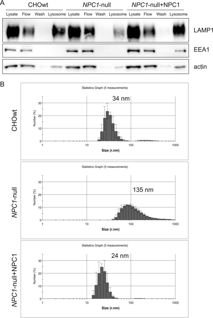
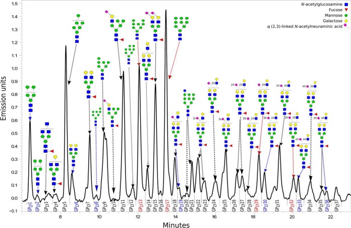
Increasing evidence implicates lysosomal dysfunction in the pathogenesis of neurodegenerative diseases, including the rare inherited lysosomal storage disorders (LSDs) and the most common neurodegenerative diseases, such as Alzheimer's and Parkinson's disease (AD and PD). Although the triggers of the lysosomal impairment may involve the accumulated macromolecules or dysfunction of the lysosomal enzymes, the role of the lysosomal glycocalyx in the lysosomal (dys)function has not been studied. The goal of this work was to analyze whether there are changes in the lysosomal glycocalyx in a cellular model of a LSD Niemann-Pick type C disease (NPC). Using the ferrofluid nanoparticles we isolated lysosomal organelles from NPC1-null and CHOwt cells. The magnetically isolated lysosomal fractions were enriched with the lysosomal marker protein LAMP1 and showed the key features of NPC disease: 3-fold higher cholesterol content and 4-5 fold enlarged size of the particles compared with the lysosomal fractions of wt cells. These lysosomal fractions were further processed to isolate lysosomal membrane proteins using Triton X-114 and their N-glycome was analyzed by HILIC-UPLC. N-glycans presented in each chromatographic peak were elucidated using MALDI-TOF/TOF-MS. We detected changes in the N-glycosylation pattern of the lysosomal glycocalyx of NPC1-null versus wt cells which involved high-mannose and sialylated N-glycans. To the best of our knowledge this study is the first to report N-glycome profiling of the lysosomal glycocalyx in NPC disease cellular model and the first to report the specific changes in the lysosomal glycocalyx in NPC1-null cells. We speculate that changes in the lysosomal glycocalyx may contribute to lysosomal (dys)function. Further glycome profiling of the lysosomal glycocalyx in other LSDs as well as the most common neurodegenerative diseases, such as AD and PD, is necessary to better understand the role of the lysosomal glycocalyx and to reveal its potential contribution in lysosomal dysfunction leading to neurodegeneration.


Figures

Fig. 1.
Fig. 1.
Characterization of the lysosomal fractions isolated using the magnetic beads from NPC1-null and CHOwt cells. A, Western blot analysis of the isolated fractions: LAMP1 - lysosomal marker, EEA1 - early endosomal marker, actin - loading control. Lysosomal fractions are positive for LAMP1 and are negative for EAA1 early endosomal markers; B, N-Acetyl-β-d-glucosaminidase (NAG) activity shows a significant increase in the isolated lysosomal fractions versus the cells lysate, * p < 0.05; C, cholesterol levels in the isolated lysosomal fractions from NPC1-null cells are significantly increased compared with CHOwt cells, ** p < 0.01; (D) GO analysis of proteins identified in lysosomal fractions (2-fold enrichment, p < 0.05).
Fig. 2.
Fig. 2.
NPC1 overexpression in NPC1-null cells reverts lysosomal isolation efficiency and the particle size distribution to that as in wild type cells. A, Western blot analysis of the isolated fractions using the “ferrofluid particles” method: LAPM1 - lysosomal marker, EEA1 - early endosomal marker, actin - loading control. Upon NPC1 overexpression in NPC1-null cells lysosomal isolation efficiency was comparable to that as in CHOwt cells; B, Particle size measurement revealed broader distribution and enlarged lysosomes in NPC1-null cells compared with CHOwt and NPC1-transfected NPC1-null cells.
Fig. 3.
Fig. 3.
Decreased lysosomal isolation efficiency in NPC1-null cells versus CHOwt is not because of different cellular uptake and distribution of ferrofluid dextran labeled particles, but rather is because of a lysosomal leakage in NPC1-null cells. A, Cells were incubated with fluorescein labeled dextran for 24 h and chased in regular medium for 0–24 h. In CHOwt cells labeled dextran accumulates in enlarged vesicles, i.e. lysosomes, whereas in NPC1-null cells signal is fading; B, After 24 h incubation with dextran (+) or without dextran (−) and 24 h chase, fluorescein signal was measured in cell lysates and in the medium. There was a non-significant trend of cellular accumulation and decreased excretion of fluorescein labeled dextran in NPC1-null cells compared with CHOwt cells.
Fig. 4.
Fig. 4.
Overlaid chromatograms of HILIC-UPLC N-glycan profiles of intraluminal, soluble fraction (dark gray) and hydrophobic membrane fraction (light gray).
Fig. 5.
Fig. 5.
Representative chromatogram of 2-AB labeled N-linked glycans released from lysosomal membrane proteins isolated from CHOwt cells and separated by HILIC-UPLC. The integration areas, together with a major structure (except for GPLY16 and GPLY37 because of the limited evidence) presented in each glycan peak are given as shown in Table I and using glucose units as well as knowledge of the biosynthetic pathway of N-glycans in the CHO cell line (26, 27). Glycan peaks (glycan peaks of lysosomal glycocalyx - GPLY) are numbered from GPLY1-GPLY37, as used in the manuscript. Glycan peaks which percentage of area increased and decreased in lysosomal glycocalyx of NPC1-null cells compared with CHOwt cells (adjusted p value < 0.05) are shown blue and red, respectively.

References

    1. Xu H., and Ren D. (2015) Lysosomal Physiology. Annu. Rev. Physiol. 77, 57–80 - PMC - PubMed
    1. Futerman A. H., and van Meer G. (2004) The cell biology of lysosomal storage disorders. Nat. Rev. Mol. Cell Biol. 5, 554–565 - PubMed
    1. Saftig P., and Klumperman J. (2009) Lysosome biogenesis and lysosomal membrane proteins: trafficking meets function. Nat. Rev. Mol. Cell Biol. 10, 623–635 - PubMed
    1. Schultz M. L., Tecedor L., Chang M., and Davidson B. L. (2011) Clarifying lysosomal storage diseases. Trends Neurosci. 34, 401–410 - PMC - PubMed
    1. Pacheco C. D., and Lieberman A. P. (2008) The pathogenesis of Niemann-Pick type C disease: a role for autophagy? Expert. Rev. Mol. Med. 1069–1075 - PMC - PubMed

Publication types