Review
. 2018 Apr;24(4):437-460.
doi: 10.1261/rna.065136.117. Epub 2018 Jan 24.

The life of U6 small nuclear RNA, from cradle to grave

Affiliations
Review

The life of U6 small nuclear RNA, from cradle to grave

Allison L Didychuk et al. RNA. 2018 Apr.

Abstract

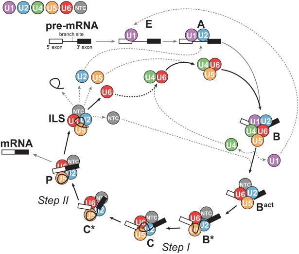
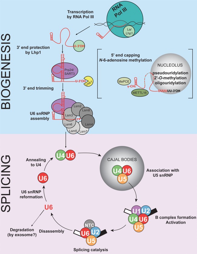
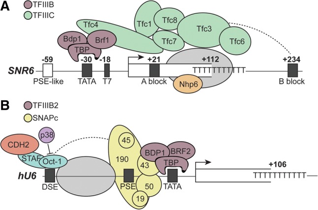
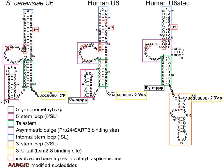
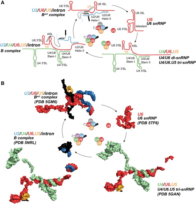
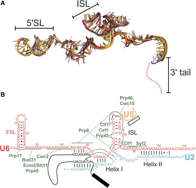
Removal of introns from precursor messenger RNA (pre-mRNA) and some noncoding transcripts is an essential step in eukaryotic gene expression. In the nucleus, this process of RNA splicing is carried out by the spliceosome, a multi-megaDalton macromolecular machine whose core components are conserved from yeast to humans. In addition to many proteins, the spliceosome contains five uridine-rich small nuclear RNAs (snRNAs) that undergo an elaborate series of conformational changes to correctly recognize the splice sites and catalyze intron removal. Decades of biochemical and genetic data, along with recent cryo-EM structures, unequivocally demonstrate that U6 snRNA forms much of the catalytic core of the spliceosome and is highly dynamic, interacting with three snRNAs, the pre-mRNA substrate, and >25 protein partners throughout the splicing cycle. This review summarizes the current state of knowledge on how U6 snRNA is synthesized, modified, incorporated into snRNPs and spliceosomes, recycled, and degraded.

Keywords: U6 gene transcription; U6 snRNA; U6 snRNP; spliceosome.


Figures

FIGURE 1.
FIGURE 1.
U6 snRNA is a core component of the spliceosome. The spliceosome is composed of small nuclear ribonucleoprotein particles (snRNPs) and a protein-only complex called the NTC, which are represented as colored circles. Additional transiently bound proteins (not shown) are also necessary for progression through the splicing cycle. The U1, U2, U4, U5, and U6 snRNPs consist of the small nuclear RNA (snRNA) for which they are named and associated proteins. The snRNPs and NTC undergo ordered assembly on the pre-mRNA and experience both conformational and compositional changes throughout the cycle. After splicing is complete, the snRNPs and NTC are released and reused (dotted lines).
FIGURE 2.
FIGURE 2.
Summary of the U6 lifecycle. Key steps in U6 snRNA biogenesis and assembly (top) and incorporation into the splicing cycle (bottom) are conserved in eukaryotes. Several additional modification steps, represented in brackets, occur in S. pombe and humans.
FIGURE 3.
FIGURE 3.
U6 snRNA gene promoter structure is divergent in eukaryotes. (A) U6 gene promoter structure in S. cerevisiae. The U6 gene is under the control of a Pol III Type II promoter, with an upstream TATA box, an internal A block, and downstream B block. The TFIIIC complex recognizes the A and B blocks and directs binding of the TFIIIB complex to the TATA box. Nhp6 also promotes transcription, but its binding site is uncertain. A possible nucleosome is indicated by a gray oval. (B) U6 promoter structure in humans. U6 synthesis is under the control of a Pol III Type III promoter, where promoter elements (in black) are exclusively upstream of the transcription start site. The TATA box is recognized by the TFIIIB2 complex, while the PSE is recognized by the SNAPc complex. Factors including OCT1, STAF, and CDH2 interact with the DSE. p38 inhibits Oct-1 binding to the DSE. A nucleosome between the DSE and PSE enhances Oct-1 and SNAPc binding.
FIGURE 4.
FIGURE 4.
Sequence and putative secondary structure of S. cerevisiae U6, human U6, and U6atac. The secondary structure of U6 from S. cerevisiae (left) is based on the structure of the U6 snRNP core (Montemayor et al. 2014), and includes the 5′SL, telestem, asymmetric bulge, ISL, and 3′ tail. The secondary structure of human U6 and U6atac within the U6 snRNP has not been experimentally determined, and is shown with secondary structure to mimic that of yeast U6. Human U6 contains a 5′SL, the ISL, and 3′ tail, and may contain an asymmetric bulge and telestem region. U6atac lacks a 5′SL but contains an additional 3′SL. Constitutively modified nucleotides are highlighted in red. Residues involved in base triples in the catalytic spliceosome (U2/U6.U5) are boxed in red.
FIGURE 5.
FIGURE 5.
U6 undergoes large conformational changes during the splicing cycle. (A) Cartoon of base-pairing throughout the splicing cycle. For simplicity, minimal interacting sequences of U2 and U5 snRNAs are shown. (B) Structure of U6 and its partners in different splicing complexes.
FIGURE 6.
FIGURE 6.
S. cerevisiae U6 does not undergo conformational changes during the transitions from Bact to ILS complexes. (A) Superimposition of U6 snRNA structures from the following S. cerevisiae spliceosomal complexes: Bact (PDB 5GM6, red and PDB 5LQW, salmon), C (PDB 5GMK, orange and PDB 5LJ5, light orange), C* (PDB 5WSG, lemon and PDB 5MQ0, lime), and the S. pombe ILS (PDB 3JB9, blue). (B) Secondary structure of U2/U6 in C* complex (PDB 5MQ0). Protein contacts to U6 are shown in green. Base triple interactions are shown by dashed lines.

References

    1. Abelson J, Blanco M, Ditzler MA, Fuller F, Aravamudhan P, Wood M, Villa T, Ryan DE, Pleiss JA, Maeder C, et al. 2010. Conformational dynamics of single pre-mRNA molecules during in vitro splicing. Nat Struct Mol Biol 17: 504–512. - PMC - PubMed
    1. Absmeier E, Wollenhaupt J, Mozaffari-Jovin S, Becke C, Lee CT, Preussner M, Heyd F, Urlaub H, Lührmann R, Santos KF, et al. 2015. The large N-terminal region of the Brr2 RNA helicase guides productive spliceosome activation. Genes Dev 29: 2576–2587. - PMC - PubMed
    1. Absmeier E, Santos KF, Wahl MC. 2016. Functions and regulation of the Brr2 RNA helicase during splicing. Cell Cycle 15: 3362–3377. - PMC - PubMed
    1. Achsel T, Brahms H, Kastner B, Bachi A, Wilm M, Lührmann R. 1999. A doughnut-shaped heteromer of human Sm-like proteins binds to the 3′-end of U6 snRNA, thereby facilitating U4/U6 duplex formation in vitro. EMBO J 18: 5789–5802. - PMC - PubMed
    1. Afroz T, Cienikova Z, Cléry A, Allain FH. 2015. One, two, three, four! How multiple RRMs read the genome sequence. Methods Enzymol 558: 235–278. - PubMed

Publication types

MeSH terms

LinkOut - more resources