. 2018 Jan 25;3(2):e98555.
doi: 10.1172/jci.insight.98555.

Mutations in Hnrnpa1 cause congenital heart defects

Affiliations

Mutations in Hnrnpa1 cause congenital heart defects

Zhe Yu et al. JCI Insight. .

Abstract

Incomplete penetrance of congenital heart defects (CHDs) was observed in a mouse model. We hypothesized that the contribution of a major genetic locus modulates the manifestation of the CHDs. After genome-wide linkage mapping, fine mapping, and high-throughput targeted sequencing, a recessive frameshift mutation of the heterogeneous nuclear ribonucleoprotein A1 (Hnrnpa1) gene was confirmed (Hnrnpa1ct). Hnrnpa1 was expressed in both the first heart field (FHF) and second heart field (SHF) at the cardiac crescent stage but was only maintained in SHF progenitors after heart tube formation. Hnrnpa1ct/ct homozygous mutants displayed complete CHD penetrance, including truncated and incomplete looped heart tube at E9.5, ventricular septal defect (VSD) and persistent truncus arteriosus (PTA) at E13.5, and VSD and double outlet right ventricle at P0. Impaired development of the dorsal mesocardium and sinoatrial node progenitors was also observed. Loss of Hnrnpa1 expression leads to dysregulation of cardiac transcription networks and multiple signaling pathways, including BMP, FGF, and Notch in the SHF. Finally, two rare heterozygous mutations of HNRNPA1 were detected in human CHDs. These findings suggest a role of Hnrnpa1 in embryonic heart development in mice and humans.

Keywords: Cardiovascular disease; Development; Genetics; Heart failure; Organogenesis.


Conflict of interest statement

Conflict of interest: The authors have declared that no conflict of interest exists.

Figures

Figure 1
Figure 1. Identification of a de novo CT deletion in Hnrnpa1.
(A) Chromosome 15–linked congenital heart defect (CHD) locus. The x axis represents the relative location of markers on a chromosome, and the y axis represents the –log(P) for nonparametric linkage–ALL (NPL-ALL) scores and BLOCK recessive scores. (B) Transmission disequilibrium test (TDT) for the combined data set from E9.5 and P0. We genotyped 40 more SNP markers for the region of chr15:90.7–100.6 Mb. Significant association with the heart defect phenotype was found starting from chr15:98.95 Mb, consistent with the NPL analysis in the embryonic lethality data set. We hypothesized the new CHD locus should be located beyond 98.95 Mb, where alleles from the C57BL/6N mutant were first shown to be predominately transmitted to affected individuals. We defined this segment, which independently assorted with the IIA genotype, as a breakpoint between Col2a1 and the new CHD locus. The breakpoint was shown to be flanked by markers rs8277842 at 98.6 Mb and rs3708604 at 98.95 Mb on chromosome 15. The green arrow pointing to the left indicates the position of Col2a1 and the blue arrow pointing to the right indicates the breakpoint. For both A and B, P < 0.001 was considered as significant. (C) Haplotype analysis in affected litters. The same region of the TDT analysis presented in chr15:90.8–100.6 Mb is shown. The haplotype information (154.4) is mainly inferred from rs6284372. In this pedigree, there are 8 F1, all bearing a heterozygous configuration for this segment. F2 collected from intercrossing F1 provide 3 main haplotypes: 154.4/154.4, 154.4/ICR-C14, and ICR-C14/ICR-C14. Among 53 individuals in F2, all 11 affected individuals was found to be carrying the 154.4/154.4 haplotype; while the unaffected individuals have a distribution of 5:24:13 for the 3 mentioned haplotype configurations, respectively. Markers do not appear to segregate in chr15:90.8–98.6 Mb, probably due to our selection of the IIA-null allele for a procollagen IIA–deficient mutant mouse line. Therefore, the haplotype for this region was inferred with an assumption that no recombination happened in the region with the pedigree under study. (D) Targeted sequencing of the 1.2-Mb candidate region detected a 2-base deletion in Hnrnpa1 gene. A screen shot from the IGV browser shows the deletion of CT in Hrnrnpa1 in a heterozygous mouse. (E) Sanger sequencing confirmed the presence of CT deletion in Hnrnpa1.
Figure 2
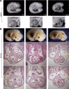
Figure 2. Hnrnpa1ct/ct mutant mice display severe congenital cardiac malformations at E9.5 and E13.5.
Hearts from 5 wild-type littermate controls, 5 Hnrnpa1+/ct heterozygous mutants, and 3 Hnrnpa1ct/ct homozygous mutants were sectioned and stained with hematoxylin and eosin. (A) A truncated and incomplete looped heart tube in Hnrnpa1ct/ct homozygous mutants at E9.5. Scale bar: 500 μm. Original magnification, ×5.1. (B) The E13.5 Hnrnpa1ct/ct homozygous mutant heart with PTA and VSD. RA, right atrium; LA, left atrium; RV, right ventricle; LV, left ventricle; AO, aorta; PA, pulmonary artery; IVS, interventricular septum; PTA, persistent truncus arteriosus; VSD, ventricular septal defect. Scale bar: 2 mm.
Figure 3
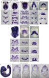
Figure 3. Whole-mount in situ analysis of wild-type embryos stained for Hnrnpa1 mRNA at different embryonic days.
Wild-type embryos were stained for Hnrnpa1 mRNA, and corresponding sections are shown from arterial pole to venous pole. At E8 and E8.25, staining for Isl1 and Nkx2.5 was also performed. Isl1 labels SHF cardiac progenitors, whereas Nkx2.5 labels both the FHF and SHF lineages. At each stage, results from 1 of 3 representative experiments are displayed. (A) E7; (B) E7.5; (C–E) E8 (2 somite pairs); (F–H) E8.25 (6 somite pairs); and (I) E9.5 (20 somite pairs). (A and B) Expression of Hnrnpa1 in the pre–cardiac crescent stages. Hnrnpa1 mRNA is detected in the ALPM (short arrow). (C) During the cardiac crescent stage, Hnrnpa1 mRNA is expressed in both differentiated myocardium (thin long arrow) and splanchnic mesoderm (wide long arrow). (F) After the formation of primitive heart tube, Hnrnpa1 mRNA maintains its strong expression in the splanchnic mesoderm but with much lower expression level in the differentiated myocardium. (I) In E9.5 wild-type embryo, Hnrnpa1 mRNA continues to be strongly expressed in the splanchnic mesoderm but not in the looped heart tube. FHF, first heart field; SHF, second heart field. Scale bar: 100 μm.
Figure 4
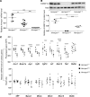
Figure 4. Expression of Hnrnpa1 and relative cardiac genes in Hnrnpa1ct/ct mutant embryos at E9.5. GAPDH was used as the internal control for qRT-PCR, and β-Actin was used as the loading control for Western blot, respectively.
(A) qRT-PCR results for E9.5 embryos. The Hnrnpa1 mRNA level in all 3 genotypes is shown. Six wild-type littermate controls, nine heterozygous mutants, and eight homozygous mutants were used. (B) Three wild-type littermate controls, three homozygous mutants, and four heterozygous mutants were used for Western blot analysis. (C) Total RNA was extracted from isolated pharyngeal region or heart tube respectively at E9.5. qRT-PCR was performed to monitor the expression of both the second heart field (SHF) and heart tube–specific cardiac genes. The number of Hnrnpa1+/+, Hnrnpa1+/ct, and Hnrnpa1ct/ct embryos in the SHF: 8, 5, and 9 for Fgf8, Fgf10, Isl1, Mef2c, and Tbx1; 8, 5, and 8 for Nkx2.5; 8, 6, and 9 for Acvr1, Bmpr1a, and Jag1, respectively. The number of Hnrnpa1+/+, Hnrnpa1+/ct, and Hnrnpa1ct/ct embryos in the heart tube: 8, 8, and 9 for Mlc2a, Mlc2v, and Nkx2.5; 11, 9, and 9 for Mef2c; 11, 11, and 9 for Myocd and SRF, respectively. The values represent mean ± SD in independent samples. **P < 0.01, ***P < 0.001, ****P < 0.0001 by unpaired 2-tailed t tests with Bonferroni correction.
Figure 5
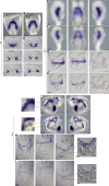
Figure 5. In situ analysis for Nkx2.5 demonstrates that Hnrnpa1ct/ct mutation leads to severe defects in both the FHF and SHF lineages.
Detection of Nkx2.5 mRNA by whole-mount in situ hybridization in both wild-type littermate controls and Hnrnpa1ct/ct homozygous mutants. Wild-type littermates and homozygous mutants are indicated by +/+ and ct/ct, respectively. Sections shown below are from arterial pole to venous pole. At each stage, results from 1 of 3 representative experiments are displayed. (A–D) E8 (3 somite pairs) embryos are shown in the ventral view, with corresponding sections. (E–H) E8.5 (9 somite pairs) embryos are shown from the right, ventral, and left, with sections from anterior to posterior poles. (I–L) E9.5 (25 somite pairs) embryos are shown from right to left lateral views, with corresponding sections. The arrows in each photo represent the regions with lower Nkx2.5 mRNA levels in homozygous mutants. A markedly changed expression pattern of Nkx2.5 was detected in differentiated myocardium (thin long arrow), foregut endoderm (short arrow), and splanchnic mesoderm (SHF, wide long arrow). FHF, first heart field; SHF, second heart field. Scale bar: 100 μm.
Figure 6
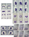
Figure 6. Altered expression pattern of Isl1 shows SHF-related defects in Hnrnpa1ct/ct mutants.
Whole-mount in situ hybridization analysis of Isl1 mRNA in Hnrnpa1ct/ct homozygous mutants and wild-type littermate controls. Wild-type embryos and Hnrnpa1 homozygous mutants are labeled by +/+ and ct/ct, respectively. At each stage, results from 1 of 3 representative experiments are displayed. (A, B, E, F, I, and J) Whole-mount in situ hybridization analysis for embryos at E8 (2 somite pairs), E8.5 (9 somite pairs), and E9.5 (25 somite pairs). (I and J) The expression of Isl1 in a small subset of SAN progenitors is lost in Hnrnpa1ct/ct homozygous mutants. (C, D, G, H, K, and L) Corresponding sections are shown below at different levels from arterial pole to venous pole. (C and D) Sections at the cardiac crescent stage (E8). (G and H) Sections are shown at OFT region, middle portion of the heart tube, and sinus venosus (E8.5). (K and L) Sections are shown from OFT to sinus venous at E9.5. The expression of Isl1 decreases markedly in the truncated DM of Hnrnpa1ct/ct homozygous mutants. (M and N) Decreased Isl1 mRNA or Isl1-expressing cardiac progenitors can be found in both splanchnic mesoderm (SHF, wide long arrow) and ventral foregut endoderm (short arrow). DM, dorsal mesocardium; OFT, outflow tract; SAN, sinoatrial node; SHF, second heart field. Scale bar: 100 μm.
Figure 7
Figure 7. KO of Hnrnpa1 impaired cardiomyocyte differentiation in vitro.
(A) Hnrnpa1 KO 1 and KO 2 mESC lines were established by the CRISPR/Cas9 method. PCR products of exon 6 in Nkx2.5-EGFP-mESCs (lane 2, 3 and 5, 425 bp), Hnrnpa1 KO 1 (lane 4, 343 bp), and KO 2 (lane 6, 404 bp) were shown. Sequencing and blasting results demonstrate that 82-bp and 21-bp fragments of exon6 of Hnrnpa1 are deleted in Hnrnpa1 KO 1 and KO 2, respectively, by the CRISPR/Cas9 technique. (B) For each group, in vitro differentiation was performed 3 times. mESCs were induced into cardiomyocytes. Embryonic bodies (EBs) formed 2–3 days later. Loosely aggregated EBs were observed in Hnrnpa1 KO 1 and KO 2 groups (day 3–5). After attachment of EBs to the plate at day 6, outgrowths were impaired in both Hnrnpa1 KO 1 and KO 2 groups (day 7–day 9). Scale bar: 200 μm. (C) The percentage of Nkx2.5-EGFP–positive cells was analyzed by flow cytometry at day 7. Compared with the Nkx2.5-EGFP-mESC group, significantly reduced Nkx2.5-EGFP–positive cells were detected in both Hnrnpa1 KO 1 and KO 2 groups. Data in C are presented as mean ± SD, with n = 3 per group. **P < 0.01; ***P < 0.001 by unpaired 2-tailed t tests with Bonferroni correction.
Figure 8
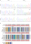
Figure 8. HNRNPA1 mutations in human congenital heart disease patients.
(A) Mutations in 2 trio families. (B) Both Gly203 and Gly283 locate in the conserved region of HNRNPA1 protein. CHD, congenital heart disease.

Similar articles

Cited by

References

    1. Fahed AC, Gelb BD, Seidman JG, Seidman CE. Genetics of congenital heart disease: the glass half empty. Circ Res. 2013;112(4):707–720. doi: 10.1161/CIRCRESAHA.112.300853. - DOI - PMC - PubMed
    1. Hoffman JI, Kaplan S. The incidence of congenital heart disease. J Am Coll Cardiol. 2002;39(12):1890–1900. doi: 10.1016/S0735-1097(02)01886-7. - DOI - PubMed
    1. Sifrim A, et al. Distinct genetic architectures for syndromic and nonsyndromic congenital heart defects identified by exome sequencing. Nat Genet. 2016;48(9):1060–1065. doi: 10.1038/ng.3627. - DOI - PMC - PubMed
    1. Nora JJ. Multifactorial inheritance hypothesis for the etiology of congenital heart diseases. The genetic-environmental interaction. Circulation. 1968;38(3):604–617. doi: 10.1161/01.CIR.38.3.604. - DOI - PubMed
    1. Lalani SR, Belmont JW. Genetic basis of congenital cardiovascular malformations. Eur J Med Genet. 2014;57(8):402–413. doi: 10.1016/j.ejmg.2014.04.010. - DOI - PMC - PubMed

Publication types

MeSH terms

Substances