. 2018 Jan 25;3(2):e96591.
doi: 10.1172/jci.insight.96591.

Lymphatic function measurements influenced by contrast agent volume and body position

Affiliations

Lymphatic function measurements influenced by contrast agent volume and body position

Echoe M Bouta et al. JCI Insight. .

Abstract

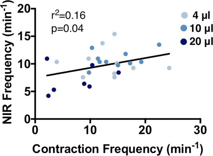
Several imaging modalities have been used to assess lymphatic function, including fluorescence microscopy, near-infrared fluorescence (NIRF) imaging, and Doppler optical coherence tomography (DOCT). They vary in how the mouse is positioned, the invasiveness of the experimental setup, and the volume of contrast agent injected. Here, we present how each of these experimental parameters affects functional measurements of collecting lymphatic vessels. First, fluorescence microscopy showed that supine mice have a statistically lower contraction frequency compared with mice sitting upright. To assess the effect of different injection volumes on these endpoints, mice were injected with 4, 10, or 20 μl of dye. The lowest frequencies were observed after 20-μl injections. Interestingly, lymph-flow DOCT revealed that although there was lower contraction frequency in mice injected with 20 μl versus 4 μl, mice showed a higher volumetric flow with a 20-μl injection. This indicates that contraction frequency alone is not sufficient to understand lymphatic transport. Finally, NIRF revealed that removing the skin reduced contraction frequency. Therefore, this study reveals how sensitive these techniques are to mouse position, removal of skin, and dye volume. Care should be taken when comparing results obtained under different experimental conditions.

Keywords: Lymph; Vascular Biology.


Conflict of interest statement

Conflict of interest: The authors have declared that no conflict of interest exists.

Figures

Figure 1
Figure 1. Mice in the supine position demonstrated lower lymphatic contraction frequency compared with mice sitting upright by fluorescence imaging of lymphatic contraction.
Mice were injected with 3 μl of FITC-dextran and underwent lymphatic contraction analysis both while sitting upright and in the supine position. The order of the positioning was randomized. Subsequently, analysis of ejection fraction (A) and contraction frequency (B) was performed. Notice that the contraction frequency is lower when mice are in the supine position versus sitting upright (B, *P < 0.05 by a paired t test, n = 6).
Figure 2
Figure 2. Injection volume and skin removal affects frequency by NIRF imaging.
Mice injected with a 50:50 mixture of FITC-dextran and indocyanine green (ICG) underwent near-infrared fluorescence (NIRF) imaging with subsequent fluorescence contraction imaging while sitting upright or supine. (AC) Intact representative NIRF images are shown after mice were injected with (A) 4 μl, (B) 10 μl, or (C) 20 μl (Supplemental Videos 1–3, respectively). (D) NIR frequency is affected by volume injected. In general, removing the skin and superfusing with saline decreases the contraction frequency. Data presented as the mean ± SEM. *P < 0.05 by 2-way ANOVA with a Tukey’s multiple comparison test; #P < 0.05 by a paired t test; n = 7–11. Scale bar: 5 mm.
Figure 3
Figure 3. Injection volume affects contraction frequency.
The same mice in Figure 2 underwent fluorescence contraction imaging following near-infrared fluorescence (NIRF) imaging. Representative FITC images of lymphatic vessels from mice injected with 4 μl, 10 μl, or 20 μl of dye are shown (AC, respectively). (D) Mice injected with 20 μl showed larger lymphatic vessel diameters. (E) Ejection fraction and (F) contraction frequency were quantified from intravital videos of lymphatic wall motion (Supplemental Videos 4–6). Mice injected with 20 μl showed lower contraction frequency compared with mice injected with 4 and 10 μl. Data presented as the mean ± SEM. *P < 0.05 by ANOVA with a Fisher’s LSD post-hoc test; #P < 0.05 by Mann-Whitney test; n = 6–11. Scale bar: 50 μm.
Figure 4
Figure 4. NIR and direct measurement of lymphatic contraction are correlated.
Analysis was performed on mice from Figures 2 and 3. A weak correlation was found between near infrared (NIR) with intact skin and fluorescence intravital contraction frequency measurements with the mouse sitting upright (P value found by linear regression, n = 6–11). These conditions are the ones most commonly found in the literature for each technique.
Figure 5
Figure 5. Larger injection volume increases volumetric flow in lymphatic vessels.
(AD) Mice injected with 4 μl or 20 μl of a 50:50 mixture of FITC-dextran and indocyanine green (ICG) in the footpad 1 hour prior to Doppler optical coherence tomography (DOCT) imaging. Representative images of mice injected with (A and B) 4 μl or (C and D) 20 μl during low volumetric flow (A and C) versus a spike in flow (B and D). Mean volumetric flow over the cross section for (E) 4 μl and (F) 20 μl averaged over 300 seconds to give the average volumetric flow (G, n = 4–5, *P < 0.05 by Mann-Whitney test). Note the larger volumetric flow in mice injected with 20 μl, even between lymphatic contractions (C, quantified in F and G). However, no difference was found in the frequency of peaks of volumetric flow (H). Scale bars: 20 μm. Data presented as the mean ± SEM.

References

    1. Iliff JJ, et al. A paravascular pathway facilitates CSF flow through the brain parenchyma and the clearance of interstitial solutes, including amyloid β. Sci Transl Med. 2012;4(147):147ra111. - PMC - PubMed
    1. Li J, et al. Efficacy of B cell depletion therapy for murine joint arthritis flare is associated with increased lymphatic flow. Arthritis Rheum. 2013;65(1):130–138. doi: 10.1002/art.37709. - DOI - PMC - PubMed
    1. Aspelund A, et al. The Schlemm’s canal is a VEGF-C/VEGFR-3-responsive lymphatic-like vessel. J Clin Invest. 2014;124(9):3975–3986. doi: 10.1172/JCI75395. - DOI - PMC - PubMed
    1. Wiig H, et al. Immune cells control skin lymphatic electrolyte homeostasis and blood pressure. J Clin Invest. 2013;123(7):2803–2815. doi: 10.1172/JCI60113. - DOI - PMC - PubMed
    1. Klotz L, et al. Cardiac lymphatics are heterogeneous in origin and respond to injury. Nature. 2015;522(7554):62–67. doi: 10.1038/nature14483. - DOI - PMC - PubMed

Publication types