Review
. 2017 Dec 10;4(4):23.
doi: 10.3390/jcdd4040023.

The Role of Cerl2 in the Establishment of Left-Right Asymmetries during Axis Formation and Heart Development

Affiliations
Review

The Role of Cerl2 in the Establishment of Left-Right Asymmetries during Axis Formation and Heart Development

José A Belo et al. J Cardiovasc Dev Dis. .

Abstract

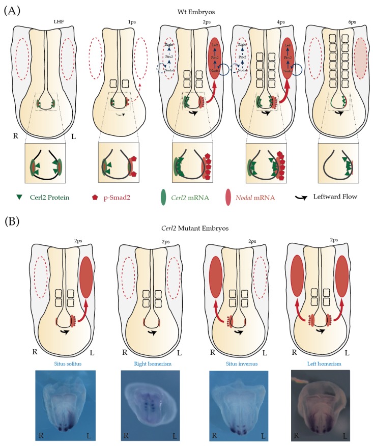
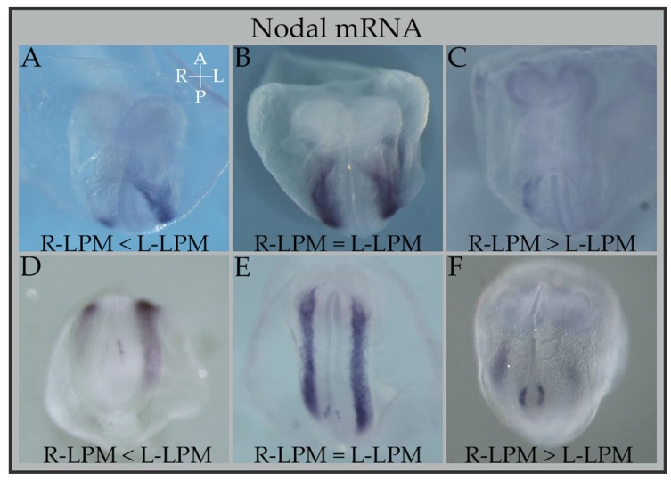
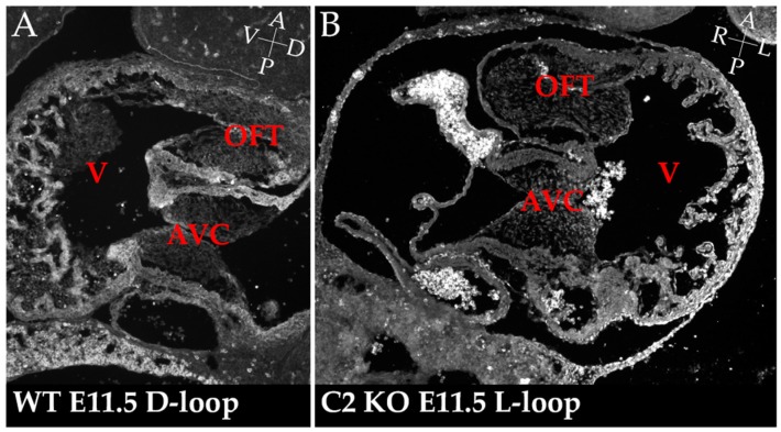
The formation of the asymmetric left-right (LR) body axis is one of the fundamental aspects of vertebrate embryonic development, and one still raising passionate discussions among scientists. Although the conserved role of nodal is unquestionable in this process, several of the details around this signaling cascade are still unanswered. To further understand this mechanism, we have been studying Cerberus-like 2 (Cerl2), an inhibitor of Nodal, and its role in the generation of asymmetries in the early vertebrate embryo. The absence of Cerl2 results in a wide spectrum of malformations commonly known as heterotaxia, which comprises defects in either global organ position (e.g., situs inversus totalis), reversed orientation of at least one organ (e.g., situs ambiguus), and mirror images of usually asymmetric paired organs (e.g., left or right isomerisms of the lungs). Moreover, these laterality defects are frequently associated with congenital heart diseases (e.g., transposition of the great arteries, or atrioventricular septal defects). Here, reviewing the knowledge on the establishment of LR asymmetry in mouse embryos, the emerging conclusion is that as necessary as is the activation of the Nodal signaling cascade, the tight control that Cerl2-mediates on Nodal signaling is equally important, and that generates a further regionalized LR genetic program in the proper time and space.

Keywords: Cerl2; LR-asymmetry; Nodal signaling; congenital heart diseases.


Conflict of interest statement

The authors declare no conflict of interest.

Figures

Figure 1
Figure 1
Sequential Nodal activity in left-right asymmetry at the mouse node during early somitogenesis. Nodal expression is represented in light red oval, and Cerl2 expression in light green oval. Cerberus-like 2 (Cerl2) protein is illustrated in green triangles, and the readout of Nodal signaling, pSmad2/3, is indicated in red pentagons. The expression of Nodal in the lateral plate mesoderm (LPM) of mouse embryos is represented by the filled red oval. Dashed to thicker lines indicate increase in intensity. (A) At the one-somite stage, as soon as the cilia at the node began to rotate generating a weak leftward flow, the asymmetric expression of Cerl2 (L < R) is established. This local reduction of Cerl2 mRNA (and Cerl2 protein) in the perinodal cells on the left side of the node results in an increase of active Nodal signal, i.e., pSmad2/3. At the 2-somite stage, Cerl2 protein (green triangles) localizes and prevents the activation of Nodal genetic circuitry on the right side of the embryo (dashed red oval). Later, due to nodal flow, Cerl2 right-to-left translocation shutdowns Nodal activity in the node and consequently affects the activity of Nodal in the LPM (dashed red oval). (B) In the absence of Cerl2 in the node, the expression of Nodal in LPM becomes randomized.
Figure 2
Figure 2
Differential Anterior-posterior bilaterality of Nodal expression in Cerl2 knock-out (KO) embryos. Bilateral expression of Nodal in the lateral plate mesoderm of Cerl2 KO embryos (50% display bilateral expression: left isomerism) at about E8.0–E8.5 (from 2 to 7 somite pairs). This bilateral expression can be equal in the left or right LPM occupying the totality of both LPMs (panels B, E); or it can be bilateral only in a more posterior or anterior area (panels A, C, D, F). This partial bilaterality can be either more preponderant on the R-LPM (panels C and F) or on the L-LPM (panels A, D). Partial bilaterality is set early on as can be seen in panel F (2–3 somite embryo), and will last until the end of Nodal’s expression in the LPM, at about 7 somites (as can be seen in panel C). Embryo axes coordinates are present in panel A, in white letters: A: anterior; P: posterior; R: Right; L: Left. R-LPM (right—Lateral Plate Mesoderm); L-LPM (left—Lateral Plate Mesoderm).
Figure 3
Figure 3
Incorrectly looped hearts in Cerl2 mutant embryos. Sagittal section of a wild-type (WT) (A) and coronal section of Cerl2 mutant (B) hearts after looping stages, at developmental stage E11.5. These sections made in different orientations (sagittal and coronal) show exactly the same anatomical features: the common ventricular chamber (V) and span the outflow tract (OFT) and atrioventricular canal (AVC) cushions of E11.5 mouse embryos. This shows that in the WT there was a correct rightward (D-loop) (A), and in the mutant the looping occurred at a different angle, a leftward one (L-Loop) (B). Embryo axes coordinates are present in panels A and B, in white letters: A: anterior; P: posterior; R: Right, L: Left; D: Dorsal; V: Ventral.

References

    1. Cooke J. Developmental mechanism and evolutionary origin of vertebrate left/right asymmetries. Biol. Rev. 2004;79:377–407. doi: 10.1017/S1464793103006298. - DOI - PubMed
    1. Kilner P.J., Yang G.Z., Wilkes A.J., Mohiaddin R.H., Firmin D.N., Yacoub M.H. Asymmetric redirection of flow through the heart. Nature. 2000;404:759–761. doi: 10.1038/35008075. - DOI - PubMed
    1. Ramsdell A.F. Left-right asymmetry and congenital cardiac defects: Getting to the heart of the matter in vertebrate left-right axis determination. Dev. Biol. 2005;288:1–20. doi: 10.1016/j.ydbio.2005.07.038. - DOI - PubMed
    1. Hamada H., Tam P.P. Mechanisms of left-right asymmetry and patterning: Driver, mediator and responder. F1000Prime Rep. 2014;6:110. doi: 10.12703/P6-110. - DOI - PMC - PubMed
    1. Levin M. Left-right asymmetry in embryonic development: A comprehensive review. Mech. Dev. 2005;122:3–25. doi: 10.1016/j.mod.2004.08.006. - DOI - PubMed

LinkOut - more resources