. 2018 Jan 24;8(1):1513.
doi: 10.1038/s41598-018-19419-6.

An optimised protocol for platelet-rich plasma preparation to improve its angiogenic and regenerative properties

Affiliations

An optimised protocol for platelet-rich plasma preparation to improve its angiogenic and regenerative properties

Julia Etulain et al. Sci Rep. .

Abstract

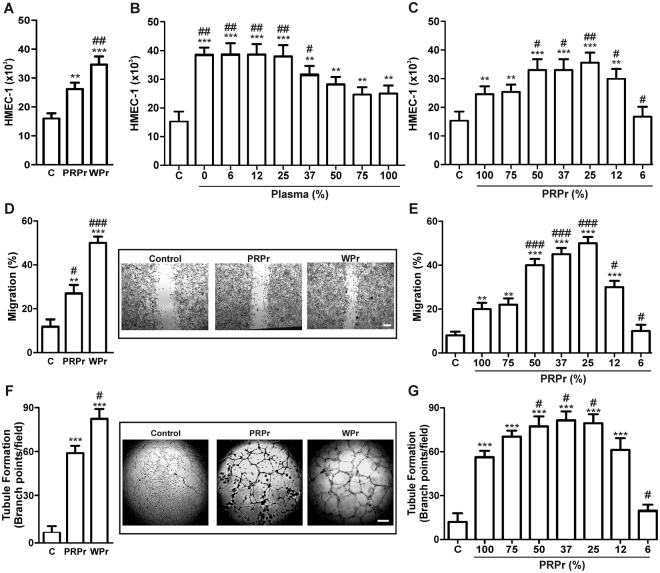
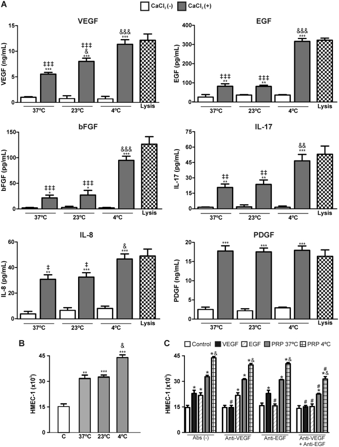
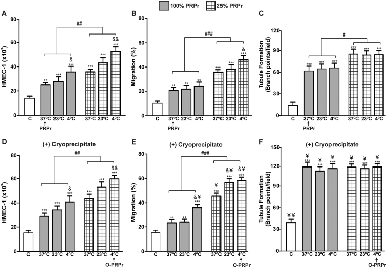
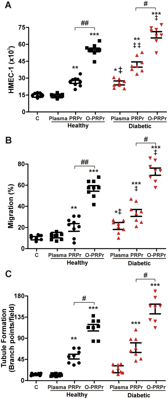
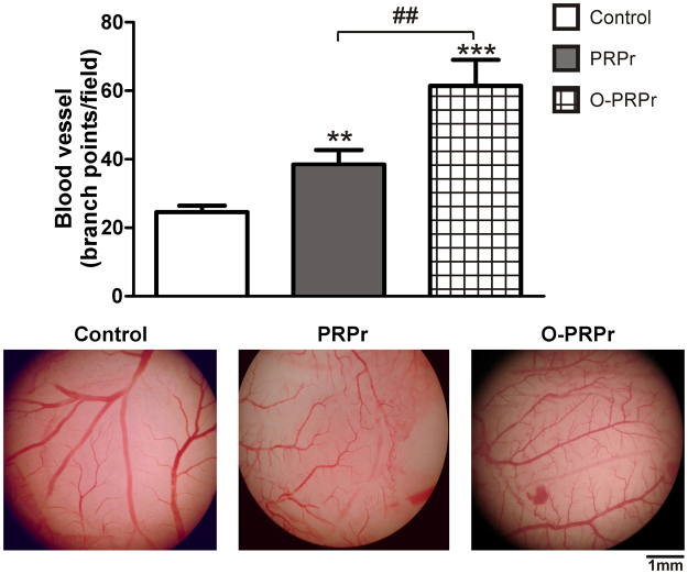
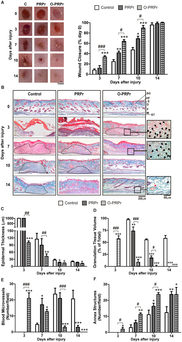
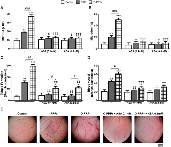
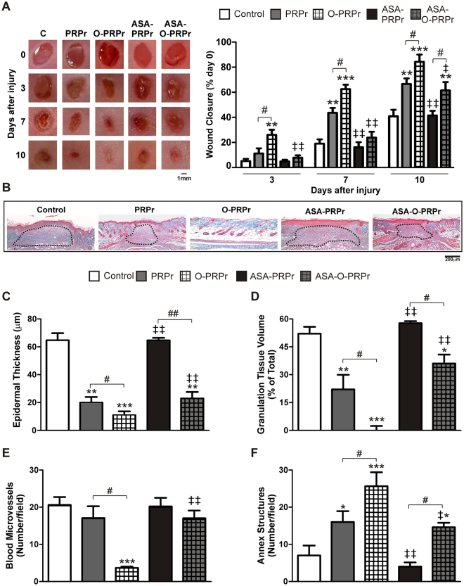
Although platelet-rich plasma (PRP) is used as a source of growth factors in regenerative medicine, its effectiveness remains controversial, partially due to the absence of PRP preparation protocols based on the regenerative role of platelets. Here, we aimed to optimise the protocol by analysing PRP angiogenic and regenerative properties. Three optimising strategies were evaluated: dilution, 4 °C pre-incubation, and plasma cryoprecipitate supplementation. Following coagulation, PRP releasates (PRPr) were used to induce angiogenesis in vitro (HMEC-1 proliferation, migration, and tubule formation) and in vivo (chorioallantoic membrane), as well as regeneration of excisional wounds on mouse skin. Washed platelet releasates induced greater angiogenesis than PRPr due to the anti-angiogenic effect of plasma, which was decreased by diluting PRPr with saline. Angiogenesis was also improved by both PRP pre-incubation at 4 °C and cryoprecipitate supplementation. A combination of optimising variables exerted an additive effect, thereby increasing the angiogenic activity of PRPr from healthy donors and diabetic patients. Optimised PRPr induced faster and more efficient mouse skin wound repair compared to that induced by non-optimised PRPr. Acetylsalicylic acid inhibited angiogenesis and tissue regeneration mediated by PRPr; this inhibition was reversed following optimisation. Our findings indicate that PRP pre-incubation at 4 °C, PRPr dilution, and cryoprecipitate supplementation improve the angiogenic and regenerative properties of PRP compared to the obtained by current methods.


Conflict of interest statement

The authors declare that they have no competing interests.

Figures

Figure 1
Figure 1
Plasma inhibits angiogenesis mediated by platelets. (A) HMEC-1 (15 × 103) were incubated with saline plus FBS 2% (control), PRPr or WPr; (B) WPr supplemented with increasing % of plasma; or (C) PRPr diluted with saline, and endothelial cell proliferation was determined after 24 h by acid phosphatase activity assay. (D,E) Migration into scratched monolayers of endothelial cells and (F,G) tubule formation in Matrigel-coated wells was induced, as indicated, with WPr and PRPr (diluted or not). Images are representative of four to seven independent experiments (magnification 40X, scale bars: 200 μm). (n = 4–7; **P < 0.01, ***P < 0.001 vs. control; #P < 0.05, ##P < 0.01, ###P < 0.001 vs. PRP 100%).
Figure 2
Figure 2
Cold preconditioning promotes the release of growth factors and cytokines from platelets. (A) PRP was incubated at 37 °C, 23 °C, or 4 °C for 30 min, and the levels of VEGF, EGF, bFGF, IL-17, IL-8, and PDGF in releasates of PRP before clotting (CaCl2−) or after clotting (CaCl2+) were determined by ELISA. Total intra-platelet levels were measured in platelet lysates. (B) PRP was incubated at 37 °C, 23 °C, or 4 °C for 30 min, clotted with CaCl2 (22 mM) for 40 min, and releasates were used to induce HMEC-1 (15 × 103) proliferation. Saline plus FBS 2% was used as control. (C) HMEC-1 (15 × 103) were incubated, individually or in combinations, with anti-human VEGFR2/KDR/Flk-1 (10 µg/ml), anti-human-EGFR (40 µg/ml) or irrelevant IgG, for 30 min. Next, endothelial proliferation was induced by addition of recombinant VEGF (20 ng/ml), EGF (20 ng/ml) or with PRPr preincubated at 37 °C or 4 °C and then clotting was induced with CaCl2 (22 mM). Saline plus FBS 2% was used as control (n = 4–5, *P < 0.05, **P < 0.01, ***P < 0.001 vs. unstimulated; &P < 0.05, &&P < 0.01, &&&P < 0.001 vs. 37 °C; P < 0.05, ‡‡P < 0.01, ‡‡‡P < 0.001 vs. lysis; #P < 0.05 vs. without neutralizing antibodies (Abs)). VEGF: vascular endothelial growth factor; EGF: epidermal growth factor; bFGF: basic fibroblast growth factor; PDGF: platelet derived growth factor; IL: interleukin.
Figure 3
Figure 3
PRP cold preconditioning, PRPr dilution, and plasma cryoprecipitate supplementation enhance angiogenesis mediated by PRP. PRP was incubated at 37 °C, 23 °C, or 4 °C for 30 min and then clotted by the addition of CaCl2 (22 mM) for 40 min. HMEC-1 (15 × 103) were incubated with PRPr pure (100%) or diluted with saline (25%). Saline containing 2% FBS was used as the control. Endothelial proliferation, migration, and tubule formation were determined in vitro without (A–C) or with plasma cryoprecipitate supplementation (D–F). (n = 5; **P < 0.01, ***P < 0.001 vs. control; #P < 0.05, ##P < 0.01, ###P < 0.001 vs. PRP 100%; &P < 0.05; &&P < 0.01 vs. 37 °C; ¥P < 0.05 vs. without plasma cryoprecipitate).
Figure 4
Figure 4
Angiogenesis mediated by PRPr from diabetic patients is increased by optimisation. Platelet-poor-plasma (plasma) and PRP were obtained from blood samples obtained from healthy or diabetic patients. PRPr was prepared as non-optimised (PRPr: PRP pre-incubated at 37 °C; PRPr without dilution or supplements) or optimised PRP (O-PRPr: PRP pre-incubated at 4 °C; PRPr diluted and supplemented with plasma cryoprecipitate). Saline containing 2% FBS was used as the control. (A) Endothelial proliferation, (B) migration, and (C) tubule formation were determined in vitro (n = 8 per group; **P < 0.01, ***P < 0.001 vs. control; #P < 0.05, ##P < 0.01, ###P < 0.001 vs. PRPr; P < 0.05, ‡‡&P < 0.01 vs. healthy same treatment).
Figure 5
Figure 5
Optimisation of PRP increases angiogenesis in vivo. Paper. filters embedded with PRPr or O-PRPr were placed on the CAM. Saline was used as the control. After 24 h, filters were removed and photographed under 2X magnification. The number of blood vessel branch points per field was analysed using the ImageJ software (n = 35–46; **P < 0.01, ***P < 0.001 vs. control; ##P < 0.01 vs. PRPr).
Figure 6
Figure 6
PRP optimisation accelerates mouse skin regeneration. Four round full-thickness excisional wounds, 3 mm in diameter, were generated in the back skin of female BALB/c mice (8–10 weeks old). PRPr or O-PRPr obtained from other mice was injected subcutaneously in the periphery of the wounds. Saline was used as the control. Healing was analysed at 3, 7, 10, and 14 days post-injury. (A) Wounds were photographed, and the perimeter of the wound area was determined using the ImageJ software and expressed as a percentage of the area on day 0. (B) Skin biopsies were stained with Masson’s trichrome. Images were captured using an inverted microscope. (C) Epidermal thickness, (D) granulation tissue volume (dotted lines), (E) immature blood microvessels containing intraluminal erythrocytes, and (F) annex structures (hair follicles and sebaceous glands) were quantified using the ImageJ software. SG, sebaceous gland; HF, hair follicle; E, epidermis; D, dermis; F, fat layer; M, muscle layer; ST, subcutaneous tissue. (Magnification 100× and 200× , n = 4–15; *P < 0.05, **P < 0.01, ***P < 0.001 vs. control; #P < 0.05, ##P < 0.01, ###P < 0.001 vs. PRPr).
Figure 7
Figure 7
Angiogenesis mediated by PRPr is inhibited by ASA and partially reversed by optimisation. PRP was incubated with ASA (0.1 or 0.5 mM) for 30 min, and then PRPr or O-PRPr was obtained and used to induce angiogenesis in vitro: (A) Endothelial proliferation, (B) migration, (C) tubule formation, and in vivo (D) blood vessel ramification over the CAM. (E) Microscopic CAM images represent one independent experiment. (n = 7–25, *P < 0.05, **P < 0.01, ***P < 0.001 vs. control; #P < 0.05, ##P < 0.01, ###P < 0.001 vs. PRPr; P < 0.05, ‡‡P < 0.01, ‡‡‡P < 0.001 vs. the same treatment without ASA).
Figure 8
Figure 8
Inhibition of PRP-regenerative activity by ASA is partially compensated for by optimisation. Four round full-thickness excisional wounds, 3 mm in diameter, were generated in the back skin of female BALB/c mice (8–10 weeks old). PRPr or O-PRPr obtained from ASA- or non-ASA- treated mice was injected into the wounds generated in ASA-treated mice. Injection with saline was used as the control. The healing process was analysed at 3, 7, and 10 days post-injury. (A) Wounds were photographed, and the perimeter of the wound area was determined using the ImageJ software and expressed as a percentage of the area on day 0. (B) Skin biopsies were obtained at day 10 and stained with Masson’s trichrome. Images were captured using an inverted microscope. (C) Epidermal thickness, (D) granulation tissue volume (dotted lines), (E) immature blood microvessels containing intraluminal erythrocytes, and (F) annex structures (hair follicles and sebaceous glands) were quantified using the ImageJ software. (Magnification 100X, n = 7; *P < 0.05, **P < 0.01, ***P < 0.001 vs. control; #P < 0.05, ##P < 0.01 vs. PRPr; P < 0.05, ‡‡P < 0.01 vs. the same treatment without ASA).

References

    1. Rittie L. Cellular mechanisms of skin repair in humans and other mammals. J Cell Commun Signal. 2016;10:103–120. doi: 10.1007/s12079-016-0330-1. - DOI - PMC - PubMed
    1. Xue M, Jackson CJ. Extracellular Matrix Reorganization During Wound Healing and Its Impact on Abnormal Scarring. Adv Wound Care (New Rochelle) 2015;4:119–136. doi: 10.1089/wound.2013.0485. - DOI - PMC - PubMed
    1. Briquez PS, Hubbell JA, Martino MM. Extracellular Matrix-Inspired Growth Factor Delivery Systems for Skin Wound Healing. Adv Wound Care (New Rochelle) 2015;4:479–489. doi: 10.1089/wound.2014.0603. - DOI - PMC - PubMed
    1. Powers JG, Higham C, Broussard K, Phillips TJ. Wound healing and treating wounds: Chronic wound care and management. J Am Acad Dermatol. 2016;74:607–625. doi: 10.1016/j.jaad.2015.08.070. - DOI - PubMed
    1. De Pascale MR, Sommese L, Casamassimi A, Napoli C. Platelet derivatives in regenerative medicine: an update. Transfus Med Rev. 2015;29:52–61. doi: 10.1016/j.tmrv.2014.11.001. - DOI - PubMed

Publication types