. 2018 Jan 24;9(1):353.
doi: 10.1038/s41467-017-02763-y.

T cells specific for post-translational modifications escape intrathymic tolerance induction

Affiliations

T cells specific for post-translational modifications escape intrathymic tolerance induction

Bruno Raposo et al. Nat Commun. .

Abstract

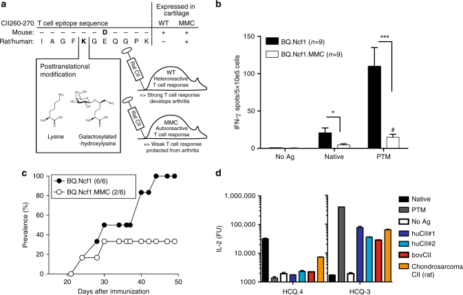
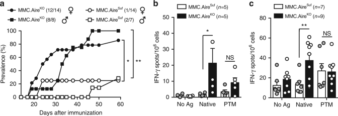
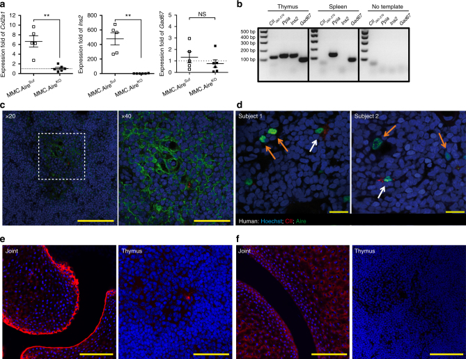
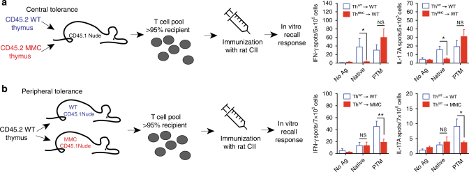
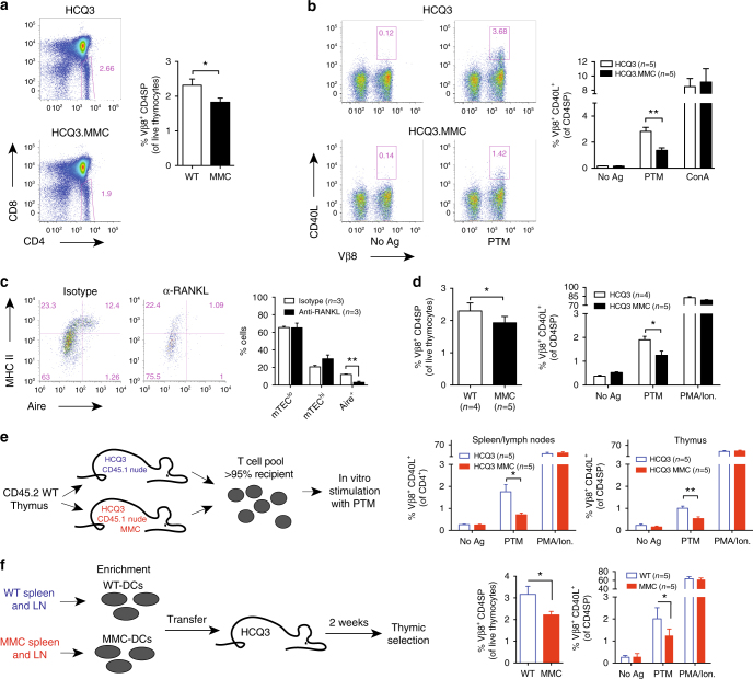
Establishing effective central tolerance requires the promiscuous expression of tissue-restricted antigens by medullary thymic epithelial cells. However, whether central tolerance also extends to post-translationally modified proteins is not clear. Here we show a mouse model of autoimmunity in which disease development is dependent on post-translational modification (PTM) of the tissue-restricted self-antigen collagen type II. T cells specific for the non-modified antigen undergo efficient central tolerance. By contrast, PTM-reactive T cells escape thymic selection, though the PTM variant constitutes the dominant form in the periphery. This finding implies that the PTM protein is absent in the thymus, or present at concentrations insufficient to induce negative selection of developing thymocytes and explains the lower level of tolerance induction against the PTM antigen. As the majority of self-antigens are post-translationally modified, these data raise the possibility that T cells specific for other self-antigens naturally subjected to PTM may escape central tolerance induction by a similar mechanism.


Conflict of interest statement

The authors declare no competing financial interests.

Figures

Fig. 1
Fig. 1
Summary of the autologous CIA model. a The MMC mouse expresses the immunodominant T cell epitope of heterologous (rat/human) CII as a self-antigen in the cartilage. A D266E amino acid substitution is the only difference between mouse and heterologous CII. A lysine at position 264 can become post-translationally modified by hydroxylation (not shown) and subsequent glycosylation with a monosaccharide. These modifications are recognized by distinct T cell clones (shown in d). MMC mice are immunized with heterologous CII (in this study rat CII) in order to induce an autoreactive T cell response. b The Ncf1 mutation was inserted onto the BQ background in order to render arthritis susceptibility in MMC mice. IFN-γ ELISPOT of lymph node cells isolated 10 days after immunization with rat CII and following re-stimulation with the non-modified (native) or glycosylated (PTM) CII peptide are shown. Bars indicate the mean number of spots ± SEM. #Statistical significance (p = 0.0195) between native and PTM responses in BQ.Ncf1.MMC mice, using a Wilcoxon's test. c Arthritis susceptibility of MMC mice in the BQ.Ncf1 background is reduced but not completely abrogated, when compared to non-MMC littermates. Number in brackets indicates cumulative number of animals that developed arthritis over total number of animals. d IL-2 production of T cell hybridoma clones specific to either the non-modified (HCQ.4) or PTM (HCQ.3) variants of the CII260–270 epitope after stimulation with the different peptides as well as whole CII protein obtained from different species (hu, human (two independent preparations/samples); bov, bovine (calf) CII from joint cartilage. The chondrosarcoma is a rat tumor line that was used as a positive control as it produces CII that is very heterogeneous in terms of PTMs). Mann–Whitney U test was used in ELISPOT assays. *p < 0.05; ***p < 0.001
Fig. 2
Fig. 2
Tolerance to the non-modified CII260–270 epitope is lost after Aire deficiency. Loss of tolerance to CII occurs in an Aire-dependent way and specifically affects T cells specific for the non-modified CII260–270 epitope. a Arthritis prevalence in MMC.AireSuf and MMC.AireKO male and female mice after immunization with rat CII. b, c ELISPOT data from in vitro recall responses of pooled lymph node and spleen cells from indicated number of MMC.AireSuf and MMC.AireKO mice 10 days (b) or 10 weeks (c) after CII immunization. Cells were stimulated with the non-modified and PTM CII260–270 peptide or left unstimulated (No Ag). Values shown are the mean ± SEM number of spots recorded for IFN-γ producing cells. Fisher′s exact test was used to calculate significance of arthritis prevalence, whereas Mann–Whitney U test was used in ELISPOT assays. *p < 0.05; **p < 0.01; NS not significant
Fig. 3
Fig. 3
The CII epitope is expressed in thymic stromal cells of mice and humans. a cDNA derived from thymi of MMC.AireSuf (n = 5) and MMC.AireKO (n = 6) was prepared and expression of CII (Col2a1) was determined by quantitative RT-PCR. Aire-dependent (Ins2) and Aire-independent (Gad67) genes were used as controls. Data were normalized to the expression of cyclophilin A (Ppia) and calibrated with one MMC.AireKO sample. Mean ± SEM is shown. b Qualitative expression analysis of the immunodominant T cell epitope CII260–270 by RT-PCR on cDNA prepared from whole thymi of 3-week-old mice. As negative control, cDNA was prepared from spleen cells of wild-type mice. Ins2 and Gad67 were used as controls for genes expressed either in the thymus alone or in both thymus and spleen, respectively. Ppia was used as housekeeping gene, whereas no template samples were used as negative controls. c MMC thymus stained for DNA (Hoechst, in blue), keratin 5 (in green), and CII (mAb cocktail, in red). Scale bar in left panel, 100 μm; in right panel, 50 μm. d Human thymus sections from two different subjects stained with Hoechst (in blue), anti-AIRE (in green), and anti-CII (in red). Orange arrows indicate thymic epithelial cells positive for AIRE alone. White arrows indicate thymic epithelial cell positive for both AIRE and CII. Scale bar indicates 20 μm. (e) Stain of joint and thymus with Hoechst (blue) and anti-CII (mAb cocktail, red) or (f) Hoechst (blue) and anti-PTM CII (T8 mAb, red), from an MMC mouse. Scale bars indicate 100 μm for left panel of e and both panels of f, and 50 μm in right panel of e. p values were calculated using Mann–Whitney U test. **p < 0.01; NS not significant
Fig. 4
Fig. 4
mTEC-mediated central tolerance to self-CII is limited to the native epitope. a Four-week-old nude mice were grafted with neonate thymi from either WT or MMC donors (n = 5/group). Eleven weeks later, mice were immunized with rat CII and draining lymph node cells were re-stimulated in vitro with different CII peptides. b MMC-positive (n = 8) and MMC-negative (n = 6) nude mice were grafted with neonate thymi from wild-type donors. Eleven weeks later, recipients were immunized with rat CII and draining lymph node cells were re-stimulated in vitro with different CII peptides. Values shown are the mean ± SEM number of spots recorded for IFN-γ-producing and IL-17A-producing cells. p values were calculated using Mann–Whitney U test. *p < 0.05; **p < 0.01; NS not significant
Fig. 5
Fig. 5
Peripheral antigen migration mediates thymic tolerance to the PTM epitope. Frequency of (a) CD4 single-positive and (b) CD4+CD40L+ thymocytes from naive HCQ3 and HCQ3.MMC mice, after antigen stimulation in vitro. Cells left unstimulated (No Ag) or concanavalin A (ConA) stimulated were used as negative and positive controls, respectively. c Treatment with anti-RANKL antibody induces selective depletion of Aire-expressing mTECs in the thymus. Indicated number of mice was treated with 100 μg of anti-RANKL or an isotype control antibody every second day for 2 weeks, and thymi were collected and analyzed the following week. Dot plots show representative examples of outcome following antibody treatment. d Treatment with anti-RANKL antibody does not alter central tolerance of HCQ3 transgenic T cells in HCQ3.MMC mice as determined by frequencies of CD4SP thymocytes (left) or up-regulation of CD40L following ex vivo stimulation with the PTM CII260–270 peptide. Cells cultured in the absence of antigen (No Ag) and in the presence of PMA/ionomycin (PMA/Ion) were used as negative and positive controls, respectively. e Three- to four-week-old HCQ3-nude and HCQ3.MMC-nude mice were grafted with neonate thymus from wild-type mice. Thymus and pooled spleen and lymph nodes from individual mice were recovered 14–21 weeks after transplantation, when recipient-derived T cells constituted >95% of the peripheral T cell pool (as determined by CD45.1 expression) and investigated for up-regulation of CD40L, as described in d. f CD11c+ cells were enriched from spleens and lymph nodes of naive MMC or WT donors and transferred to the indicated number of naive HCQ3 mice. Two weeks later, recipient mice were sacrificed and thymocytes were prepared and investigated ex vivo for frequency of CD4SP cells (left) and up-regulation of CD40L, as described for in d. Data from two pooled experiments are shown. p values were calculated by unpaired t test. *p < 0.05; **p < 0.01. Gating strategies used for analysis of flow cytometry data are shown in Supplementary Fig. 8

References

    1. Klein L, Hinterberger M, Wirnsberger G, Kyewski B. Antigen presentation in the thymus for positive selection and central tolerance induction. Nat. Publ. Group. 2009;9:833–844. - PubMed
    1. Derbinski, J. & Kyewski, B. How thymic antigen presenting cells sample the body′s self-antigens. Curr. Opin. Immunol. 22, 592-600 (2010). - PubMed
    1. Anderson MS, et al. The cellular mechanism of Aire control of T cell tolerance. Immunity. 2005;23:227–239. doi: 10.1016/j.immuni.2005.07.005. - DOI - PubMed
    1. Nagamine K, et al. Positional cloning of the APECED gene. Nat. Genet. 1997;17:393–398. doi: 10.1038/ng1297-393. - DOI - PubMed
    1. Ramsey C, et al. Aire deficient mice develop multiple features of APECED phenotype and show altered immune response. Hum. Mol. Genet. 2002;11:397–409. doi: 10.1093/hmg/11.4.397. - DOI - PubMed

Publication types