. 2018 Apr;37(15):1961-1975.
doi: 10.1038/s41388-017-0089-8. Epub 2018 Jan 25.

Hypoxia induces miR-153 through the IRE1α-XBP1 pathway to fine tune the HIF1α/VEGFA axis in breast cancer angiogenesis

Affiliations

Hypoxia induces miR-153 through the IRE1α-XBP1 pathway to fine tune the HIF1α/VEGFA axis in breast cancer angiogenesis

Huichun Liang et al. Oncogene. 2018 Apr.

Abstract

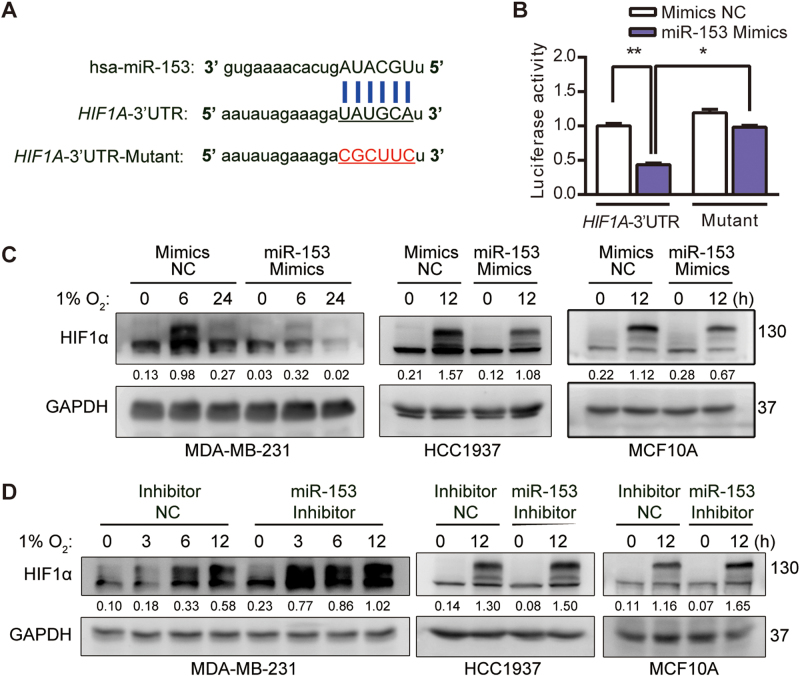
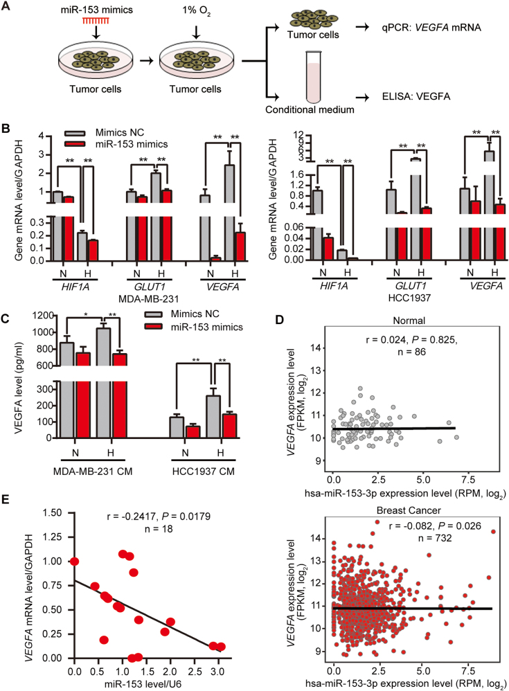
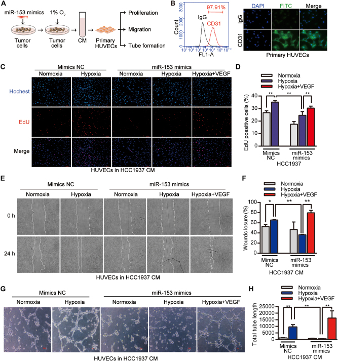
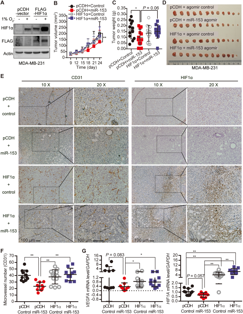
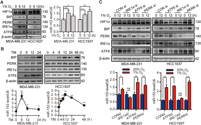
It is well documented that hypoxia activates the hypoxia-inducible factor 1-alpha (HIF1α)/vascular endothelial growth factor A (VEGFA) axis to promote angiogenesis in breast cancer. However, it is unclear how this axis is negatively regulated. In this study, we demonstrated that miR-153 directly inhibits expression of HIF1α by binding to the 3'UTR of HIF1A mRNA, as well as suppresses tube formation of primary human umbilical vein endothelial cells (HUVECs) and breast cancer angiogenesis by decreasing the secretion of VEGFA. Importantly, expression of miR-153 was induced by hypoxia-stimulated ER stress, which activates IRE1α and its downstream transcription factor X-box binding protein 1 (XBP1). X-box binding protein 1 directly binds to the promoter of the miR-153 host gene PTPRN and activates transcription. These results indicate that hypoxia induces miR-153 to fine tune the HIF1α/VEGFA axis in breast cancer angiogenesis and miR-153 could be used for breast cancer anti-angiogenesis therapy.


Conflict of interest statement

The authors declare that they have no competing interests.

Figures

Fig. 1
Fig. 1
miR-153 inhibits the HIF1α protein expression. a The predicted miR-153 binding sequence and the mutant sequence at the 3′-UTR of HIF1A mRNA. b miR-153 suppressed the luciferase activity of the HIF1A-3′-UTR but not the HIF1A-3′-UTR-Mutant. HEK293T cells were transfected with the miR-153 mimics (50 nM) and pMIR-HIF1A-3′-UTR or pMIR-HIF1A-3′-UTR-Mutant (800 ng/ml) together with pCMV-Renilla control (8 ng/ml). Following transfection for 48 h, the cell lysate were collected for a dual-luciferase report assay. *P < 0.05 and **P < 0.01, t-test. c miR-153 mimics (50 nM) inhibited the increase in hypoxia-induced HIF1α protein expression in the MDA-MB-231, HCC1937, and MCF10A cell lines. The HIF1α protein level was detected by using western blot analysis. d The inhibitor of miR-153 (50 nM) increased expression of HIF1α induced by hypoxia in the MDA-MB-231, HCC1937, and MCF10A cell lines. The HIF1α protein level was detected by using western blot analysis
Fig. 2
Fig. 2
miR-153 inactivates the HIF1α/VEGFA axis in breast cancer. a Schematic illustration of the sample treatment, collection, and detection. MDA-MB-231 and HCC1937 cell lines were transfected with miR-153 mimics (50 nM) for 48 h and were then exposed to 1% O2 for 24 h. The cells were collected for real-time PCR to detect the HIF1A, GLUT1, and VEGFA mRNA levels. The conditional media (CM) were harvested for the enzyme-linked immunosorbent assay (ELISA) to detect the VEGFA protein levels. b The miR-153 mimics decreased the mRNA levels of HIF1A and inhibited the transcription of GLUT1 and VEGFA under hypoxia. 'N' means normoxia, and 'H' means hypoxia. **P < 0.01, t-test. c The miR-153 mimics inhibited the secreted VEGFA protein levels from the hypoxia-treated breast cancer cells. *P < 0.05, **P < 0.01, t-test. d There is a negative correlation trend between miR-153 and VEGFA mRNA in 732 breast cancer patients from the TCGA database. e There is a negative correlation trend between miR-153 and VEGFA mRNA in 18 clinical breast cancer samples
Fig. 3
Fig. 3
miR-153 suppresses the proliferation, migration, and tube formation of primary HUVECs in a paracrine manner. a Schematic illustration of the sample treatment and collection. The breast cancer cells were transfected with miR-153 mimics (50 nM) for 48 h and were then exposed to 1% O2 for 24 h. The conditional medium (CM) was harvested for culturing the primary HUVECs. b CD31 expression of primary HUVECs was detected using flow cytometry and immunofluorescence assays. The representative images for immunofluorescence are shown. c The DNA synthesis of primary HUVECs was inhibited by CM collected from hypoxia-treated HCC1937 cells transfected with miR-153 mimics. The exogenous VEGFA (20 ng/ml) could rescue the phenotype. DNA synthesis was detected by using the Click-iTTM EdU Alexa Fluor® 647 Imaging Kit. Representative images are shown. d The quantitative results of c. **P < 0.01, t-test. e The migration of primary HUVECs was inhibited by CM collected from hypoxia-treated HCC1937 cells transfected with miR-153 mimics in the wound-healing assay. The exogenous VEGFA (20 ng/ml) could rescue the phenotype. Representative images are shown. f The quantitative results of e. *P < 0.05 and **P < 0.01, t-test. g The tube formation of primary HUVECs was inhibited by CM collected from hypoxia-treated HCC1937 cells transfected with miR-153 mimics. The exogenous VEGFA (20 ng/ml) rescued the phenotype. Representative images are shown. Scale bar, 50 μm. h The quantitative results of g. **P < 0.01, t-test
Fig. 4
Fig. 4
miR-153 inhibits the tumor angiogenesis by downregulating the HIF1α/VEGFA axis. a Overexpression of HIF1α in the MDA-MB-231 cell lines, as detected by western blot. b–d The miR-153 agomir suppressed the growth of the MDA-MB-231-pCDH-vector tumors but not that of the MDA-MB-231-pCDH-HIF1α tumors in a nude mouse xenograft model. Tumor growth was measured every 3 days. All tumors were collected and weighed on the last day of the experiment. Data were represented as the mean ± s.d. of six mice for each group (12 tumors). *P < 0.05, **P < 0.01, t-test. e, f miR-153 decreased the number of microvessels, as measured by the CD31 immunohistochemical staining, in the MDA-MB-231-pCDH-vector xenograft tumors, but not in the MDA-MB-231-pCDH-HIF1α xenograft tumors. Represented images are shown. Scale bar, 100 μm. **P < 0.01, t-test. g miR-153 decreased the expression levels of HIF1A and VEGFA at the mRNA level in the MDA-MB-231-pCDH-vector xenograft tumors, but not in the MDA-MB-231-pCDH-HIF1α xenograft tumors, as detected using RT-qPCR. *P < 0.05, **P < 0.01, t-test
Fig. 5
Fig. 5
Hypoxia upregulates miR-153 expression by triggering ER stress protein IRE1α. a Hypoxia (1% O2) triggered ER stress and upregulated miR-153 expression in MDA-MB-231 and HCC1937 cell lines. The HIF1α, BIP, PERK, IRE1α, and ATF6 protein levels were detected by western blot analysis, and the miR-153 level was detected by the real-time polymerase chain reaction. *P < 0.05, **P < 0.01, t-test. b Tunicamycin (TM, 5 μg/ml) upregulated miR-153 expression in MDA-MB-231 and HCC1937 cell lines. The BIP, PERK, IRE1α, and ATF6 protein levels were detected by western blot analysis, and the miR-153 level was detected by RT-qPCR. *P < 0.05, **P < 0.01, t-test, compared with the value of the TM 0-h treatment. c Knockdown of IRE1α specifically blocked hypoxia-induced miR-153 expression in MDA-MB-231 and HCC1937 cell lines. The HIF1α, BIP, PERK, IRE1α, and ATF6 protein levels were detected by western blot analysis, and the miR-153 level was detected by the real-time polymerase chain reaction. *P < 0.05, **P < 0.01, t-test
Fig. 6
Fig. 6
X-box binding protein 1 induces miR-153 expression by binding to the PTPRN promoter. a Knockdown of XBP1 inhibited the upregulation of miR-153 induced by hypoxia in the MDA-MB-231 and HCC1937 cell lines. **P < 0.01, t-test, compared with the value of CONsi with the 1% O2 0-h treatment. b Knockdown of XBP1 inhibited the upregulation of miR-153 induced by TM in MDA-MB-231 and the HCC1937 cell lines. **P < 0.01, t-test, compared with the value of CONsi with the TM 0-h treatment. c Illustration of the miR-153 location in two host genes, PTPRN and PTPRN2. The XBP1 binding sites in the promoter region of these two host genes are indicated. d Hypoxia induces expression of miR-153 and PTPRN, but not PTPRN2, at the mRNA level in the MCF10A, MDA-MB-231, and HCC1937 cell lines, as detected by RT-qPCR. e Hypoxia induced PTPRN protein expression, as detected by western blot analysis. f Hypoxia-induced XBP1 bound to the promoter of PTPRN, as determined by the chromatin immunoprecipitation (ChIP) assay. g The PTPRN promoter containing the XBP1 binding motif was significantly activated by hypoxia in HEK293T cells, as determined by dual-luciferase assay. **P < 0.01, t-test. h The work model of this study

Similar articles

Cited by

References

    1. Folkman J. How is blood vessel growth regulated in normal and neoplastic tissue? G.H.A. Clowes memorial Award lecture. Cancer Res. 1986;46:467–73. - PubMed
    1. Urra H, Hetz C. A novel ER stress-independent function of the UPR in angiogenesis. Mol Cell. 2014;54:542–4. doi: 10.1016/j.molcel.2014.05.013. - DOI - PubMed
    1. Albini A, Indraccolo S, Noonan DM, Pfeffer U. Functional genomics of endothelial cells treated with anti-angiogenic or angiopreventive drugs. Clin Exp Metastas- 2010;27:419–39. doi: 10.1007/s10585-010-9312-5. - DOI - PubMed
    1. Senger DR, Galli SJ, Dvorak AM, Perruzzi CA, Harvey VS, Dvorak HF. Tumor cells secrete a vascular permeability factor that promotes accumulation of ascites fluid. Sci (New Y, NY) 1983;219:983–5. doi: 10.1126/science.6823562. - DOI - PubMed
    1. Senger DR, Van de Water L, Brown LF, Nagy JA, Yeo KT, Yeo TK, et al. Vascular permeability factor (VPF, VEGF) in tumor biology. Cancer Metastas- Rev. 1993;12:303–24. doi: 10.1007/BF00665960. - DOI - PubMed

Publication types

MeSH terms