

Proliferation and differentiation of stem cells in contact with eluate from fibrin-rich plasma membrane
- PMID: 29367906
- PMCID: PMC5771793
- DOI: 10.1016/j.rboe.2017.12.004
Proliferation and differentiation of stem cells in contact with eluate from fibrin-rich plasma membrane
Abstract
Objective: To evaluate the ability of the eluate from fibrin-rich plasma (FRP) membrane to induce proliferation and differentiation of isolated human adipose-derived stem cells (ASCs) into chondrocytes.
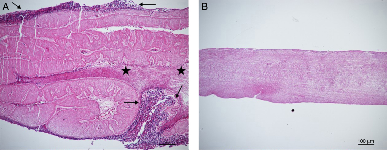
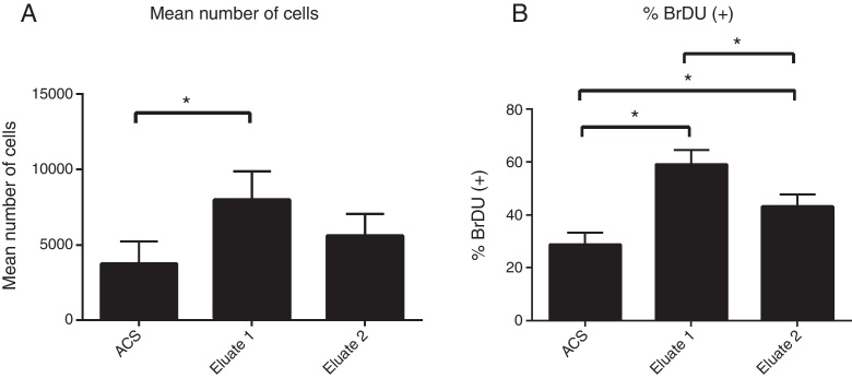
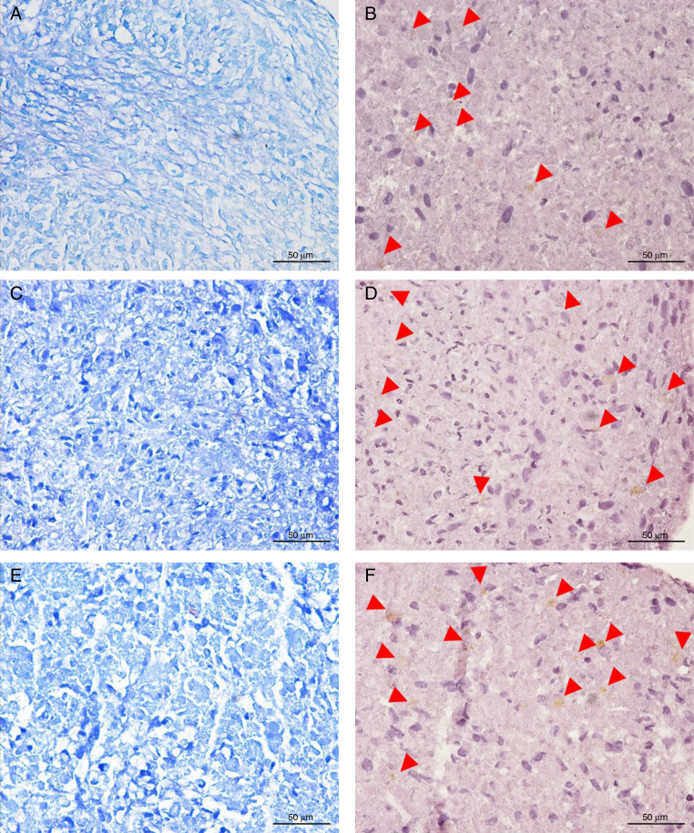
Method: FRP membranes were obtained by centrifugation of peripheral blood from two healthy donors, cut, and maintained in culture plate wells for 48 h to prepare the fibrin eluate. The SCATh were isolated from adipose tissue by collagenase digestion solution, and expanded in vitro. Cells were expanded and treated with DMEM-F12 culture, a commercial media for chondrogenic differentiation, and eluate from FRP membrane for three days, and labeled with BrdU for quantitative assessment of cell proliferation using the High-Content Operetta® imaging system. For the chondrogenic differentiation assay, the SCATh were grown in micromass for 21 days and stained with toluidine blue and aggrecan for qualitative evaluation by light microscopy. The statistical analysis was performed using ANOVA and Tukey's test.
Results: There was a greater proliferation of cells treated with the eluate from FRP membrane compared to the other two treatments, where the ANOVA test showed significance (p < 0.001). The differentiation into chondrocytes was visualized by the presence of mucopolysaccharide in the matrix of the cells marked in blue toluidine and aggrecan.
Conclusions: Treatment with eluate from FRP membrane stimulated cell proliferation and induced differentiation of the stem cells into chondrocytes, suggesting a potential application of FRP membranes in hyaline cartilage regeneration therapies.
Objetivo: Avaliar a capacidade do eluato proveniente da membrana de plasma rico em fibrina (PRF) de induzir proliferação e diferenciação das células-tronco humanas isoladas de tecido adiposo (CTDAh) em condrócitos.
Método: As membranas de PRF foram obtidas por centrifugação de sangue periférico de dois indivíduos saudáveis, cortadas, colocadas em poços de placa de cultivo por 48 h para obtenção do eluato de fibrina. As CTDAh foram isoladas do tecido adiposo por digestão com solução de colagenase e expandidas in vitro. As células foram expandidas e tratadas com meio de cultivo DMEM-F12, meio comercial para diferenciação condrocítica, e eluato de fibrina durante três dias e marcadas com BrdU para avaliação quantitativa da proliferação celular com o uso do sistema de imagens High-Content Operetta®. Para o ensaio de diferenciação condrogênica, as CTDAh foram cultivadas em micromassa por 21 dias e coradas com azul de toluidina e agrecana para avaliação qualitativa em microscópio óptico. As avaliações estatísticas foram feitas por meio dos testes Anova e Tukey.
Resultados: Houve uma maior proliferação das células tratadas com o eluato de fibrina comparativamente com os outros dois tratamentos, nos quais o teste Anova apontou significância (p < 0,001). A diferenciação em condrócitos foi visualizada pela presença de mucopolissacarídeos na matriz das células tratadas com meio de diferenciação ou eluato e marcação positiva para agrecana.
Conclusões: O tratamento com o eluato da membrana de fibrina estimulou a proliferação celular e induziu a diferenciação das células-tronco em condrócitos, o que sugere uma potencial aplicação da membrana de PRF nas terapias de regeneração de cartilagem hialina.
Keywords: Cartilage; Membranes; Platelet-rich plasma; Regeneration.
Figures
Similar articles
-
Functional tissue-engineered microtissue derived from cartilage extracellular matrix for articular cartilage regeneration.Acta Biomater. 2018 Sep 1;77:127-141. doi: 10.1016/j.actbio.2018.07.031. Epub 2018 Jul 18. Acta Biomater. 2018. PMID: 30030172
-
Human platelet lysate successfully promotes proliferation and subsequent chondrogenic differentiation of adipose-derived stem cells: a comparison with articular chondrocytes.J Tissue Eng Regen Med. 2015 Jul;9(7):808-18. doi: 10.1002/term.1649. Epub 2013 Jan 9. J Tissue Eng Regen Med. 2015. PMID: 23303715
-
Assessment of chondrogenic differentiation potential of autologous activated peripheral blood stem cells on human early osteoarthritic cancellous tibial bone scaffold.Musculoskelet Surg. 2014 Jun;98(1):35-43. doi: 10.1007/s12306-013-0303-y. Epub 2013 Nov 1. Musculoskelet Surg. 2014. PMID: 24178764
-
Centrifugal gravity-induced BMP4 induces chondrogenic differentiation of adipose-derived stem cells via SOX9 upregulation.Stem Cell Res Ther. 2016 Dec 8;7(1):184. doi: 10.1186/s13287-016-0445-6. Stem Cell Res Ther. 2016. PMID: 27931264 Free PMC article.
-
Regeneration of articular cartilage by adipose tissue derived mesenchymal stem cells: perspectives from stem cell biology and molecular medicine.J Cell Physiol. 2013 May;228(5):938-44. doi: 10.1002/jcp.24255. J Cell Physiol. 2013. PMID: 23042088 Review.
Cited by
-
Platelet-Rich Fibrin Scaffolds for Cartilage and Tendon Regenerative Medicine: From Bench to Bedside.Int J Mol Sci. 2019 Apr 5;20(7):1701. doi: 10.3390/ijms20071701. Int J Mol Sci. 2019. PMID: 30959772 Free PMC article. Review.
-
Evolution and Clinical Advances of Platelet-Rich Fibrin in Musculoskeletal Regeneration.Bioengineering (Basel). 2023 Jan 3;10(1):58. doi: 10.3390/bioengineering10010058. Bioengineering (Basel). 2023. PMID: 36671630 Free PMC article. Review.
-
Therapeutic Potential of Dental Pulp Stem Cells and Leukocyte- and Platelet-Rich Fibrin for Osteoarthritis.Cells. 2020 Apr 15;9(4):980. doi: 10.3390/cells9040980. Cells. 2020. PMID: 32326610 Free PMC article.
References
-
- Fuller R. Osteoartrose. In: Greve J.M.D.A., editor. Tratado de medicina de reabilitação. Roca; São Paulo: 2007. pp. 889–904.
-
- Pei M., He F., Li J., Tidwell J.E., Jones A.C., McDonough E.B. Repair of large animal partial-thickness cartilage defects through intraarticular injection of matrix-rejuvenated synovium-derived stem cells. Tissue Eng Part A. 2013;19(9–10):1144–1154. - PubMed
LinkOut - more resources
Full Text Sources
Other Literature Sources
Miscellaneous