

doi: 10.21769/BioProtoc.2652.
Ex vivo Trophoblast-specific Genetic Manipulation Using Lentiviral Delivery
Affiliations
- PMID: 29367940
- PMCID: PMC5777579
- DOI: 10.21769/BioProtoc.2652
Item in Clipboard
Ex vivo Trophoblast-specific Genetic Manipulation Using Lentiviral Delivery
Bio Protoc.
.
Abstract
In this protocol report, we describe a lentiviral gene delivery technique for genetic modification of the rat trophoblast cell lineage. Lentiviral packaged gene constructs can be efficiently and specifically delivered to the trophoblast cell lineage of the blastocyst. The consequences of 'gain-of-function' and 'loss-of-function' blastocyst manipulations can be evaluated with in vitro outgrowth assays or following transfer to pseudopregnant rats.
Keywords: Blastocyst; Lentiviral vector; Placenta; Trophoblast.
Conflict of interest statement
Authors declare no conflict of interest or competing interests.
Figures
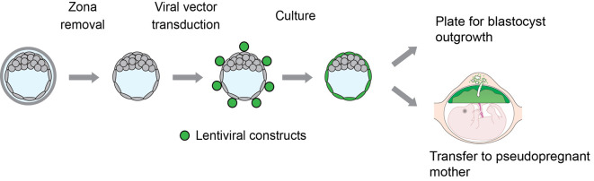
Blastocysts are transduced with lentiviral particles (shown in green) expressing specific ‘gain-of-function’ or ‘loss-of-function’ constructs and cultured for 72 h for outgrowth assays or directly transferred to day 3.5 pseudopregnant animals for in vivo analyses.
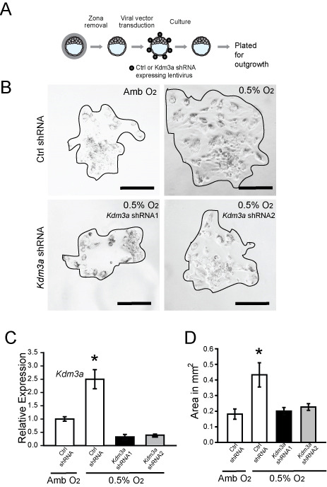
A. Schematic showing experimental plan for lentiviral transduction of blastocysts and outgrowth assay. Blastocysts were transduced with control (Ctrl) or Kdm3a shRNA and cultured for 72 h to allow hatching from the zona pellucida. The attached blastocysts were exposed to ambient (Amb) or low oxygen (0.5% O2) for 24 h and analyzed. B. Representative images of blastocyst outgrowths from Ctrl shRNA and exposed to Amb, Ctrl shRNA and exposed to 0.5% O2, and Kdm3a shRNAs and exposed to 0.5% O2. C. Measurement of Kdm3a transcripts in control and knockdown cultures was measured by qRT-PCR. Asterisks indicate significant differences among groups (n = 6/group; *P < 0.05). D. The bar graph shows quantification of outgrowth area in square millimeters. The area of the outgrowth was measured using ImageJ software (Ctrl shRNA + Amb, n = 6; Ctrl shRNA + 0.5% O2, Kdm3a shRNA1 + 0.5% O2, n = 10; Kdm3a shRNA2 + 0.5% O2, n = 10; *P < 0.05). Data presented in C and D were analyzed with ANOVA and Student-Newman-Keuls test. This figure appeared in
Chakraborty et al. (2016)
.
References
-
- Ain R., Canham L. N. and Soares M. J.(2003). Gestation stage-dependent intrauterine trophoblast cell invasion in the rat and mouse: novel endocrine phenotype and regulation. Dev Biol 260(1): 176-190. - PubMed
-
- Ain R., Konno T., Canham L. N. and Soares M. J.(2006). Phenotypic analysis of the rat placenta. Methods Mol Med 121: 295-313. - PubMed
-
- Chakraborty D., Cui W., Rosario G. X., Scott R. L., Dhakal P., Renaud S. J., Tachibana M., Rumi M. A., Mason C. W., Krieg A. J. and Soares M. J.(2016). HIF-KDM3A-MMP12 regulatory circuit ensures trophoblast plasticity and placental adaptations to hypoxia. Proc Natl Acad Sci U S A 113(46): E7212-E7221. - PMC - PubMed
Grants and funding
LinkOut - more resources
Full Text Sources
Other Literature Sources
Research Materials
