. 2018 Jul;75(14):2613-2625.
doi: 10.1007/s00018-018-2752-9. Epub 2018 Jan 24.

Analysis of novel endosome-to-Golgi retrieval genes reveals a role for PLD3 in regulating endosomal protein sorting and amyloid precursor protein processing

Affiliations

Analysis of novel endosome-to-Golgi retrieval genes reveals a role for PLD3 in regulating endosomal protein sorting and amyloid precursor protein processing

Aamir S Mukadam et al. Cell Mol Life Sci. 2018 Jul.

Abstract

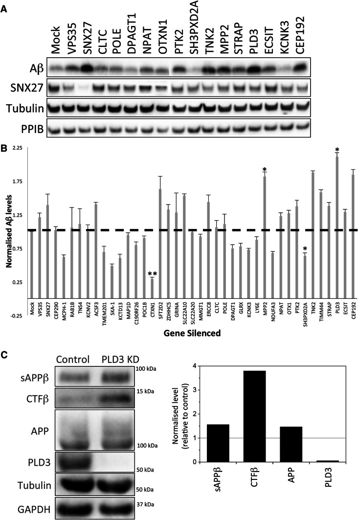
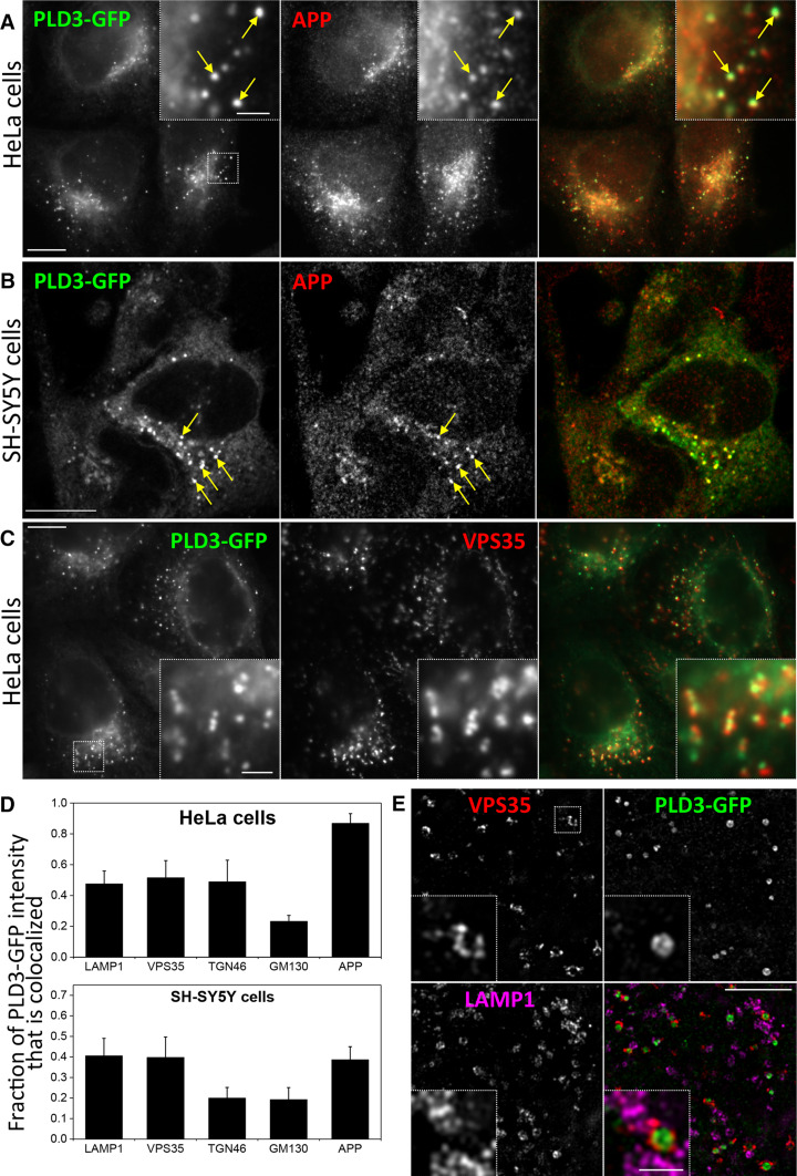
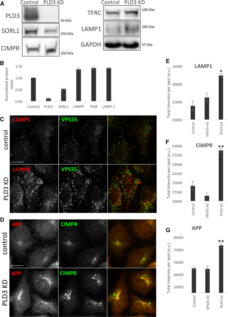
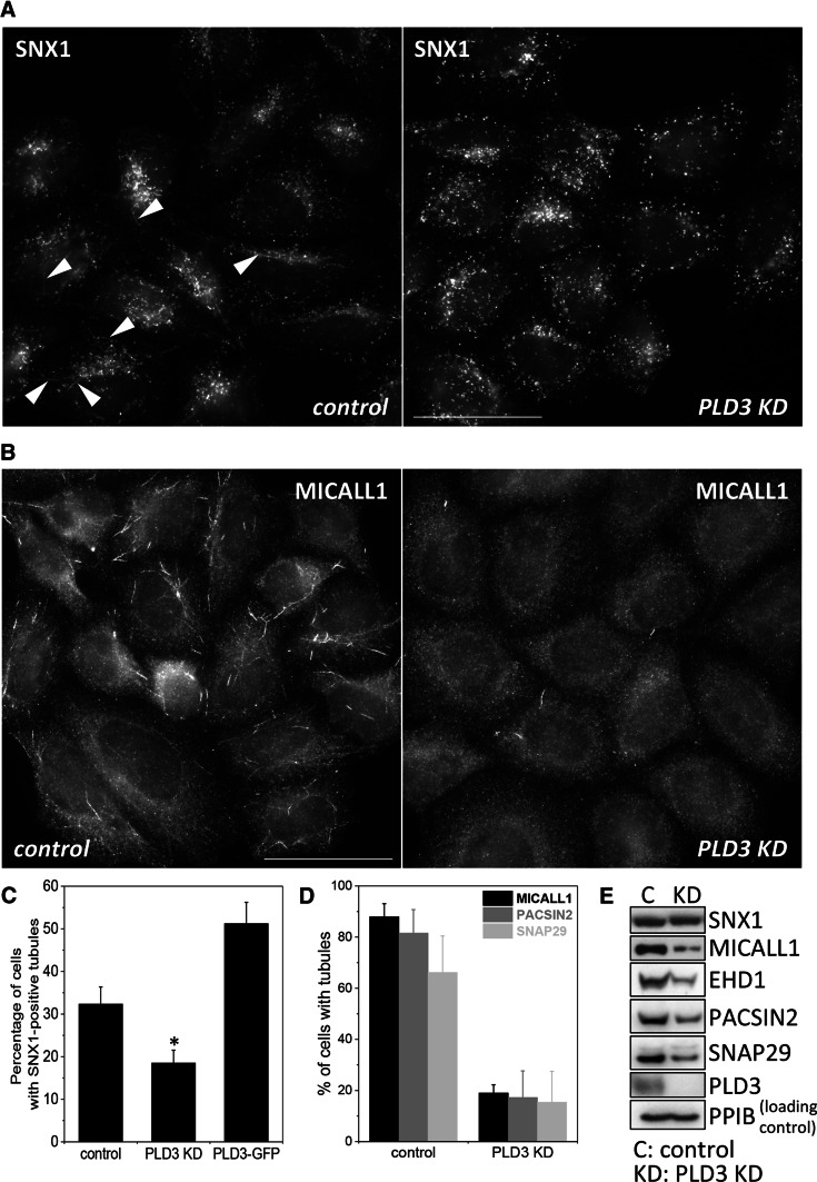
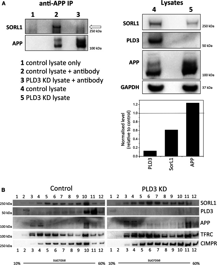
The processing of amyloid precursor protein (APP) to the neurotoxic pro-aggregatory Aβ peptide is controlled by the mechanisms that govern the trafficking and localisation of APP. We hypothesised that genes involved in endosomal protein sorting could play an important role in regulating APP processing and, therefore, analysed ~ 40 novel endosome-to-Golgi retrieval genes previously identified in a genome-wide siRNA screen. We report that phospholipase D3 (PLD3), a type II membrane protein, functions in endosomal protein sorting and plays an important role in regulating APP processing. PLD3 co-localises with APP in endosomes and loss of PLD3 function results in reduced endosomal tubules, impaired trafficking of several membrane proteins and reduced association of sortilin-like 1 with APP.

Keywords: Alzheimer disease; Amyloid precursor protein; Endosome; Phospholipase D; SorL1.


Figures

Fig. 1
Fig. 1
PLD3 knockdown increases secreted Aβ levels. a Cell culture supernatants were collected from control or siRNA-treated HEK293 cells stably expressing swAPP and analysed by Western blotting for Aβ. Corresponding cell lysates were also analysed by Western blotting for SNX27, tubulin, PPIB and GAPDH (not shown). b Quantitation of Aβ levels detected by Western blotting as in a and normalised to the three loading controls. Error bars indicate SD of three experiments. Statistical significance was determined by Student’s t test in Microsoft Excel (**P < 0.01, *P < 0.05). c Additional Western blotting of APP, Aβ and CTFβ levels in lysates from control and PLD3 knockdown cells. Tubulin and GAPDH are loading controls. The band intensities have been quantified and are shown in graphical form next to the blot
Fig. 2
Fig. 2
PLD3–GFP co-localises with APP in the endo-lysosomal system. HeLa cells (a) or SH-SY5Y (b) cells stably expressing PLD3–GFP were fixed and stained for GFP and APP and imaged using full-field (a) or confocal (b) microscopy. Colocalisation was observed and is highlighted in the enlarged inset areas. c HeLa cells stably expressing PLD3–GFP were fixed and stained for GFP and VPS35 and imaged using full-field microscopy. Colocalisation was observed and is highlighted in the enlarged inset areas. d The colocalisation of PLD3–GFP with markers of the Golgi and post-Golgi endo/lysosomal system was quantified using the M1 coefficient of colocalisation. e The localisation of PLD3–GFP was analysed in detail by structured illumination microscopy. A single 110-nm-thick section is shown. GFP signal is observed on the lumenal side of VPS35 endosomes, consistent with the predicted type II membrane topology of PLD3–GFP. Scale bars ac 10 μm, insets 2 μm, e 5 μm, inset 1 μm
Fig. 3
Fig. 3
Silencing of PLD3 perturbs endosomal protein sorting. a Control SH-SY5Y cells and cells treated with PLD3 siRNA were lysed and analysed by Western blotting. Levels of SorL1 appear reduced whilst other proteins such as the transferrin receptor (TFRC) are modestly increased. b Western blot data from three independent experiments are shown graphically. c, d Control and PLD3-silenced HeLa cells were fixed and stained for various endosomal proteins, lysosomal markers and transmembrane proteins. Scale bars = 20 μm. Quantitation of the changes in immunofluorescent localization of LAMP1 (e), CIMPR (f) and APP (g) upon PLD3 knockdown using automated microscopy. Statistical significance was determined by Student’s t test in Microsoft Excel (**P < 0.01, *P < 0.05)
Fig. 4
Fig. 4
Loss of PLD3 impairs endosomal tubule formation or stability. a HeLa cells were treated with siRNA to abolish PLD3 expression and then fixed and labelled with anti-Snx1. Arrow heads indicate tubules that were rarely observed in PLD3 knockdown cells. Scale bar = 50 μm. b As in a, but cells were labelled with antibodies against the MICALL1 protein. Scale bar = 50 μm. c Snx1-decorated endosomal tubules were imaged in control HeLa cells, PLD3 siRNA-treated HeLa cells and HeLa cells stably expressing PLD3–GFP. Tubules were quantified. The results of two independent experiments are shown (average ± SD) with more than 50 cells counted in each condition in each experiment. Statistical significance was determined by Student’s t test in Microsoft Excel (*P < 0.05). d MICALL1 tubules were counted in three independent experiments, scoring more than 75 cells each time for each condition. Tubules that were PACSIN2- or SNAP29 positive were also counted in more than 100 cells and the data presented graphically. e Cell lysates were analysed by Western blotting. Loss of PLD3 expression does not affect SNX1 levels but does result in reduced levels of MICALL1 and the associated proteins (i.e. EHD1, PACSIN2 and SNAP29)
Fig. 5
Fig. 5
Silencing of PLD3 perturbs SorL1 association with APP and alters SorL1 subcellular distribution. a Control or PLD3 siRNA-treated SH-SY5Y cells were lysed and treated with monoclonal anti-APP antibody to immunoprecipitate (IP) APP. Lysates (right panel) and co-immunoprecipitated proteins (left panel) were analysed by Western blotting, the SorL1 protein is indicated by an arrow. The band intensities have been quantified and are shown in graphical form next to the blot. b Control and PLD3-silenced SH-SY5Y cells were lysed and subjected to centrifugation on a 10–60% sucrose gradient. Fractions (1–12) were collected and analysed by Western blotting. The fractionation profile of SorL1 is markedly altered upon PLD3 knockdown

References

    1. Takahashi RH, Nagao T, Gouras GK. Plaque formation and the intraneuronal accumulation of β-amyloid in Alzheimer’s disease. Pathol Int. 2017;67(4):185–193. doi: 10.1111/pin.12520. - DOI - PubMed
    1. Toh WH, Gleeson PA. Dysregulation of intracellular trafficking and endosomal sorting in Alzheimer’s disease: controversies and unanswered questions. Biochem J. 2016;473(14):1977–1993. doi: 10.1042/BCJ20160147. - DOI - PubMed
    1. Rajendran L, Annaert W. Membrane trafficking pathways in Alzheimer’s disease. Traffic. 2012;13(6):759–770. doi: 10.1111/j.1600-0854.2012.01332.x. - DOI - PubMed
    1. Rajendran L, Schneider A, Schlechtingen G, Weidlich S, Ries J, Braxmeier T, Schwille P, Schulz JB, Schroeder C, Simons M, Jennings G, Knölker HJ, Simons K. Efficient inhibition of the Alzheimer’s disease beta-secretase by membrane targeting. Science. 2008;320(5875):520–523. doi: 10.1126/science.1156609. - DOI - PubMed
    1. Small SA, Gandy S. Sorting through the cell biology of Alzheimer’s disease: intracellular pathways to pathogenesis. Neuron. 2006;52(1):15–31. doi: 10.1016/j.neuron.2006.09.001. - DOI - PMC - PubMed

Publication types

LinkOut - more resources