

Bilateral muscular slips between superior and inferior rectus muscles: case report with discussion on classification of accessory rectus muscles within the orbit
- PMID: 29368252
- PMCID: PMC5996005
- DOI: 10.1007/s00276-018-1976-6
Bilateral muscular slips between superior and inferior rectus muscles: case report with discussion on classification of accessory rectus muscles within the orbit
Abstract
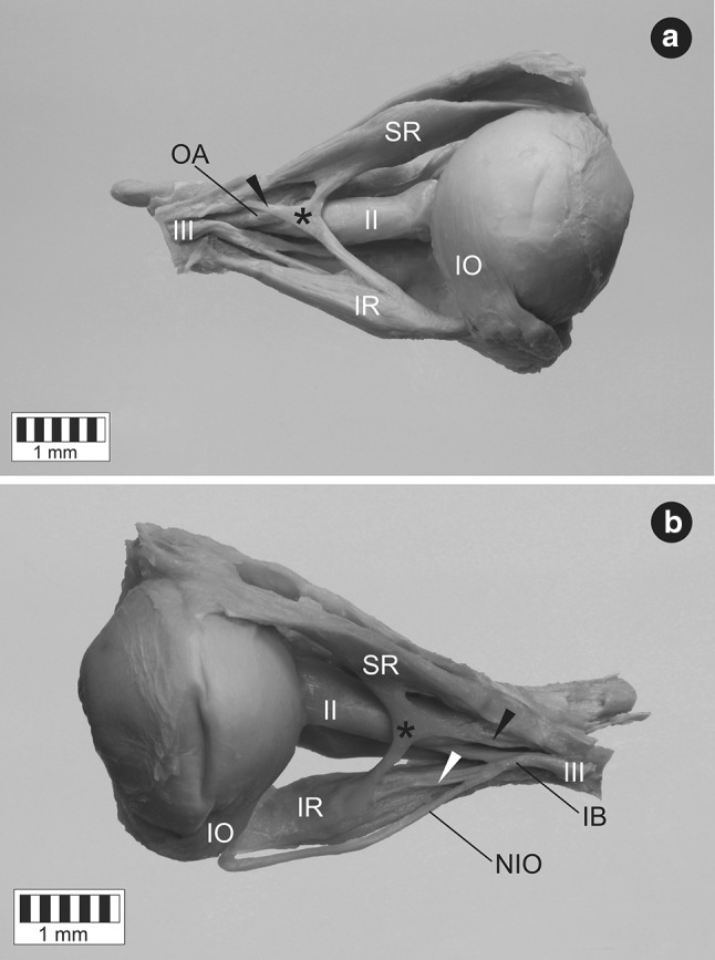
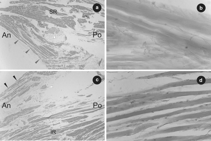
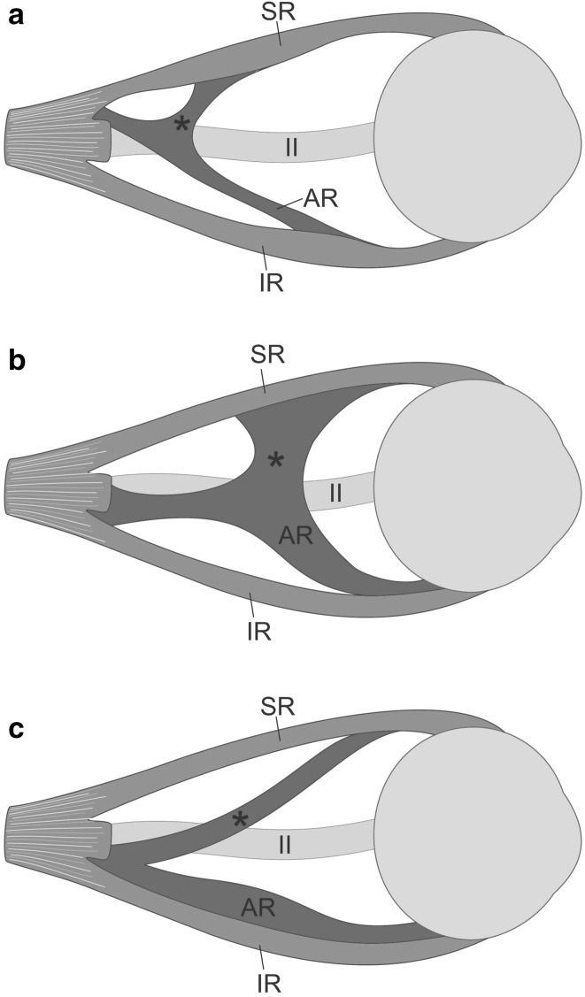
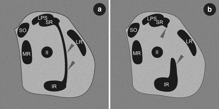
Accessory rectus muscles have rarely been reported as muscular 'bands' or 'slips' originating from the common tendinous ring (annulus of Zinn) and inserting in atypical location. This group of muscles is innervated by the inferior branch of the oculomotor nerve, lies on lateral side of the optic nerve and inserts in rectus muscles. Since there are only few descriptions of such unusual findings in the medical literature, the anatomical data on accessory rectus muscles is limited. Furthermore, existing reports vary in terms of studied objects (cadavers or living subjects), medical history (absence or presence of ocular movement disorders or eye movement abnormalities) and details of anatomical description. This report complements earlier publications and provides complete anatomical description of the accessory rectus muscle observed bilaterally during the dissection of a 68-year-old male cadaver with no eye movement abnormalities reported in the medical history. The accessory rectus muscle was divided into two 'slips' or 'heads'-superior and inferior-running in the sagittal plane (laterally to the optic nerve and the main trunk of the ophthalmic artery) and attached to the superior and inferior rectus muscles. Noticeable thickening of both superior and inferior rectus muscles at the insertion point of the accessory muscle heads was observed only in the sagittal plane. On both sides, the inferior head of the accessory rectus muscle was innervated by one of sub-branches derived from the inferior branch of the oculomotor nerve. No sub-branches to the superior head were macroscopically observed during the dissection. The classification, embryological background and clinical relevance of this variation have been discussed.
Keywords: Anatomic variation; Extraocular muscles; Inferior rectus muscle; Orbit; Superior rectus muscle.
Conflict of interest statement
The authors declare no conflict of interest.
Figures


References
-
- Bergman RA, Afifi AK, Miyauchi R (2015) Illustrated encyclopedia of human anatomic variation: opus I: muscular system: alphabetical listing of muscles. http://www.anatomyatlases.org/AnatomicVariants/MuscularSystem/Text/R/15R.... Accessed 5 Sept 2017
-
- Bohnsack BL, Gallina D, Thompson H, Kasprick DS, Lucarelli MJ, Dootz G, Nelson C, McGonnell IM, Kahana A. Development of extraocular muscles require early signals from periocular neural crest and the developing eye. Arch Ophthalmol. 2011;129:1030–1041. doi: 10.1001/archophthalmol.2011.75. - DOI - PMC - PubMed
-
- Demer JL, Miller JM. Orbital imaging in strabismus surgery. In: Rosenbaum AL, Santiago AP, editors. Clinical strabismus management: principles and techniques. Philadelphia: Saunders; 1999. pp. 84–98.
Publication types
MeSH terms
LinkOut - more resources
Full Text Sources
Other Literature Sources
